










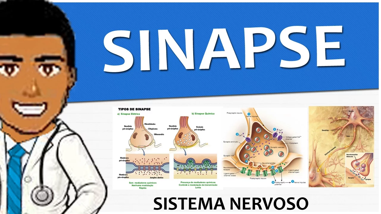











A synapse is the junction where one neuron communicates with another, consisting of a pre-synaptic terminal, synaptic cleft, and post-synaptic terminal; there are two main types—electrical synapses (gap junctions) that allow direct ionic current transfer through connexin channels across a 3nm gap for fast signaling, and chemical synapses that use neurotransmitters released from vesicles in the pre-synaptic terminal to bind to receptors on the post-synaptic terminal, with glutamate serving as the primary excitatory neurotransmitter and GABA as the primary inhibitory neurotransmitter, working together to regulate neural activity and prevent excessive signaling.
Understanding Synapses: Electrical and Chemical Transmission
Added:Basic anatomy of a neuron, including the cell body (soma), dendrites, axon, and axon terminal.
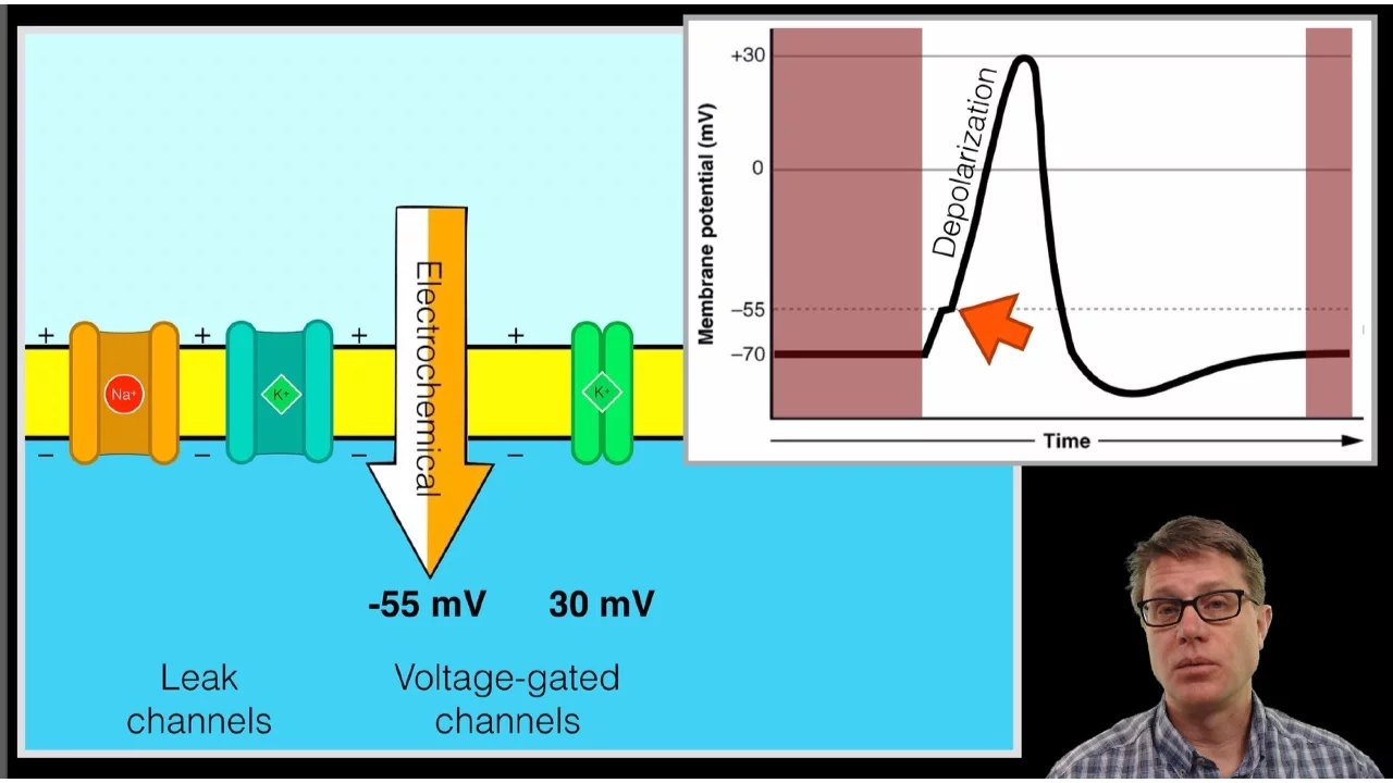
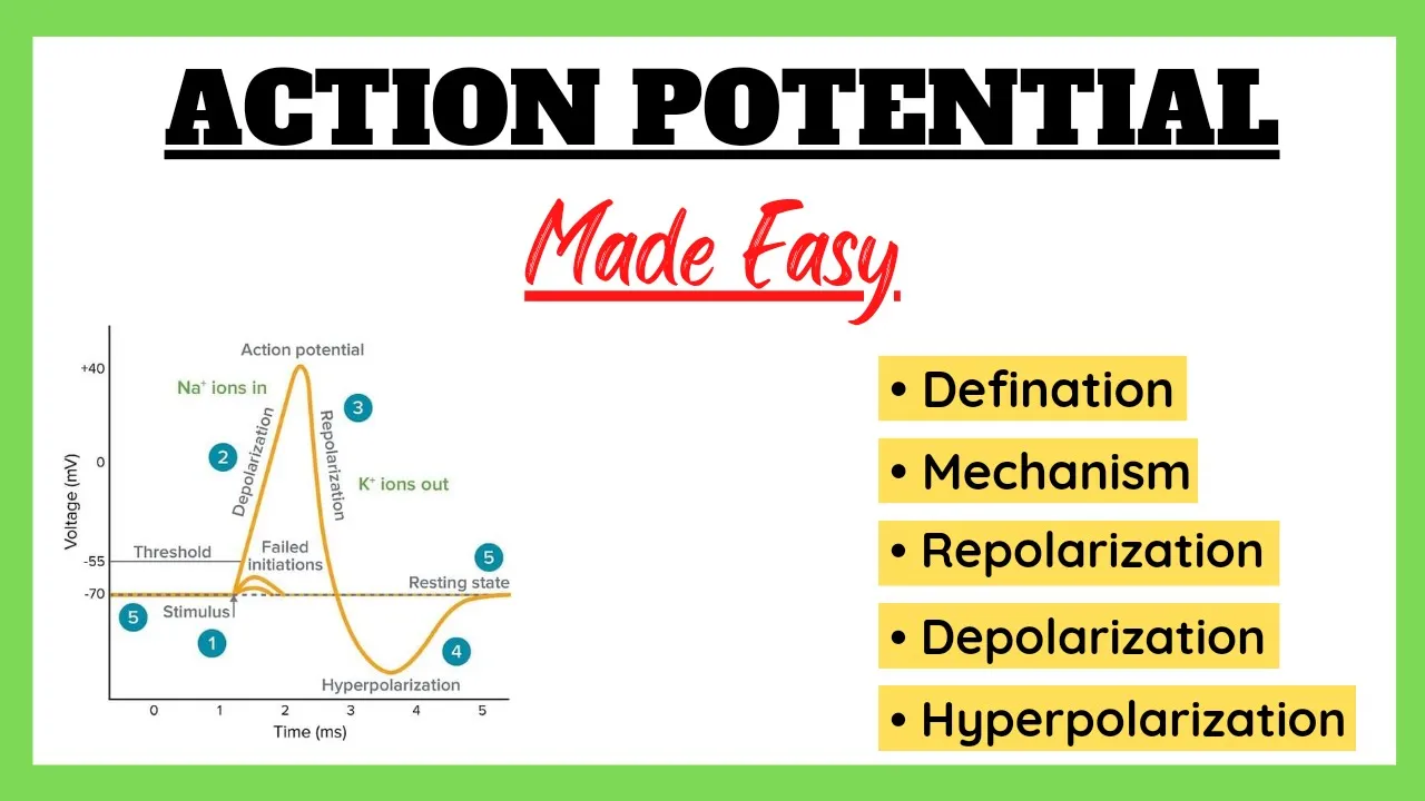
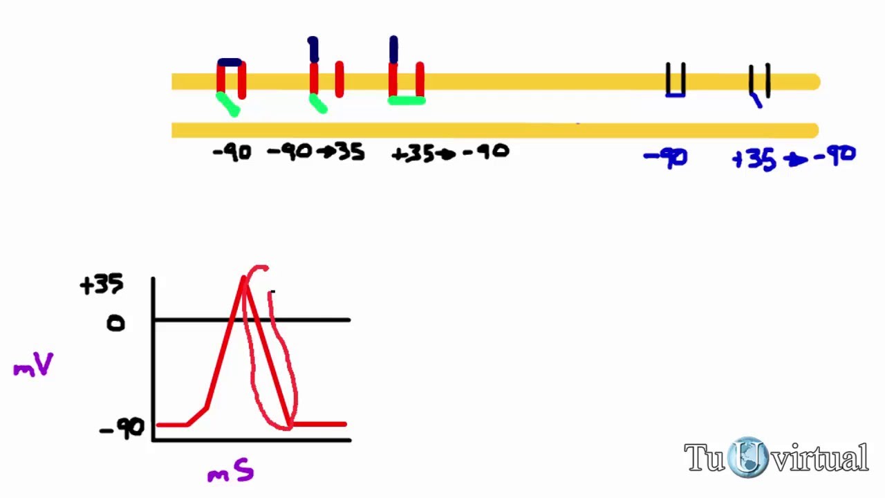
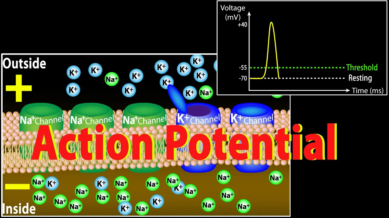
The generation and propagation of an action potential, including depolarization and repolarization phases.
The concept of electrochemical gradients and the function of voltage-gated ion channels.
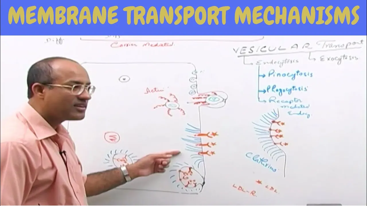
Basic cellular biology principles, specifically cell membrane structure and the concept of exocytosis.
Synaptic integration, specifically how postsynaptic potentials (EPSPs and IPSPs) undergo spatial and temporal summation.
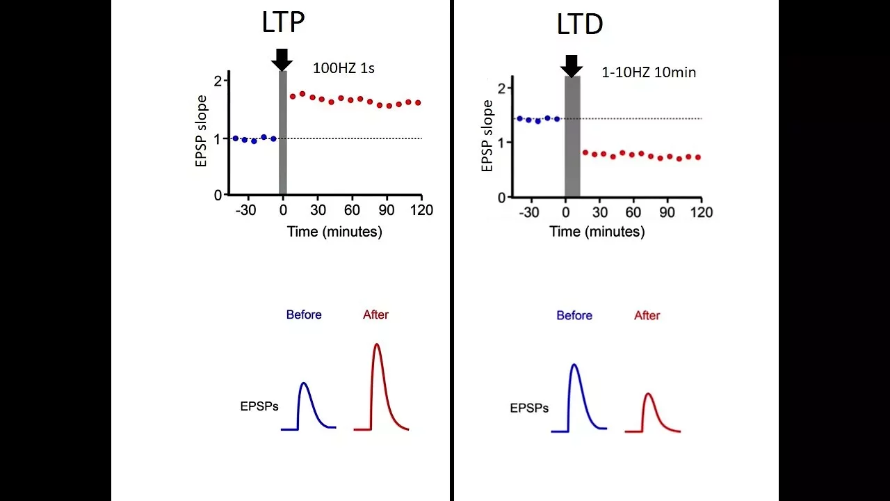
The mechanisms of synaptic plasticity, including Long-Term Potentiation (LTP) and Long-Term Depression (LTD) as the basis for learning and memory.
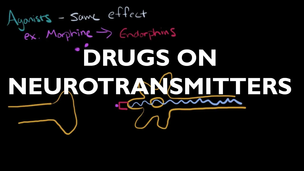
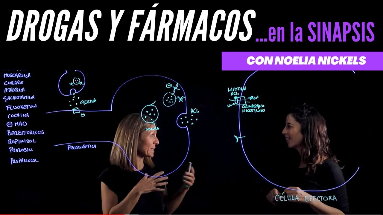
Neuropharmacology, exploring how agonists, antagonists, and reuptake inhibitors modulate synaptic transmission.
The pathophysiology of synaptic diseases (synaptopathies) such as epilepsy, schizophrenia, and Parkinson's disease.
63.8K views1.3Klikes6:48Original Release: 2015-11-15
Hey there! I’m Alie Astrocyte, and you’re tuned in to Neuro Transmissions!
In our last few episodes, we talked about resting potentials and action potentials, the language used by neurons to communicate their signals from one end of the cell to the other.
But how does that message get passed from one neuron to the next?
Each neuron is separated from its neighbors. How do they do it?
Now I get to talk about my favorite part of the neuron - the synapse!
Before we go on, if you haven’t already, you may want to brush up on the anatomy of the neuron with this video.
And you might also want to watch the videos about resting and action potential.
As we discussed last week, your neurons use ions and electrical potential differences to transmit signals from one end to the other.
As that signal reaches the end of the axon, it comes to a structure called the “synaptic terminal”.
This structure has several parts.
The side of the cell sending the signal is called the “pre-synaptic terminal”.
The gap in the middle is called the synaptic cleft.
And finally, on the other cell receiving the signal is the “post-synaptic terminal”.
There are two basic categories of synapses: The electrical synapse (or “gap junction”) and the chemical synapse.
The electrical synapse is the more simple of the two, allowing direct ionic current transfer between cells to pass the action potential across the synaptic cleft.
These types of synapses have very little space between the pre and post synaptic terminals.
About three nanometers.
and use specialized proteins called connexins to form channels that allow the ionic current to pass between cells.
It’s sort of like a skywalk between two buildings.
They're still two distinct buildings, but it’s easy to send a message across to the next building because you can just walk across the bridge. Because the gap is so small, electrical synapses allow cells to respond very quickly to signals, and mean that the cells connected tend to react together.
Because of this, they are important for tasks that require very fast signaling.
Like defensive reflexes in animals.
The other type of synapse, the chemical synapse, is wider and more complex, with slower signalling.
In these synapses, the synaptic cleft can be 20-50 nanometers wide.
Still pretty tiny, but a lot bigger than a gap junction!
Because of this, using ions to directly signal on the next cell isn’t practical.
There's no skybridges connecting these two buildings!
Instead, chemical synapses came up with a different way to send their messages.
Remember the pre-synaptic terminal? Let’s think of it as a cellular post-office.
In the pre-synaptic terminal, you can find these itty bitty spherical structures, called vesicles.
Think of them like envelopes.
These vesicles are little pockets of membrane that hold the information to be transmitted, keeping the message contained until the “post office” of the synapse is told to release the information.
Inside each vesicle are chemicals known as a “neurotransmitters”.
Quite literally, these molecules transmit information between neurons.
These are like the letters contained within the envelope.
As the signal arrives in the pre-synaptic terminal, it tells these vesicles to move to the edge of the cell, where the vesicle membrane fuses with the cell membrane and opens up, dumping its contents into the synaptic cleft.
Once the neurotransmitters reach the post-synaptic terminal, they attach to receptors.
You can think of these receptors like a mailbox.
The neurotransmitter binds to the portion of the receptor that sticks out of the cell membrane, and the information they contain is interpreted by the receptor into some kind of a response.
Sometimes that response means opening an ion channel attached to the receptor itself letting ions flow and changing the electrical potential of the cell, like we talked about in our last video.
Often, the receptor passes the message along to other proteins that rush off into the cell to make other kinds of changes that will affect the electrical potential.
There are many different kinds of neurotransmitters, but two of the most common are glutamate (found at glutamatergic synapses) and gamma-aminobutyric acid, or GABA (found at GABAergic synapses).
These two neurotransmitters usually have opposite effects on the cells they are signaling.
Glutamate is considered “excitatory”, while GABA is considered “inhibitory”.
Let’s talk about each of these separately.
There are two main types of glutamate receptors: the N-methyl-D-aspartate receptor (NMDAR for short) and the α-amino-3-hydroxy-5-methyl-4-isoxazolepropionic acid receptor (or, AMPAR for short.
Thank goodness.
When glutamate binds to an NMDAR or AMPAR, it opens a channel in the cell membrane attached to the receptor, letting positively charged ions move in and out of the cell.
Because these receptors are part of an ion channel, the binding of glutamate results in a very fast response, which lets the cell rapidly depolarize and start transmitting the action potential down to the other end.
This is why they are called “excitatory” synapses.
Because glutamate binding to the receptor excites the cell to spark an action potential, making the post-synaptic cell spike.
Inhibitory synapses have the opposite effect.
The GABA binding to the receptor actually represses signalling, blocking the post-synaptic cell from firing.
GABA can bind to two kinds of receptors.
The GABAa receptor is much like the AMPARs and NMDARs, opening a pore that lets negative ions into the cell, which makes it harder for the cell to fire an action potential, because the inside of the cell becomes even more negative than normal.
GABAb receptors are called “metabotropic” receptors. They’re the kind I mentioned before where the receptor sends off a messenger protein to signal elsewhere in the cell, generally resulting in the same thing: making the inside of the cell more negative, to prevent it from firing a signal.
Hang on. If your brain has to fire action potentials in order to send a signal, why would an inhibitory signal be useful?
Well, it can help prevent your brain from sending too many signals at once.
Too much signaling can be just as bad as too little!
It helps your cells fine-tune their firing.
For example, a cell might receive an excitatory signal followed quickly by an inhibitory signal, so it turns off very quickly, and can’t fire again right away.
So now you have a broad overview of the synapse.
It’s crazy to think that this structure gives your brain the precise tuning and communication it needs to let you walk, talk, breathe, eat, sleep, think, and be who you are while moving through the environment around you.
But it’s true, our brains wouldn’t be much good without the synapse.
Thanks for watching! If you liked this video, give this video a thumbs up and subscribe to become a Brainiac.
Tune in next week as we talk about all of the different major structures of the brain in our episode on neuroanatomy.
Until our next transmission, I’m Alie Astrocyte. Over and out!
Up Next

12:04
Nervous System Anatomy & Physiology Explained | Action Potential & Neurons | Crash Course Biology #26
@crashcourse
2.8M views•2012-07-23

50:12
Bacterial Communication: Quorum Sensing and Biofilm Formation Explained
@JHUAAP
12.6K views•2011-06-28
6:41
The Brain's Waste Removal System: Glymphatic & Lymphatic Discoveries
@neurotransmissions
81.9K views•2017-11-12

7:08
Bacteriophages: Earth's Deadliest Killers and Future Antibiotics
@kurzgesagt
34.6M views•2018-05-13
Related Study Plans & Knowledge Roadmaps
Structured learning paths in Biology
biology
RNAi: Biogenesis, RISC & Gene Silencing
01Molecular Biology Foundations: DNA to Protein
02Introduction to RNA Interference (RNAi)
03Molecular Biogenesis of miRNA and siRNA
+ 2 more learning modules
10h5 modules
Explore Roadmap
biology
Mycorrhizae: Signaling, Biophysics & Ecology
01Foundations of Mycorrhizal Symbiosis
02Molecular Signaling and Root Colonization
03Biophysics and Bioenergetics of Nutrient Exchange
+ 1 more learning modules
11h4 modules
Explore Roadmap
biology
Circadian Clocks: TTFL, SCN & Periphery
01Foundations of Molecular Biology & Gene Expression
02Introduction to Chronobiology and Actograms
03The Core TTFL: Molecular Architecture of the Clock
+ 3 more learning modules
14h6 modules
Explore Roadmap
biology
Population Genetics: Math, Drift & Selection
01Genetic & Probability Foundations
02The Hardy-Weinberg Principle and Baseline Models
03Mathematical Modeling of Natural Selection
+ 3 more learning modules
18h6 modules
Explore Roadmap