

















The dorsal neural tube serves as a dynamic environment where neural crest progenitors sequentially traverse from ventral to dorsal positions before delamination, with fate restriction occurring earlier than previously thought; this process is governed by a molecular network of transcription factors (Snail, Sox9, and FoxD3) that act as a switch between neural/melanocytic fates, with melanocyte specification requiring the downregulation of these neural-promoting factors.
Dorsal Neural Tube Dynamics in Neural Crest Fate Decisions
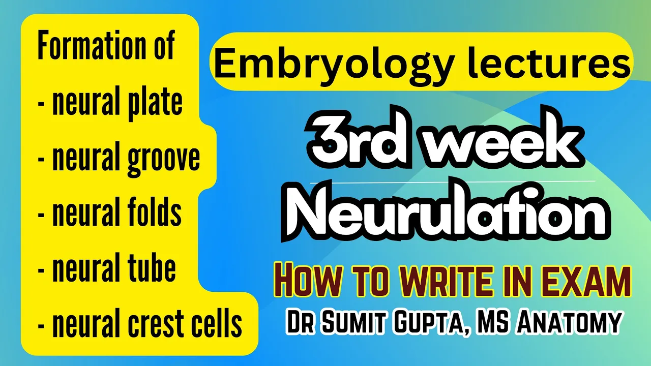
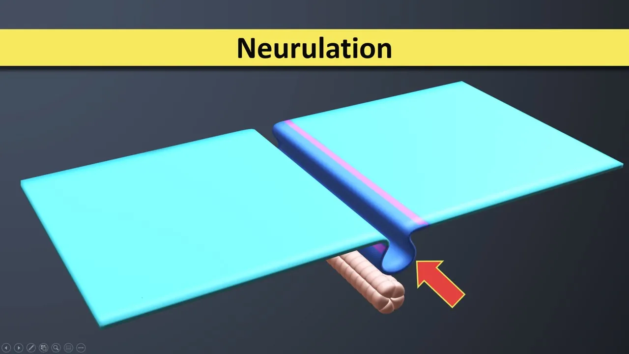
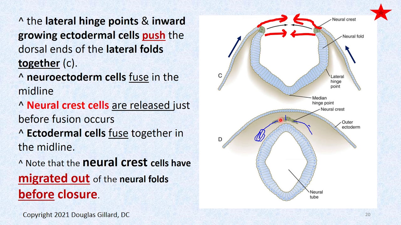
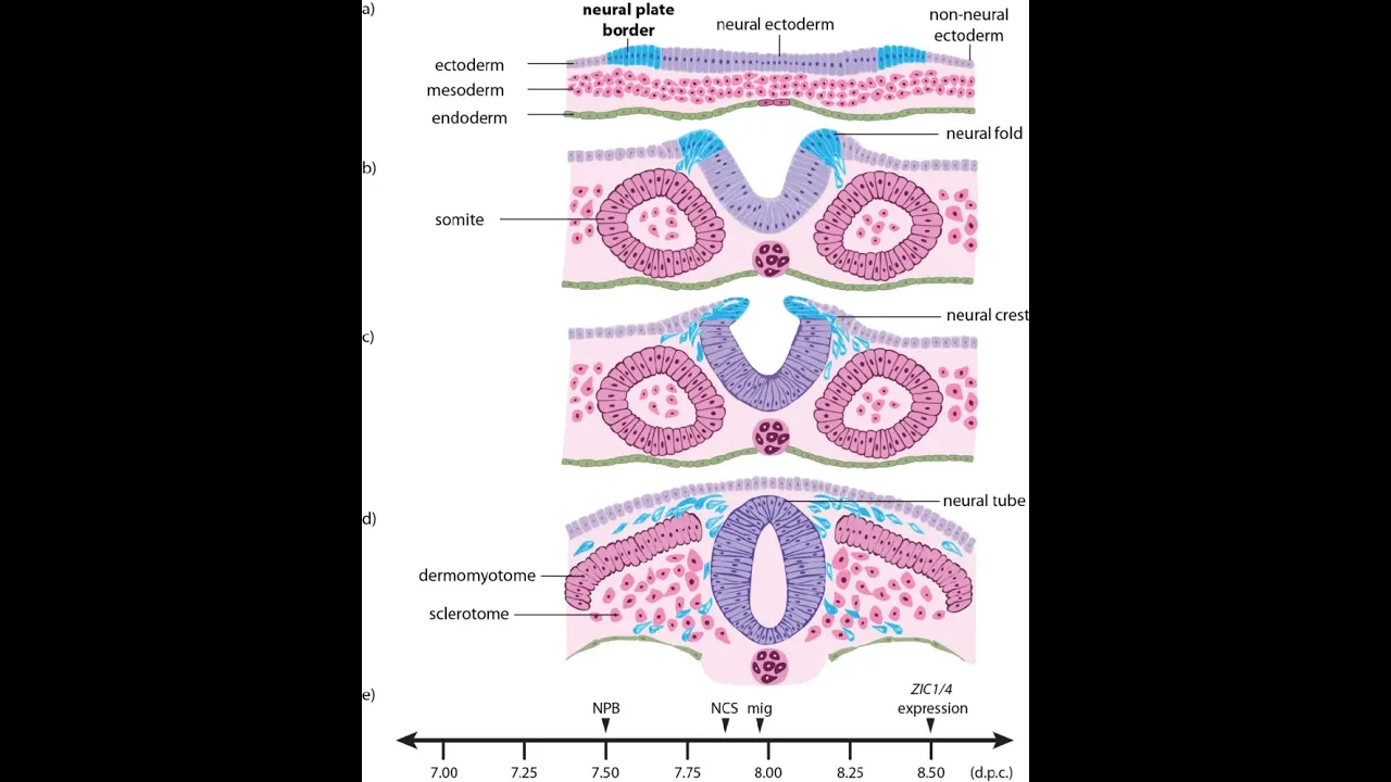
Added:The process of neurulation and the structural organization of the early embryonic neural tube.
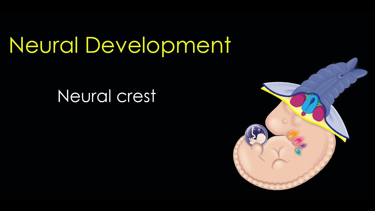
The origin and multipotent nature of neural crest cells at the boundary of the neural plate and ectoderm.
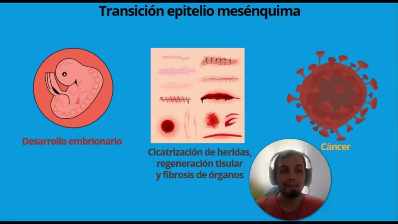
Fundamental mechanisms of Epithelial-to-Mesenchymal Transition (EMT) and cell migration.
Key developmental signaling pathways such as BMP, Wnt, and FGF signaling in ectodermal patterning.
Specific migratory pathways and molecular guidance cues (e.g., ephrins, semaphorins) directing neural crest cells.
Terminal differentiation of neural crest cells into diverse lineages, including melanocytes, craniofacial cartilage, and peripheral neurons.
Pathophysiology of neurocristopathies, which are congenital disorders resulting from abnormal neural crest development (e.g., Hirschsprung's disease).
The evolutionary significance of the neural crest as a defining feature of vertebrate evolution (the 'fourth germ layer').
397 views2likes1:19:38Original Release: 2011-12-29
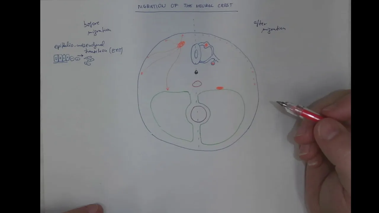
good evening we would like to start today we have K khim who a professor at the department of medical neurobiology and also this is the IM I think Institute for institute for medical research Israel Canada and a member of our new brain Center El who is working at the faculty of medicine since I remember myself I think a little bit later because she's younger and I'm very happy to have you here and she will talk about has a little bit concern about much we know about development so so I ask her to to give more background for those of you are experts please don't expect today the Deep science but more why not speak for yourself I I will try to follow it if you don't follow it don't let us continue try to the doal neural tube Dynamic setting for faith and decision self Faith decision okay so let me let me immediately introduce you into a romantic atmosphere and uh um I thank you for inviting me to H to the Safa Center to tell you a little bit about what we are doing our thoughts and Views can you hear me mikaell I can this is this is the closest I can okay I'll try to speak loud good so um I I'm happy two folds first because this is a center to which I belong as well but second of all because it's I think an opportunity to introduce some of you to some of the concepts and a little bit of data that I'm going to present regarding the development of a very interesting structure in development of the nervous system which is called the neural Cris and uh without any further Ado before entering into more theoretical Concepts regarding developmental neurobiology where we are headed to and what uh interests us let me just uh give you a little bit of a background to those of you who are tabasa in this respect okay so um again just as an introduction uh the entire nervous system in vertebrates uh develops from one of the germ layers that is called the ectoderm and the O and the ectoderm that you can see here under go in the midline of the embryo namely look at your spinal cord and brain this would be your midline under goes a process of specification into a structure that is called the neuroepithelium meaning that this structure now which is thicker than the adjacent exoderm will be spaced to generate exclusively components of the nervous system and not of other systems of the body so there is a certain restriction in the developmental potential of this portion of your ectoderm that from this time on we know that it is going to generate spinal cord brain and also the neural crest about which we are going to talk a lot today so uh in addition to undergoing this process of specification the neural ectoderm which is this structure here under goes a morphogenetic change and all that I'm telling you today is the truth but not all the truth okay because because I'm going to simplify things a little bit so one mode by which the nervous system is formed is by folding off this neuroepithelium and in fact the neural folds come together they approach each other until they close and fuse in the midline to generate something which is very simple a neural tube in fact this is a mean by which many tissues in the embryo are formed and the neural tube or the nervous system is not an extraordinary feature here your gut the entire uh respiratory and digestive system is formed via a very similar uh mechanism namely the formation of a very primitive tube from which many organs are going then cell types and so on and so forth are going to develop later on so what we can see here is a folding neural epithelium as you see this is thicker than this region on both sides this is going to become the exoderm or the epidermis the skin that is going to cover the nervous system and this is going to become the neural tube now I would like from uh This Moment On to uh Point your attention to a structure or to a group of cells which morphologically is completely indistinguishable at this time point from the rest of the epithelium a group of cells that are sitting here in the interface between The Superficial ectoderm faded to become a epidermis and the neural ectoderm that is going to become the nervous system and these are the neural crests now uh once these neural folds come and oppose each other they fuse in the midline the Ecto regenerate you can still see some Scar Tissue here this is scanning electron micrograph and you have a new neural tube which as I mentioned before is going to generate the entire spinal cord neural crest and your brain now also everything looks the same because the neural tube is what we call a pseudo stratified epithelium it's never mind an epithelium with very particular characteristics what we can say is that molecularly speaking there is already a segregation a diversification of some fate within this structure and the neural crest which is our issue today are these particular cell in dark blue which transcribe a factor a transcription factor which is a DNA binding protein in this case named Snail 2 or slug uh in previous years we know of about more than 30 transcription factors and different receptors that are expressed and characterized this particular region of the dorsal neural tube now as you may know the dorsal neural tube after the formation production and as you will see in a minute delamination and exit of these cells which will form the entire peripheral nervous system the dorsal tube forms the roof plate which is which is a an organizing Center very important to uh determine different interneuronal types in the nervous system both brain as well as spinal cord so the dorsal neural tube has to be seen as a kind of very Dam damic structure which changes with time and this is what I'm going to try and convey to you during this talk so just uh without entering obviously into any detail just notice what are the derivatives of the neural crest this is a very very discrete cell population uh which comprises a very few number of cells at the beginning of its ontogeny but later on diversifies both qualitatively as well as quantitatively into an enormous amount of cell types just to mention some of them which are relevant to us obviously neurons and glea of the sensory ganglia the satellite cells within those ganglia the autonomic nervous system which comprises all the neurons and G sympathetic ganglia parasympathetic ganglia the entire innervation of your intestines and the entire Digestive and respiratory system comes from the neural cres so many many neurons and gal cells an additional uh cell of Interest are the Schwan cells lining along peripheral nerves endocrine and Par Endocrine cells of which adreno medary cells the chromatin cells which are responsible for the fight or flight response through caming come from the neural crest pigment cells all the pigment cells of your body except for the retinal pigment are a neural crest derivative interestingly not a neural derivative however uh in malformations or diseases of the neural Christ uh generically called neurocristopathies we can very often see uh the involvement of pigment cells one of them is hung's Disease an neural Mega colon It's the the the distal intestine which is not inated because of a lack of migration of neural crests into the side and in people with hpr's disease in particular it is a an autosomal dominant uh disease in particularly uh in particular of Ashkenazi juice um uh what we can see is also patches of pigmentation in the skin so it is something which um involves both neural derivatives as well as pigment cells another one would be neurofibromatosis which are tumors of Schwan cells Schwan Nomas in which you can see also a very active involvement of pigment cells this is just to illustrate and we'll come back but this is going to be quite a central point in the talk and then something which is evolutionary important and I'm just going to mention it very um in very general terms we know that the entire nervous system derives from ectoderm we also know that the [Music] entire the entire connective tissue the bones of your body and your muscles derive from another layer which is called the mism however however in the head of all the vertebrates it appears that all the bones of your head of your neck of your maxila and mandibula are of neural crest origin something which is called the Mis exoderm a kind of chimera between mism and ectoderm which has an evolutionary significance and we'll come to that in a minute so the message from this slide is not to overwhelm you with data the message is that you take a very very tiny population of embryonic cells and it diversifies enormously to give rise to many many derivatives in the embryo in a very stereotypic Manner and the neural crest is an invention of the vertebrate embryo and therefore it has become during the last decade one of the most interesting and appealing systems to study developmental biology of development neurobiology because one of our main interest in this field is to understand how a homogeneous population of cells during early development is able to segregate into so many different derivatives what are the signals what are what is the nature of the interactions between the cells what is the amount of information that is carried down by the cell sometimes from maternal information sometimes from uh cell intrin processes and what is the amount of information that the cell gets from its environment and how does it integrate all this information to ensure such an output so these are the main questions we ask and one of the model systems that we use in the laboratory is in in fact the neural crest and let me then try to conceptualize a little bit on what I said before we enter into the crest and saying that although you all of you deal with the complexity of cont ious of physiology of plasticity in the adult brain what we do is we deal with actually the generation of complexity this is why I think that and as we talked with Mona or Alon before this talk it's very important that people at elen in general biologists and people in general concerned with uh function of the nervous system should get some course at least in developmental neurobiology I think that it it clarifies some of your ideas a little bit put them may put them also in place so in terms of generation of complexity I would like to obviously address development but let me just uh convey you a few things about Evolution uh as I said one of the uh important things that the crest does is it creates misal uh derivatives such as bone muscle which is smooth muscle in particular and little bit of adipose tissue in the region of the head but why is this so there is a very beautiful theory that is called the new head hypothesis emitted in the 80s by guns and norcat and what they say is the following during Evolution we witnessed the calization or the concentration of neural structures from a first diffused type of uh information processing that takes place throughout the body in invertebrates to something that both physically and in terms of information processing takes now place within our brain so the brain has grown most of the senses have concentrated to the region to the seic and fascial regions and together with this Evolution tells us that predation became one of the main features namely animals turned from vegetarians into carnivores they needed very strong job to masticate and to digest their prey so one of the inventions of uh evolution in the transition between invertebrata and vertebrata uh that is theoson is in fact the neural CEST so why is it so I will not enter into detail but the theory would claim that with the process of calization the brain which is a newly emerging huge structure that concentrates with so many inputs uh needed a kind of protection now the Miso because of reasons I'm not going to detail now which normally generates bone is unable to generate bone in the head of the embryo completely unable for those of you aisos there is no Sonic hog there is no notore there is no induction of PX one PX 9 in mmal tissue so there is a complete inability of the formation of bone and cartilage in that so Evolution says that there is a cooption of different functions and in this in this sense the neural crest has undertaken the role of creating the um the skull of these embryos this is a cheek okay a scull of a cheek and what you see in red is the contribution of the neural crest to the scull of a cheek okay so it's enormous it's a huge amount with very very few bones coming basically from what we called the paraxial mism and if we bring this to the human what we see is that many of the bones of the human being except for the Bas oyot and except for another occipital bone are all derived from the neural crest including the malul and the maxila so this is a very important issue very difficult to demonstrate but with very nice um evolutionary correlate so let me uh just go back if I may to develop a little bit the issue of ontogeny and of development so Evolution uh contributes to the generation of complexity but also during development this is exactly what we are dealing with and when I'm talking about development I'm basically talking about the generation of cellular diversity you saw this huge list of derivatives of the neural crest we talk about the mechanisms underlying the specification of a cell to decide what is going to be its fate during adulthood and this is a decision which I'm going to show you today is taken much earlier than previously thought uh then pattern formation cells having decided or not yet decided what they will be uh doing in life have somehow to form a form a pattern we have bilateral symmetry we have a liver in one side we have a ganglia segmentally organized throughout the body these are patterns the cells do create patterns by migrating stereotypically and we have to understand how does a cell Migrate how does a cell know when and where to stop migrating how does a cell know that it has to coales within the peripheral nervous system into a gangon or into a nucleus within the brain etc etc these are the questions we are trying to understand finally these cells are going to differentiate namely they are going to uh Express the fact the um the the um the entire amount of of transcription factors or the entire degree of specification that they have for example if we take a muscle cell it would Express myod and misi these are Master genes for myogenesis however the cell is still a myoblast and it is only when it becomes a cell expressing tropomyosin desine actomyosin Etc and it becomes a fiber that we can call it a differentiated muscle cell so there is a huge difference obviously between fate specification and differentiation these are two consecutive and sequential events and finally morphogenesis I think this is Trivial the formation of a form which is distinct in different um Aggregates of cells in particular in the nervous system when every aggregate is composed of different cell types different neuronal types and obviously also different satellite cell types or Gad cell types so this is all about and this is a crush course basically in the velopment which in which developmental biology and neurobiology are obviously coming together here developmental neurobiology is only a branch of biology and the amount of interactions between the tissues is enormous so you cannot be a good developmental neurobiology if you don't understand uh development in general this is one lesson that I um learned years ago this is why we also work with other systems not only on Crest or or or CNS neurons okay so once the H basic feature of the dorsal neural crest has been established in the neural tube and you have to look at the tube as a kind of baguette that is pointing to you so this is a crosssection it's like I'm slicing the baguette into different slices and you see here the pseudostratified epithelium these are epithelium cells that you can see also here attached by cadherin and so on and so forth and then begins a very dramatic event for the lifetime of a neural crestal which is an epithelial to meenal transition mind you this is a very very widespread phenomenon in development in general and this is one of the highlights that uh transforms a cell which is very rigid and very connected to its neighbor that only proliferates and expands into a cell that is able to engage in movement in migration and what do these cells do they begin migrating they leave the neural tube they invade the entire embryo through Pathways that are highly stereotypic so years ago I wrote with my former mentor of during my post many years ago a book and I I thought that I knew everything about the neural crest and once uh writing the different chapters I became acquainted with the fact that nothing was known about the actual process of em epical to meenal transition so I decided that we would like to uh begin understanding this process and in fact more than 10 years and Maya Gman is here she also joined us at a certain point now she joined giat tram and um we began trying to understand how are these cells undergoing this epithelial to meenal transition this is again and SCM a scanning electron micrograph the epidermis has been removed you can see the dorsal Cube you can see these little beautiful cubes called somites which are going to become your vertebra and your muscles okay and what you can see is that neural crystals these beautiful cells here begin leaving they immigrate they undergo this EMT Epal suzen Mal proces they leave the neural tube and they migrate into the somites that are the substrates for migration and organization of all the ganglia and what you can see is that this EMT is graded along the axis here as we approach the head which is up there there are many cells but their number becomes fewer and fewer as we go to the still unsegmented region of the embryo this is approaching the tail and other things are happening in this area of the embryo so we had to understand what are the factors that account for such a graded behavior and a graded delamination of cells delamination cells that leave a lamina cells that become meenal and in fact I'm not going again to uh um uh explain this this is not the center of the talk what we have found in almost I think 10 years of work is a molecular network of very intricate interactions between different molecules and the only one I'm going to mention is bmp4 and nogin so bmp4 bone morphogenetic protein 4 is a morphogen it's a molecule which acts at different concentrations leading the cells to become different pH pretty much like Sonic Hedgehog for those of you who Heard It In addition it has other other functions such as being the master regulator of uh EMT of the lamination of neural cres now it turns out that bmp4 is expressed throughout the entire axis in the dorsal neural tube however Noggin there is a molecule called nogin it's an inhibitor of BMP and these two molecules the molecule and the antim molecule live together in the very same cells however they create a gradient why because excuse me because BMP basically in transcriptional terms is constant along the axis however Noggin is extremely concentrated here thereby inhibiting activity of BMP there and no crest cells can come out however nogin this appears as the somite uh differentiate as these structures differentiate BMP is released from inhibition and a full Cascade of events begins taking place here and these events involve obviously uh breakdown of cadan intercellular interactions uh the breakdown of Ro GTP which hold the cells together wind signaling and a lot of other things that I'm not going to talk about so what we have done in the first stage is understand the molecular basis leading to this graded exit of cells from the neural primordium the question that remains open from these studies is what was the state or what is the state of commitment of the cells that laminate from the dorsal neural tube are these cells multi poent are they able at the Single Cell level to generate each and every of the derivatives of this axial level or are they already fate restricted are they already sell knowing what they will become and uh what we know about these cells is in the area of the trunk we work between the hind Limb and for limb or between the wing and the hind limb of the Asian embryos what we know is that these cells form their sympathetic ganglia Schwan cells along peripheral nerves dorsal root ganglia and then the melanocytes the pigment cells here now what you see here in green is the result of one of the systems that we use that is called electroporation it's a system by which we can deliver genes either overexpress them or delete genes from uh different structures uh using a spatial and temporally regulated um type of mechanism okay I'll come to that a little bit later basically what you do is you inject into the Lumen of the neural tube into this hole here you inject DNA in this case we have injected DNA that encodes for green fluorescent protein for a protein that fluoresence in green so DNA is a negatively charged molecule and if you put your embryo okay or you put electrodes between within the egg okay it's very easy to work with Asian embryos because you take your embryo from the incubator you open a window you have direct access to the embryo that is sitting in front of you and you can put it in an electric chair namely between a positive and a negative electrode and you zap it you apply electric current and then DNA is driven to the positive electrode the electroporation creat very little holes within cells that very are very tiny microscopic holes into which DNA can enter into the cell the holes seal immediately within seconds after electroporation and what you have is a kind of transient transgenic animal namely an embryo that will Express in itself a foreign Gene and this is the foreign Gene gfp okay so uh using this technique what we asked is whether the first question whether there is any order in the colonization of these different derivatives now just look at it the entire hem neural tube is green but the only cells that live are the neural crestal so we don't care about this ones which stay within the epithelium okay just as a kind of of remark so what we asked is whether there is any kind of order in the colonization by these green cells of the different derivatives or whether this is done at random and in fact what we found is that there is a highly patterned order in the colonization of the cells meaning that if we electroporate the neural tube at a very early stage okay this some this is somite stage this is one way in which we can determine the age of the embryo if we do it at a very early stage when the entire cohort of neural crystals is still sitting in the dorsal tube what you get is green cells in all the derivatives the electroporation technique is a mosaic technique not all the cells are electroporated you have an efficiency of about 70% if you do this at a later stage you begin losing green cells in sympathetic ganglia if you do it a little bit later you lost the sympathetics and the Schwan cels and so on and so forth in a way that shows that the colonization of the different derivatives is in fact a one in which the ventral derivatives are colonized first and then the dorsal most derivatives namely first the sympathetic ganglia then the schan cels then theg and finally the melany and if you do it at 44 somite no more neural PR cels can be stained so no peripheral structures are seen any longer so this rised the first important question if there is order what is the mechanism that accounts for the replenishment of the cells within the dorsal neural tube what does happen within the dorsal neural tube that accounts for two days of consecutive delamination of cells and colonization of derivatives and in order to answer this question schi Christin a PhD student in the lab who is now a poad dni uh emitted two alternative hypothesis the first one the first one is that in the dorsal neural tube you have stem cells or a kind of stem like mechanism by which a cell will divide into a green cell that will delaminate and a cell which is red that will remain as a kind of stem cell to account again and again for another round of uh proliferation and delamination and an alternative possibility is that at this particular time in development prior to delamination there is no more stem model but instead there are few green cells which are the first to leave the dorsal neural primordium and once this happens there are gra cells that come into the dorsal midline and will leave in turn and so on and so forth until until the entire court of neural neural press progenitors has left the neural primordium and the peripheral nervous system is formed in fact what I would like very briefly to show you is that this is the mechanism and that the stem Cil mechanism I'm not going to enter into this accounts in fact for the first expansion of the very very primitive crystals but not for the behavior that we see during the process of immigration of cells so if this would be correct then you uh should expect that if you uh Mark the dorsal midline uh with cmdi a lipophilic marker or with GSP that one cell leaves but the other cell remains in the tube so there is always residual labeling in the tube if this is the mechanism that holds and mind you this is not what happens because you mark the dorsal tube with a green or a red mark and after a few hours all the cells have left you can see the green cells in dorsal root gang also here red cells in dorsal Ro ganglia and no residual cells over there you can also Mark the cells and by this I'm trying to give you a feeling of what we do just as a kind of again feeling this area is something like 50 microns okay so we are talking about something which is very very difficult to tackle experimentally and uh you can also look at an embryo from a dorsal side from the back you mark different cells in the dorsal midline a few hours later everything is gone cells are here and no residual progeny over there any longer so obviously this source and sync model as we call it uh is likely to be the case but in order for this to be true what you have to show is that these red cells once the greens have ones have left these red cells are able to relocate into the dorsal domain and this is what in fact they do let me just go over this scheme I'll I'll skip the actual data for Simplicity purposes what we do is we Mark a ventral area within the dorsal tube and we can see with time that the cells approach the dorsal tube and begin leaving so this means this means that the dorsal neural tube is a dynamic structure nobody knew that before we all thought that the dorsal neural tube prior to the actual formation of the spinal cord is a region where cells proliferate where cells also die and where cells differentiate but nobody knew that there is a kind of funnel type of mechanism in which a cell when it leaves the neural primordium another cell replaces or takes its place in turn will leave and so on and so forth try to look at I don't know an analogy like a river in which it looks like a stable structure but if you look at individual particles they are streaming all the time everything is dynamic damic in front of your eyes this is the way I would like you to look at this structure so progenitors located at ventral sides to the midline progressively relocate dorsal work prior to leaving the neural tube this ventral to Dorsal movement not only involves crest cells but also roof plate cells I'm I'm not going to talk about this issue the main point is that the dorsal cube is a dynamic region sequentially traversed by distinct progenitors look at it as a kind of Fel so if this is true we should be able molecularly to see the process what do I mean by molecularly let's look at all those molecules or genes that characterize the neural crest and in fact we can look at three genes Fox B3 Snail 2 so 9 all of them transcription factors okay DNA binding proteins and what we see is that at a very early stage when all the cells are still in the dorsal tube the expression pattern of these three genes this is protein this is RNA is very broad this is in C2 hybridization when cels begin delaminating what we see is that the pattern of expression begins to be very narrow already and when all the neural derivatives have left and only melanocytes are still populating the dorsal tube those ones which are the last ones to leave the pigment progenitors there is no more FOC socks or snail in the DOA neural so there is a dynamic behavior of the gene that characterize the neural crest pretty much as at the cellular level the way we showed it so if everything is dynamic think of it we propose that there is a fate map within the neural tube what is a fate map in developmental biology fate map is like the hominus for those of you who work in the brain a face map means that we would be able to predict by the localization of a cell in different areas of the dorsal tube or by the time it leaves the dorsal tube we would be able to predict what its fate is going to be so we proposed that a few years ago and schoi indeed with there Nan in the laboratory demonstrated that this is the case I'm going to be a little M to go um a little faster and show you that for example if we uh Mark the dorsal neural tube clonally namely we we have developed a technique by which we inject DNA into single cells and are able to look at the progeny of single cells in Vivo within the living embryo if you do this and this is how it looks six hours later when gfp is translated into protein so if you do this very early what you find is that sells the green ones this is a magnification of this box here go go only to sympathetic ganglia if you do this two somites later namely uh 90 minutes twice to okay H later you see it in Schwan cels if you do it a little bit later it will populate the RGS if you do it again a little bit later it is going exactly and only to melanoid so there is a temporal face map I didn't show you the spatial fate map by which we are able to predict what a cell is going to be via its location in the tube and the time it leaves the neural primordium so let me just um say that what we find is that there is a continuous delamination of cells that is associated with a ventral to Dorsal cell relocation these are these arrows shown here that there is an inverse localization of crest cells Visa derivatives namely this cell which is located dorsally is going to become the ventral most cell the sympathetic cell and this cell that is located here is going to become the dorsal cell aano side so there is an inverse localization we also found that prospective roof plate cells the definitive dorsal portion of the spinal cord is in fact located very ventrally and once the cells undergo the dorsal movement the roof plate closes crests are out and this is how the dorsal uh spinal cord is formed and then that the is a spatial spatial and temporal pH Mark finally what we learned from the discrete derivatives of the clonal analysis is that crests are fate restricted what does it mean that if we Mark a single cell it will not generate sympathetic as well as Sensory neurons it will only generate either sympathetic or sensory neurons now this does not tell us if the cell is intrinsic Bally restricted or whether restriction is basically dependent on the environment so this is the question that we are facing now still all of the or these two mechanisms can account for a fate restriction that is seen in Vivo and in order to try and understand how uh this happens and this is basically the model I'm going to try and um uh present to you we did a series of different experiments that I'm going to describe very briefly so um the idea of cells already F specified prior to immigration is a novel idea because in general the accepted Dogma in the field was that most neural cels are multi poent namely that the cell leaves it has no information as to fate it migrates and then it is instructed by signals of the Homing site for example sympathetic gangion or a sensory gangion to become either a neuron or a gal cell and so on and so forth and we find that this is not the case so um I think I'm going to skip this it's a very tricky and difficult experiment I'm I'm going to go to something which I think is more plastic and uh show you that one way in which we can basically um challenge a cell and ask it are you multi poent or are fate restricted is by changing its locations this is quite a classical experiment and let me show you what is the idea if a cell is multi poent okay and I put it ectopically I take it from place a and put it in place B if it adopts the fate of the new location then I have to conclude that this C cell is multipotent that it was naive and it was instructed in the new location to become whatever okay okay however if even if a topically localized the cell still at regulates a program that is characteristic of some basic or original location then you would have to say that this cell is at least fate restricted and what uh ER did is the following what we know is that the first cells to leave the neural primordium these are the red cells migrate ventrally they migrate from the the dorsal portion into the area of the belly and these cells are all fated to generate neural derivative sympathetic dorsal ganglia Schwan a day later in the cheek embryo when these cells are already in place there is the the lamination of the last cells which are melanocytes namely pigment progenitors and these are very different neural progenitors from pigment progenitors so what Eris did is he took molecularly and you'll see how these cells when they were still in the dorsal neural tube he provided them with a receptor which is a guidance receptor that instead of letting them migrate ventrally brought them immediately into the dorsal lateral pathway and asks the question whether these cells will become melanoides or upregulate the noo markers characteristic of their original fate and this is the experiment let me just show you some highlights what you see here the the fixation of the embryos is 24 hours later before the actual normal melanocytes are appearing there so what you see is the neural tube hemit tube has been electroporated and this is what you see when you electroporate GSP this is the control all of the cells go ventrally no cells go under the skin now if you electroporate ednrb2 which is the endotel receptor B 2 that is a guidance receptor what you see is that about 35 to 40% of the cells now migrate underneath the ectoderm these are marked Now by arrowheads and you ask yourself are these melanocytes the answer is no they do not express MC1 which is a melanosomal marker they do not express melm which is a differentiation marker green cells are not red however you can see these markers here a they appearing in this nice melanocytes migrating there and they do not express tiit which is again a receptor characteristic of melanoid so these green cells here are not blue so these cells do not appear to upregulate melanocytic markers so do they upregulate neural markers the answer is yes and just look with me here at these ones these are the controls just for the sake of time this is the population that now migrates dorsal laterally with the DNB and you can see that among these cells there is expression this is the blow up of kesan kesan is the mash one homologue it's very important for autonomic differentiation IET one which is characteristic of sensory neurons as you can see here also for moton neurons but in the pns for sensory neurons FX D3 one of the characteristic markers of the Crest is not downregulated there track C which is a neurotropin receptor again expressed there and neurofilament which is the ultimate differentiation factor for neurons so what the cells did is in spite of being in a completely foreign environment they have up regulated the program which is characteristic of their original fate that was neural and not melanocytic and this is to mean that these cells are fa restricted and not naive now you can argue that if we bring them prematurely into the dorsolateral pathway The Superficial pathway maybe this pathway is not inducive or conducive to melanogenesis yet because it's a little bit too early so we challenge the system again and what I'm showing you is again the same experiment with the same result but in in a much more sensive way so what we did is We performed qu to cheek transplantations of a neural primordium of a very young embryo that contained all the crest cels and took this neural tube that we enzymatically dissected from a quail host from a quail donor and put this in a hole that was left after removal of the cheek neural tube now the cheek host is a very old one it is a host in which all the neural derivatives are already in place and what is going on is melanogenesis so we cannot use again this uh excuse we are now challenging the neural cells with a melanogenic environment and just for you to wake up this is what comes out if you let this uh little kiras hatch this is a a cheek control this is a quail control this is the Japanese uh quail and these are the night chy miras just to remind you that pigment okay comes from the neural crest and this is what has been grafted here so this is the experiment the experiment goes like that we isolated 18 somite stage donor a very young one and this is the host it has 40 somites a very much older one day older and this is the way we removed here under the microscope obviously everything is microsurgery using Avan embryos and this is the hole after removal of the neural tube and then you come and you put your tube look how how tiny the tube is Vis A or when compared with that of the host within a few hours the tube closes everything heals because the embryo is extremely extremely plastic and highly regenerative and you ask the question of what happens to these cells do they become melanoides do they become neural the same question with basically the same answer so the first part is look at this is what you do again you section here the baguette you perform a cross-section through the embryo and what you see is here this is the newal tube how do I know the graft has been nicely Incorporated because I can stain with a marker called qcn that will recognize every cell of the Quil so everything that is red comes from the donor and what you see here is that this red neural tube has produced red cells neural crest Sals that instead in of going dorsolaterally into the melanocytic environment look at it it they have formed a mini gangon that coales with the normal dorsal root gangion of the host and within this gangion some of its cells have even uh Incorporated very nicely and not regulated as you can see here I let one so some of them have even differentiated into neurons so what we can do here is again uh combine electroporation of a gene together with transplantation these are not easy experiments but uh schlom and Aris did this experiments what they basically did is they electroporated the tube in the quail donor with ednrb namely they forced the cells instead of going to the drg they told them now you have to go to the melanocytic environment which is the dermis the sub ectodermal domain and then tell me what are are you going to become and let me just show you this this is the ednrb so some of the cells now are going into the dorsal domain not only to the ganglia and what you can see is that inite of going now to the dermis to the lateral domain they will still Express and maintain Fox D3 look at this cell these green cells are also going to be neurofilament positive kesan positive this is what you see here and I let one pos okay so again in spite of being totally diverted into a foreign environment the cells know what they are so Crest progenitors keep their neural identity even if fors prematurely into the lateral pathway or if exposed to an old melanotic environment meaning that the a is early dorsal tube at plank levels is a dynamic region traversed sequentially by fi restricted and made be even also specified progenitors and this is news in the field because of the dogma of multi potency no multipotent cells at this particular stage and furthermore early fate restrictions are cell intrinsic and this is very important very important and do not depend on the nature of the migratory pathway because even if we put them in a different migratory pathway they will still behave according to their intrinsic program and not to the the program dictated by the environment what are the mechanisms let me try and uh skip this uh in favor of the last few slides that I would like to show you uh in the last few slides will address the role played by Fox Fox and snail in the premigratory neural crest what is the idea the idea is basically that if there is fate restriction and there is no dependency at least initially upon factors from the environment then the weight of the Fate decision should be taken back to the dorsal neural VI so what is special about the dorsal domain of the tube that imparts fate restriction to the cells we impli and hypothesize the existence of a molecular code a molecular Network a combination a cross do between molecules that will say if molecules a b and c operate then you will become a melanite if molecules C D and E operate you will become a sensory neuron this is the idea we are at the beginning of the road we are trying to uh first of all know all the very many factors that are present in the dorsa neural tube and what I would like to show you today is what these three factors Fox Sox and snail are doing there and I'll do it quite quickly the message I'm going to convey you is that these three factors act in the dorsal neural tube as a switch between the production of a neuron or a swan Cel mainly generically a neural derivative and a melanotic cells both of them neural Cress derivatives and I'll go very simply through this the idea is as you saw before that fox socks and snail are present in the dorsal tube as long as neural progenitors are produced however when mural progenitors left Fox socks and snail disappear from the tube and melanocytes are negative in fact what we demonstrated and I skipped is that melanocytes initially belongs to the fox lineage however they downregulated it during their Journey from the ventral to the dorsal domain of the neural tube okay so there is a lot of dynamism Dynamics within this area so if Fox Sox and snail disappear in melanoblast this means that maybe their down regulation that normally curse is necessary to enable a switch from becoming mural to becoming melanocytic so we have to inhibit these three factors in order to be able to switch and in order to test this hypothesis what we did we prevented this normal down regulation we prevented it in the cheek and what you get is a disappearance of melanoid and a reprogramming of the cells into Gia this is the message I'll go through it very briefly what you can see is that here in the normal conditions you get melanoides okay however if you apply Fox so or snail this is at later stages you only hit melanocytes you do not hit neural progenitors you see that the cells uh that are here are MC1 Negative they are not melanoid whereas here they are all red no melanoid no melanite the green cells are not uh red here and they also Dow regulate ednrb you see that these cells here are ednrb positive however these ones the green ones are ednrb negative negative and negative so they down regulate trait which are characteristic of the melanocytic pathway however they upregulate traits that are neural they are unable to generate neurons because melanocytes apparently have lost the capacity to become a neuron however they are still plastic to become swanel and in fact they are able to upregulate P0 and this is what you see here this is the normal pathway the normal melanocytic pathway P0 negative but look at this pathway with Fox you can see I hope red cells here red cells here and red cells there so continuous expression of fox nail and socks respecified melanocytes in to G namely you have to have a normal down regulation of these genes in order to have the normal switch between a neuron or a Schwan cell into a gal cell and very interestingly these three factors which do the same thing to neural crest progenitors are obviously hierarchically related we find and I'm not going to enter the details that snail acts Upstream of socks and fox is Downstream of both of these factors so there is a little minimal network of three transcription factors that are able to generate the switch between becoming neural or becoming melanocytic and this is now the last part of the talk in which you obviously have to do the opposite if you overexpress FX and clearly what is important is that if Fox is Downstream of these other two this gives us the legitimation to work on Fox okay okay because all the other two converge into folks so excuse me so uh what you do when you overexpress is you eliminate melanocytes and uh remain with neurons so the idea or the question is what happens if you eliminate a fox D3 in the system namely the converse experiment in the cheek embryo this is possible however it is much more tricky so what we did is to together with Patricia laosi from vanderville we got Mouse embryos in which Fox D3 this Gene was very specifically deleted only in the neural crest lineage not in the entire embryo because Fox is a very very um uh important molecule it is important in gastrulation so the embryos die very early so what we did is what is called a conditional knockout only in the neural Crystal and wi one as I showed you before before before is a marker that is highly expressed and specifically expressed in neural press progenitors so it was done under the regulation of the wind one uh uh okay never mind I'll go directly to the data and in fact what we see the prediction is that if over expression is melany to neural uh loss of function will lead to the entire Converse phoy namely an upregulation and over production of melano at the expense of neurons and Gia and this is what we get so the first thing is when we look at early development you see the dorsal tube this is the mutant this is a nrot what we see is mitf mitf is the mikalia gene it's a master regulator of melanogenesis in order to become a melanocyte give the cell mitf and even a neuron is going to become a melan side so this is a very important Gene and to one is a neural marker never mind what you see in the mouse embryo is the appearance of the first melanocytes at a late stage but look at the mutant the mutant up regulated mitf already in the dorsal neural tube we never ever can see this in a normal embryo namely the dorsal neural tube becomes melanogenic already very early and prematurely now what we also see is that crystals and going migration become mitf positive and in the ganglia you can see massive melanogenesis this is seen not only by mitf but with a battery of melanocytic markers that I'm not going to show you so instead of having neurons in this drg you have melanoides you never ever can see a melanoid in a normal ganglion okay this is seen also at the cranial levels namely in cranial sensory ganglia the jagular The Petal the trigeminal you can see beautiful melanogenesis at the expense of neurons Anda so it has completely reverted the programming of the cells in addition to that you can also see uh that in the peripheral nerves Schwan cells have become melanoides so this is a control this is a cranial nerve 11 the accessory nerve you can see a beautiful staining of neural markers no melanocytes however in the mutant look at this many many blue cells many many melanocytes spread on the nerve actually on the nerve this is very reminiscent of a kind of neurofibromatosis type of phenotype and in the peripheral nerve at spinal cord levels exactly the same you can see no melanocytes in a normal spinal nerve a lot of melanogenesis in the nerves so folks is important socks and snail are important for fate restriction of neural crystals and this is my summary slide that present an interim model of Fate determination in the premigratory domain of the neural tube so what we claim is that we can basically subdivide the spinal cord progenitors into two stages an early neural stage and a late melanocytic stage in the early neural stage there is a minimal Network at least and we are working now on expanding and understanding the full network of snail Soxs and fox D3 Fox acting Downstream of the other two transcription factors that has a dual type of information vis a different progenitors it promotes a neural identity however it inhibits this is the sign for inhibition it inhibits with the Advent of mitf the master regulator of melan melanogenesis and of ednrb2 which controls lateral Migration by the way these are SE SE separable uh processes I'm not going to enter into this at a later stage once the neural progenitors have left the dorsal primordium there is a factor that inhibit the expression of snail socks and fox D3 a factor whose identity we still ignore and when this happens the inhibition on mitf and EDB is released mitf induces melano site identity ednrb promotes lateral migration and we have demonstrated that it it's enough to uh misexpress in any type of cell these two genes to get a melanite so there is a switch between melanogenesis between neurogenesis and melanogenesis in the neural crest taking place before migration in a environment independent Manner and this is given at least by a minimal code of transcription factors so this is what I wanted to tell you and last but certainly not least uh the people who perform this work are mainly schoi Christin and er Nan and is still in the lab trying to cope now with the molecular Network underlying f restrictions and let me mention also Maya who is here in the audience and moved to Gat Maya was a pooc in the lab she contributed a lot to our understanding of the process of EMT of neural crystals and the involvement of R gtpases which are acting cyos skeleton modifying molecules valon and kadhir who works a little bit with schoi and as I said you cannot be a good neurobiologist at least not a good developmental neurobiologist without understanding other parts of the embryo and uh several people in the laboratory are working on the ontogeny of the somit the development of the muscular skeletal system which is not less amazing and uh parts of the work that I did not describe to you were performed together with a in our department and with Trish laosi for uh the mice for the mouse studies thank you [Applause] so uh the Joe bones and the smooth muscles come from lateral migration ER basically they come from the migration of what we call mesencephalic neural crest and um diic posterior dialon and entire medon so the Migra migrations are let's say this is your meon the migrations are both transverse as well as rostral and they go and they cover your entire brain in order to generate well to to uh produce the jaw this is not the mesencephalic crest this is the ralic crest coming from the brain from uh rmo mes from Hogs expressing rmo Ms uh for to seven but still from the Ecto absolutely absolutely and in the intestines the same and what what what the intestines are aligned with the those lateral migrations or not well the intestine is extremely interesting um what what happens in the intestine as I said is that you have uh all the enteric ination which uh is subdivided into the myenteric plexus and the sub mosal plexus mner and a Plexi is entirely formed by neural cels in addition to sympathetic and parasympathetic innervations so what happens there assume you have a the intestine which is a tube at the beginning okay very very simple one so this is the neck what we call somite uh 1 to7 something like in this area and then you have all the somites here and you have sacral somite uh let's say 28 35 so what we know is that the ination of the inner Plexi of the Gap arise exclusively from somite 1 to 7 now from 7 to 28 nothing happens and from the sacral area namely somite 28 to a little bit um uh more Cally so what the cells do at the area of your so all your in comes from your neck this is the message okay okay the ination of your your stomach your intestines your liver everything comes from your neck so what these cells do is that they uh this is the neural tube okay they leave the dorsal neural tube they migrate until they reach the gut again this is the P sign the sandwich this is the gut once they arrive into the gut the cells associate with the meesen of the gut and migrate and they migrate up to the tail and then while migrating they proliferate like crazies and they leave behind neural Anda okay and then the sacral Crest does the same however the sacral Crest does not colonize the entire length of the gut it colonizes it until the area of the umbilicus so what we have is yeah the area of the midgut okay so what you have is a double type of ination of the h gut and a single vagal ination of the entire enteric nervous system but it's very funny and very special and absolutely fascinating thing sh that cells are essentially committed for for a state before they migrate right so this would imply that their migration trajectory needs to be very accurate because they're already committed they need to get to the exact place so is there do you know how this is done and how this is I I I didn't speak about that but the the um um the intuitive approach would be and some people claim this is the case that if you know who you are okay you also know where to migrate through and to uh it appears this is not the case okay and this is what I tried to allude when I said uh that uh melano site identity and lateral migration can can be separated uh this can be basically okay taken into molecules now melano identity mitf etc etc etc lateral migration is ednrb2 at least in the cheek embryo okay in other embryos these are other molecules like C and ednrb so this would imply that if you upregulate mitf you automatically upregulate ednrb this is not the case it appears that the regulation of of mitf is cell intrinsic and the regulation of ednrb happens only when the cell emerges from the neural tube and ednrb is under the regulation of otellin of the liand of ednrb that is provided by the environment so once the cell leaves it finds the liand it upregulates the receptor and dynamically it begins responding to this ligant by um enhancing receptor expression and then crawling through the pathway so this is what we believe happens in any event we can upregulate in a neural cell mitf and transform it into a melanos site however it will migrate into a dorsal root gangon we can upregulate and you saw it ednrb bring the cell into the lateral pathway and it will still be a neural so these two events uh Contra counterintuitively are obviously a cell that is a melano will have regulate ednrb but they are separable experimentally so they are regulated differentially which is not intuitively so imag the last experiment which is so dramatic where you force neurons and dorsal gangling become Moses I forget was done by snail too done by an I'm yeah Fox Fox by folks and you forced mocy and the evidence to that was they were expressing mocy markers right if I remember right you didn't show they stopped expressing neural markers the question is is it possible that they still Express receptors for ngf and for for for neurotropins and maybe these lpes are growing a mug no no no I I didn't show I didn't show the data these are still cooking this is a a a work that we are um um preparing for submission and the answer is no uh this melanocytic basically uh in early development these are quite exclusive H traits the moment you begin upregulating mitf you inhibit folks okay so there is an in a cross inhibition between mitf and neural markers so we have tested for brain three we have tested for track C we have tested for B for different gal markers in the mouth BFA and others and they all disappear now what happens to this dorsal root ganglia believe me they don't survive okay and melanos side uh with time cannot survive within a neural structure okay so this uh the fate of these cells within a few days but this is what happens is to die tumor cells at least do very well with the brain well yes but what what you see in normal development of this mutants is that there is a 50 to 7 % decrease in the total amount of cells and these are the melanocytes which undergo apoptosis but they do so later on not at 10 and a half 11 and a half they do so from E12 and onwards you can't be melanite and eventually not but you have touched upon a fantastic idea plastically in culture you can take a swan cell and it can become a melanocyte normally you can take a melany and culture make it uh to become a Schwan Cel so the plasticity between at least Schwan cels and melanocytes exist and we know that it comes from a single cell but this is plasticity under conditions in which you remove the cell from the normal environment and from the constraints to which this cell is uh um restricted to within the embryo it happens in a dish when you give the cell with a lot of growth factors so it revert however you cannot revert a melanocyte into a neuron the neuron once and and you saw it when we apply fox or socks or snail the melanocytes the late ones become Schwan cells become pzer positive cells however they never or at least we never saw them differentiating into a neuron we can reprogram reprogram them partially this means that the melano site in the dorsal tube has irreversibly lost the neurogenic ability however it has kept the gliogenic ability and there are many examples for Gia melanos site things and half of it I didn't present to you there is a beautiful experimental Paradigm in the peripheral nerve that I would be glad to show you still unpublished data Mona so the the question now would be what in the natural environment prevents multi potency okay okay th this is obviously this is obviously the key question and the idea that I have is that there are uh cell cell interactions of an inhibitory nature uh in which a cell and well I'm not going to show you in the interest of time maybe obviously nobody knows what happens but there are very very close interactions between one cell and the other and one a cell leaves the nature of the interactions of the others begins changing what we think happens there is that all these changing interactions between cells are generated by a gradient of a morphogen and I I have to do it one just one second it will be much clearer much clearer like this I skipped this slide which is obviously very important the question is whether specification which is obviously the the other side of the coin to multi potency happens via a spatial mechanism or via a temporal mechanism namely You could argue that the cells have a color coding meaning specification already in the dorsal neural tube or you can argue that the cells are still multi poent in the dorsal neural tube and only by the time they approach the dorsal midline they become different we have excluded this we have excluded this by conditional um inhibition of dispersion of cells in which when you regain this inhibition the cells still go according to their original pathway so we don't think this is the case I I can tell you what the experiment is however we think that there is already specification here why why do we eliminate multi potency we think that there is and this is hypothetical a gradient of bmp4 in different cells along the dorso ventral axis here become exposed to different concentrations of bmp4 for example the yellow cell is exposed to high bmp4 whereas the red and blue cells will be exposed to a lesser or to a lower concentration of bmp4 and bmp4 is instructive so if you have an instructive mechanism within the dorsal tube pretty much as you have Sonic hok in the ventral tube that that determines in an instructive way the um development of ventral cells into floor plate and distinct type of moton neurons then what you end up having here is loss of multi potency and the beginning of differential specification to distinct lineages so this is hypothetical and this is basically the model that we are trying now to H H demonstrate and I call upon people who know and like to do modeling this is a very nice project to do some theoretical modeling of how many progenitors do we have how do gradients in development indeed uh end up in making differential fate specification so this this is the idea so this is why we think cells are not multipotent all right the last question in interest of time and this cell determination are very restricted to the noral pro does it happen even before in the noral place uh this is a very good question we don't know the the the the question is how early is fate segregation or at least fate restriction taking place basically you have to argue that at a certain point in development still in the neural plate when the first crest cells begin appearing uh there should be at least this is our view but again it's theoretically they have to create a critical mass of cells that begins then specifying simple one to start expressing snail andx oh well but snail Fox and socks only discriminate between neural and melanocytic they do not discriminate between subneural progenitors such as uh sensory and autonomic traits and stuff like that but it is theoretically a simple exper expent we haven't done that but what about the newer F we haven't done that it's the same question the question is how early does it happen what we did is we worked on the closed neural tube prior to delamination just because people said that long after delamination and during migration cells are still multipotent and it's much easier working here because don't forget every cell is 10 microns we have to impale single cells imagine that you go to a neural plate I mean it's it's technically extremely challeng this is very challenging it took this is a project that took more than five years just to begin impaling single cells and do horrible statistics of hundreds of embryos so this this is very difficult so go to the neural Place very difficult but the question obviously conceptually is correct how early can we Trace back uh fi restrictions okay thank you very much
Up Next

73:00
Mechanisms of Cell Migration: EMT, Adhesion & Angiogenesis
@cellmigrationseminars4300
1.2K views•2020-09-29

50:12
Bacterial Communication: Quorum Sensing and Biofilm Formation Explained
@JHUAAP
12.6K views•2011-06-28

5:35
Enteric Nervous System Explained: The Gut's Brain | Neurobiology Lecture
@alumniu6029
438 views•2018-09-12

7:08
Bacteriophages: Earth's Deadliest Killers and Future Antibiotics
@kurzgesagt
34.6M views•2018-05-13
Related Study Plans & Knowledge Roadmaps
Structured learning paths in Biology
biology
RNAi: Biogenesis, RISC & Gene Silencing
01Molecular Biology Foundations: DNA to Protein
02Introduction to RNA Interference (RNAi)
03Molecular Biogenesis of miRNA and siRNA
+ 2 more learning modules
10h5 modules
Explore Roadmap
biology
Mycorrhizae: Signaling, Biophysics & Ecology
01Foundations of Mycorrhizal Symbiosis
02Molecular Signaling and Root Colonization
03Biophysics and Bioenergetics of Nutrient Exchange
+ 1 more learning modules
11h4 modules
Explore Roadmap
biology
Circadian Clocks: TTFL, SCN & Periphery
01Foundations of Molecular Biology & Gene Expression
02Introduction to Chronobiology and Actograms
03The Core TTFL: Molecular Architecture of the Clock
+ 3 more learning modules
14h6 modules
Explore Roadmap
biology
Population Genetics: Math, Drift & Selection
01Genetic & Probability Foundations
02The Hardy-Weinberg Principle and Baseline Models
03Mathematical Modeling of Natural Selection
+ 3 more learning modules
18h6 modules
Explore Roadmap