















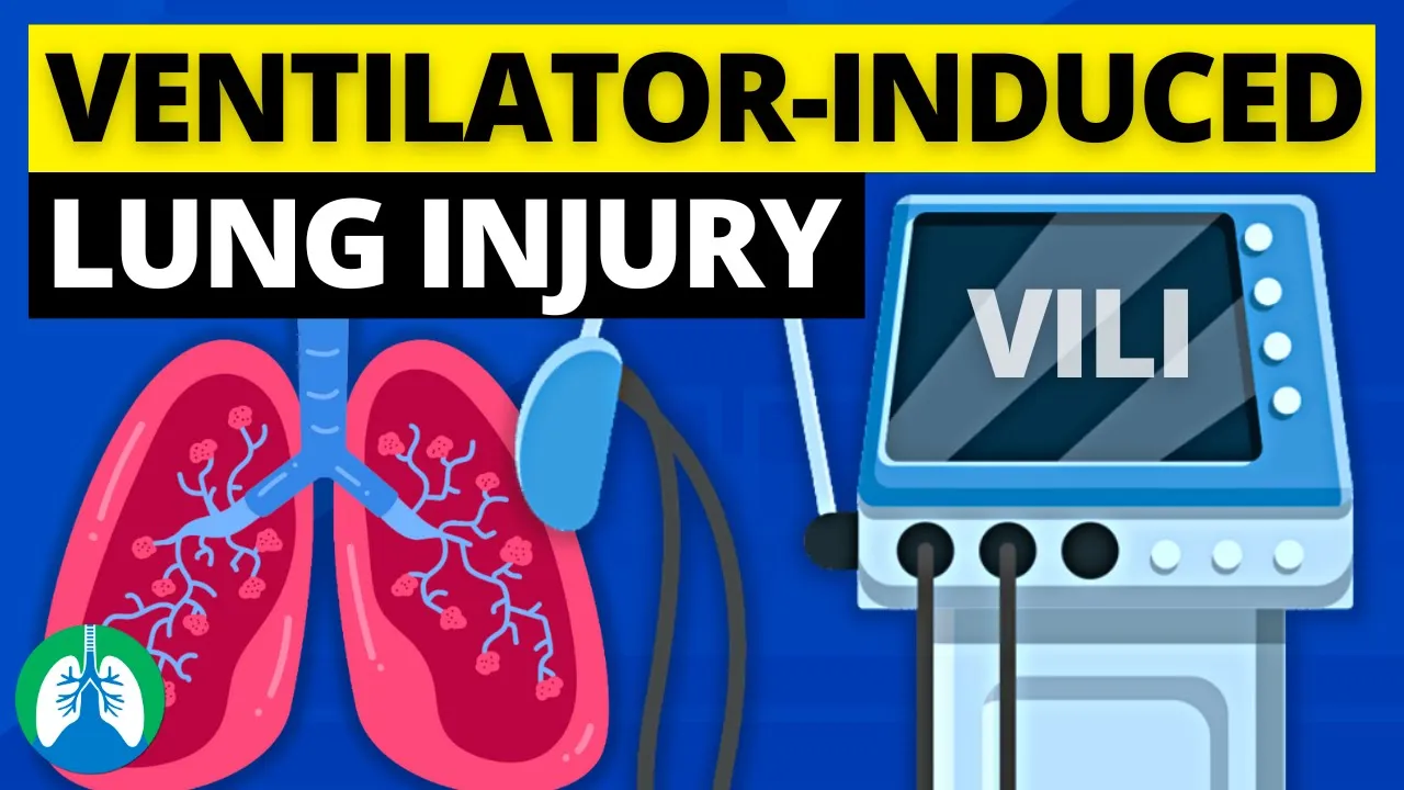
Mechanical ventilation should follow lung protective strategies to prevent ventilator-induced lung injury, including low tidal volume ventilation (4-8 ml/kg predicted body weight), plateau pressure less than 30 cm H2O, driving pressure less than 15 cm H2O, and individualized PEEP settings (8-14 cm H2O for ARDS patients), with mode selection tailored to patient conditions such as ARDS, COPD, or neuroprotective ventilation.
Setting the Ventilator Right | Mechanical Ventilation Principles | CPD Programme
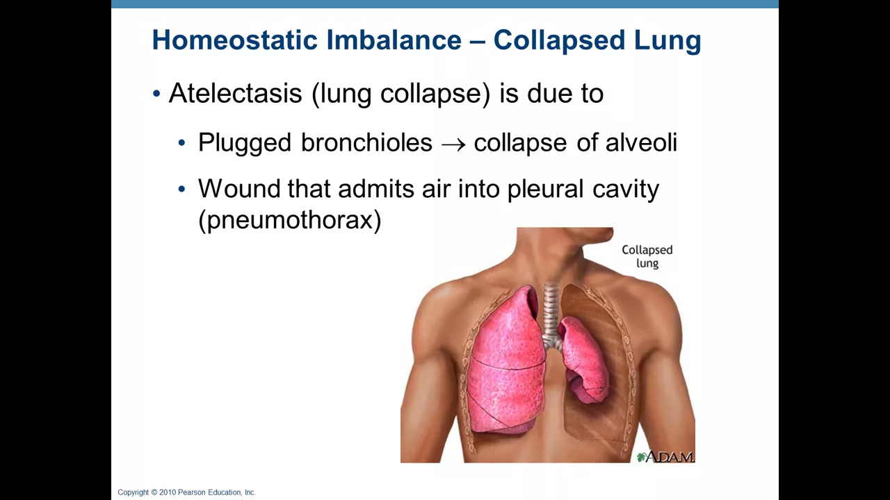
Added:Basic pulmonary physiology, including the mechanics of breathing, lung compliance, airway resistance, and the process of gas exchange.
Fundamental understanding of arterial blood gas (ABG) interpretation and how pH, PaCO2, and PaO2 reflect patient respiratory status.
The clinical indications for initiating mechanical ventilation and the differences between non-invasive and invasive respiratory support.
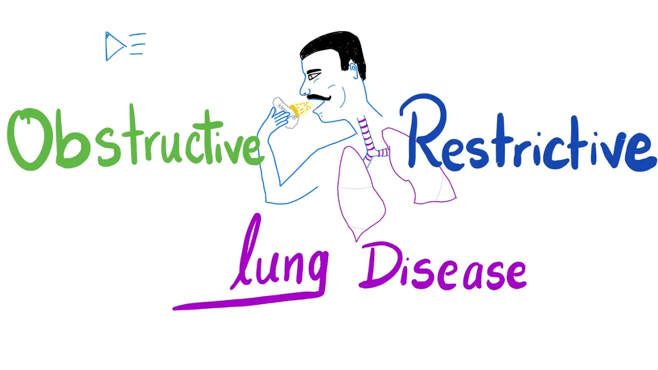
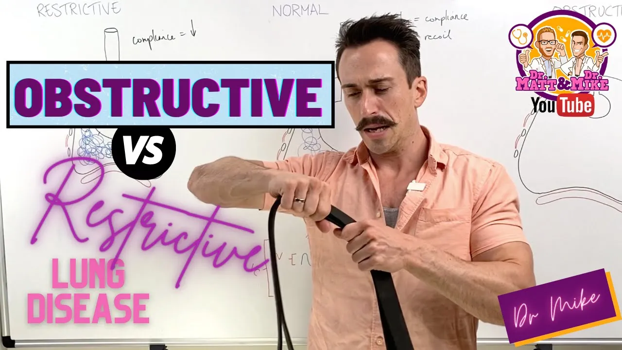
Anatomy of the respiratory system and the pathophysiological differences between restrictive lung diseases (like ARDS) and obstructive lung diseases (like COPD).
Advanced troubleshooting of patient-ventilator dyssynchrony and managing ventilator alarms in acute care settings.
Weaning protocols and liberation strategies, including spontaneous breathing trials (SBTs) and extubation readiness assessment.
Advanced rescue therapies for severe ARDS, such as prone positioning, neuromuscular blockade, and extracorporeal membrane oxygenation (ECMO).
Prevention and management of ventilator-induced lung injury (VILI) and ventilator-associated complications like ventilator-associated pneumonia (VAP).
473 views9likes1:33:10Original Release: 2026-04-02
So before starting the uh presentation proper, I invite you all to the watch this short video.
>> Hope you all that my life is a perfect example.
>> The man behind me hasn't been able to breathe on his own since 1952, but that hasn't stopped him from living a great life. So, so this >> great >> Paul Alexander was one of the last people in the world still in an iron lung. It was home keeping him alive more than 70 years. [music] In 1952, Paul contracted polio and became paralyzed from the neck down. He was 6 years old.
>> [music] >> Eventually, Paul would be able to gulp or take in air for hours [music] at a time, allowing him to leave the confines of the iron lung during the day and accomplish more than anyone thought was possible for him. College, law school, [music] and a 30-year career as a courtroom attorney. And Paul [music] wrote his autobiography.
>> I've got some big dreams.
I'm not going to try to accept from anybody their limitations on my life. I'm not going to do it. My life is incredible.
Is is a horrible disease.
I cannot move [music] period.
a lot.
All right.
All right. So, uh that is uh uh Paul Alexander. So, who lived uh most of his life inside iron lung. Uh so uh the aim of his uh I mean being in iron lung was just to breathe but uh today we have more challenges. So we we have more sophisticated ventilators which are precise and powerful but which can be potentially harmful as well.
So uh so every breath we deliver through a ventilator can be save the save the life or it can injure the lab. So we have to be clear of what we want at the end of the day. So during next one or so so I'm going to cover few things that would be beneficial I mean to have an idea regarding the ventilation.
So I'll be discussing the basic principles of mechanical ventilation, setting up a ventilator which is the core topic today and we'll see how we can apply those things into case uh practical scenarios as well as uh how we tackle the problems when we encounter problems >> during ventilation and a little bit of loops and scalers.
All right. So talking about the core goals of mechanical ventilation, what we want? So one thing we need to ensure adequate ventilation as adequate oxygenation uh as well as to facilitate carbon dioxide elimination and most importantly to protect the lung from further injury.
So our aim is not to normalize the blood gases uh but to keep the lung open and safe until the underlying condition heals like in pneumonia patient until the pneumonia settle we have to make sure the all the u strategies to keep the lung safe.
So uh this is achieved by L protective ventilation which is a a game changer in critical care settings.
Why we need a lung protective ventilation? Because ventilator can do damage. So ventilator induce lung injury.
Uh, Miran, uh, we can't hear you.
Miran, can you hear us?
Uh, a polite request from the participants.
Could you please keep your uh mics muted?
>> Uh, Miran, can you hear us? We can't hear you actually. And your screen is uh I think it has raised No.
Uh there's a problem from Mihiran's side. Just uh hold on for a second. Uh I have informed him. He will sort it.
Uh, Mihiran, still we can't hear you.
Can you hear us?
>> Uh, now can you hear me?
>> All right. Uh, >> thank you. Right now, now we can hear you.
>> All right. So, I think uh >> Can you see the sides?
>> We can see now. Uh, yeah, we can see now.
>> All right. uh so I was discussing about the ventilator in this lung injury. So basically apart from the volume trauma, biot trauma, atelum and u barot trauma oxygen per se can cause harm. So it can cause oxygen induced uh free radical formation as well as absorption at electasis. So we have to prevent this ventilator induced lung injury. So how can we prevent those? Basically we have to uh stick into few rules I mean few strategies not rocker science just uh low tidal volume ventilation 4 to 8 mm per kg predicted body weight pressure less than 30 driving pressure less than 50 and the pe should be individualized.
So it should be compatible with the pressures and compatible with the lung compliance and oxygenation and as well as the hemodynamics. So we have to adjust the things accordingly.
All right. So regarding the low tidy volume ventilation so it's the it is mainly came into the uh arena in uh year 2000 with the landmark trial uh arma study. So which has shown the clear survival benefits with low tidal volume with ARDS patient. So which is one of the most impactful intervention in critical care uh settings. All right. So which prevent the volume trauma and induce biodrama. So over last two decades which has been uh shown I mean proven over and over again this low tidal volume ventilation it has mortality benefit. All right. Okay. So let's move on to lung physiology a little bit. So imagine if you blow a balloon with the strobe. So you have to overcome the resistance of the stroke as well as the elastic work of the ban. So same applies to the lung as well. So either patient or the ventilator have to overcome the airway resistant as well as the elastic recoil elastic work of the lung. So when we increase the trans palmon pressure the lung volume increases.
All right. But we have to overcome the resistance as well. Due to resistant during inspiration it goes like that. So the work of breathing when you talk about work of breathing so it it has both resistive work as well as the elastic work which is denoted by this triangle. So what happens when in non-compliant lung diseases like ARDS, pneumonia, pulmonary edma. So those are the lung is non-compliant. So it needs more pressure to achieve the same volume. So the uh graph is more less steeper but the so so increase in the uh work of breathing but the resistive resistive work is same. So but what happens during obstructive lung disease the compliance is okay but but the resistive work will be more. So it has more needs more work compared to the op uh res uh elast uh I mean compared to ards and things and you can see the expiration is inside the elastic work because it is passive process. So but in obstruct severe obstructive lung disease even the expression can be active. So the this elastic uh I mean resistive work can go beyond the uh elastic work during expiration as well.
So when we incorporate this into pressure time curve during the inspirator phase which has achieve pressure which is called peak inflation pressure. So this peak inflation pressure determine I mean determined by the two things one thing is the airway resistant as well as the lung compliance. So and the flow ceases during the inspirated force and during that period the pressure is called tattoo pressure when there is no flow.
All right. So in resistive lung obstructive lung disease what happens is the resistance is more hence the peak inflation pressure is more but since the compliance is normal the plate pressure is normal because plate pressure is solely determined by the compliance all right so but in a patient like non-compliant lung disease both the plateau pressure and peak inflation pressure rises so you can clearly the P pressure rises. So eventually I mean the key message is peak inflation pressure doesn't reflect the barot trauma risk.
It is what ventilator sees but the plate pressure is solely determined the uh compliance and it is the pressure what alvola feels. Hence the uh it it is closely related with the barro. So you have to clearly understand that fact.
Okay. So keeping those thing is mind keep those thing in mind. So we go on to the plate pressure. So they have clearly shown if we maintain a plate pressure less than 30. So the bar trauma is less.
All right. As well as the driving pressure. So driving pressure means the pressure uh difference between pressure support and the PP maintain. So if you maintain a driving pressure less than 15 again the barot trauma risk will be less. So they have clearly shown lower the uh driving pressure lower the mortality. All right I think you uh understand the fact then we come into the PEEP. So I said PEEP should be individualized depending on several factors. So let's see what happens when we increase the PEEP. So initially it reduces the com uh component of the collapsed lung. It gradually recruit the lung at the same time it part of the lung is going to distend and eventually it is get over distended and the compliance will gradually increase and come into plateau and later come down. So you can see this part of the graph shows the over distended lung. So that is not preferable to ventilate and this part it has more collapse lung it can cause more ataloma. So hence the more pref I mean mainly in ARDS patient and uncompile lung disease we have to maintain PE between this that is moderate P. So 8 to 14 line. All right. So this is bit uh different in obstructive lung disease and neuroprotective ventilation. I'll discuss it later. All right.
All right. So I I don't think this is these are new things to you this low tidal volume low pressures. The problem with us not with us in I mean most of clinician in the world we know the facts but we don't apply. So, so one/ird of the patient I mean uh still being ventilated with uh over I mean large volumes and over pressures. So this is the problem is with the not is not with the knowledge but with the execution.
All right.
So that is uh with more theoretical stuff. So we'll move on to the bit mechanical stuff keeping those physiological stuff and the evidences aside. All right. So this is invasive mechanical ventilation. So I'm going to uh discuss more invasive mechanical ventilation. Uh I'm not going to touch the non invasive ventilation here. All right.
So for before talking about invasive mechanical ventilation we have to know about a ventilator breath. So in a breath even even if a spontaneous breath or a ventilator breath it has two components. One thing is uh one component is inspiratory phase and the expirator phase. All right. So if you take a inspirator phase that has three things. So one thing which has to trigger the breath. So it can be machine triggered or patient triggered basically. So machine triggered. So for an example if we set a if in a paralyzed patient if you ventilate in a paradise patient if you set a ventilated rate of 20 they have uh respirator cycle will be 3 seconds and every 3 seconds they initiated B. So there's no u need of patient trigger but if the patient breathes uh during I mean on the ventilator that negative pressure or flow the flow change can be detected by the machine and trigger that can induce a breath. All right, that is trigger and once the breath is triggered then the next step is what is controlled during the inspirator phase and what's the deming. So in pressure control ventilation it can be pressure or volume control ventilation it can be flow achieving uh set tidal volume. All right. So, next thing is cycle. What ends the expiratory I mean what ends the inspiration.
Okay. That can be time or flow in uh volume control v flow uh pressure control ventilation uh or even volume of pressure even pressure and volume. All right. So depending on these factors we can identify for four types of breads.
All right. So first thing is the mandatory brace. So in mandatory breast there is no patient trigger. So all is that is triggered by the machine and ended by the machine. So during the inspirator that is fully supported by the uh ventilator. All right. So in assisted breathing the breath is triggered by the patient.
All right. But it is fully supported by the machine.
All right. And ends by the machine.
Okay. That is assisted BS and uh you can see the spontaneous bits that is not supported which has inspirated P and the expirator P and the uh supported P. So these spontaneous braids are supported by the pressure support we have given.
So breads are triggered by the patient and partially supported by the machine.
uh what we have set as a pressure support and ended by the patient which is called supported breaths. Those are the four braids we can identify. All right. So then we talk about the control variables. All right.
So we talk about uh the factors uh the main thing which is controlled during the inspirator phase that can be pressure control, volume control or even it can be dual control like pressure regulated volume control volume targeted pressure control likewise. All right. So two main entities mainly the volume control ventilation where we set a uh definite tidal volume.
All right. So ventilator delivers a constant flow to achieve that set tidal volume. All right. So expiratory phase the flow uh expir phase is passive anyway. So it achieves certain peak inflation pressure to pressure. This pressure is depend depend on depends on the lung compliance. So uh the less compliant lungs have higher inflation pressures while the more compliant lung has lower pressures. All right. So this volume control ventilation is more in favor of using condition where we need a precise minute ventilation. All right.
Like in neuroprotective ventilation or ventilating metabolic dis like metabolic acidosis. But the problem is as I said the pressures can go up and cause baroma. All right. In contrast in pressure control ventilation we set the constant pressure and ventilator delivers a constant pressure during the inspiration. So volume gradually increases and the volume achieves the achieved is depend depend depends on the lung components again. So less compliant lung uh dis like ARDS achieve less volume and flow is decelerating and coming to expiratory phase. So this is due to this limitation of the peak pressures as well as decelerating flow.
This pressure control ventilation is in more favor of ventilating uh non-compliant lung diseases like ARBs. But the problem is the tidal volume can be varied. So we have to keep an eye on the title volumes okay depending on the compliance. All right.
But what evidence says so no matter the uh we discuss things so they have haven't demonstrated any mortality difference between these two volume control or pressure control ventilation as far as we maintain those lamp protective strategies. So again I have to emphasize we have to whatever the mode we have to maintain those protective strategies.
All right. So coming into the u main part of the lecture how to set the ventilator up.
Okay. Uh so we have to set certain things. We have to set the mode. We have to set the fire to tidle volume respirator rate I ratio beep and even trigger sensitivity and few more. So the basic stuffs are these. All right. So let's talk about one by one. Okay.
So how to set the FO2. Okay. So our goal is to maintain adequate oxygenation not hypoxia not hyperoxia both will be harmful. So anyway in initial stages we should start high we should start with high uh 100% oxygen then we assess the oxygen oxygenation by oxygen indices like uh partial pressure of the oxygenation saturation and uh even PF ratios and depending of those uh parameters we can adjust the things. So in most of the ICU patient the target is to maintain 90 to 96 saturation and 60 to 80 mm mercury of flash pressure of oxygen. All right. But in ARDS patient the saturation targets are lower. It's 88 to 95. It is because we we are not going to chase oxygenation in the expenses in the expense of bar trauma volume. one thing as well as with hyperoxia this insult can be worsened I mean that can cause free radical induced uh lung injury. Therefore in ARDS patient we maintain 88 to 95 saturation 55 to 80 mm mercy of partial pressure of oxygen and in as you all know the chronic hypercapnic patient like in sepd the oxygen targets are lower 88 to 92 and in postcardiac arrest patient and neuroprotective ventilation we aim for bit higher targets to minimize the secondary brain injury that would be uh 94 to 98 light. All right. So uh and other thing is you you I mean you have to always combine this FiO2 with PE. So for an example if when we ventilating ARDS patient uh when the patient desaturates no point of keep on increasing the 500. So I mean uh we have to increase the speed accordingly as well because uh uh we have to ensure the both things should be in a balance. All right. So there you you can use the uh graphs I mean uh tables which has provided by the net and uh other uh things which can uh set a optimal peak and the other important thing is you have to reassess frequently and try to get the oxygenation uh to match the those set uh set oxygen uh oxygenation targets.
So next thing is the tidle volume. So as I have been telling over and over again we have to maintain low tidal volume. So important thing is we have to uh adjust it to the predicted body weight because it the lung volume is determined more the height not the weight. All right. So suppose you have a patient with 150 cm uh patient with 100 kilos. So you are not going to ventilate uh 600 ml of tidal body rather you have to calculate the predicted body weight for 150 cm and uh calculate accordingly.
So in ARDS patient you may start with 6 ml per kg and in other patients you can start with 6 to 8 ml per kg and then you have to assess the pressures and tight accordingly. All right. Say it is the plate pressure is more than 30. What you have to do? You have to reduce the title volume by 1 ml per kg at a time. Then reassess still if it is still high you have to go down with the ventilator up to 4 ml per kg. All right. So this is the basis of low tide volume ventilation.
All right. Then the respirator rate.
Okay. So we have to uh adjust the respit rate along with the IE ratio. Okay. Uh so regarding the respit ratio we preferably we should set the respirate rate of uh 12 to uh 12 to 20 but keep in mind most of the patient we start to ventilate has high respirator drive.
they even I mean originally they have before intubation they have higher respir rate like 35 36 like that so we have to make sure we can't just convert them to 35 respir rate into 12 all right so so that in those patient we can start with a higher respirate rate like 20 24 then we can adjust uh assess the carbon dioxide status and the ventilation and adjust the respirate rate accordingly.
So in ARDS patient what happens? So we start with high high respir rate like 2024 and suppose if the carbon dioxide is high. So again so one thing again uh the one major thing I have to emphasize in ARDS patient we accept permissive hypercapnea.
All right. What do you mean by phosphere hypercapnea is we can accept high carbon dioxide level which can be 55 or 60 there is no um I mean defined value but we have to go with the pH so if the pH is less than 7.2 2 then we have to increase the respirator rate okay up to 35 okay so I ratio in non-compliant lung diseases we set is 1 to2 usually but in extreme cases we uh I mean we concentrate more on oxygenation where we can set the I ratio 1:1 or even inverse ratio 2:1 or 3:1 to increase the inspirated time. So they have our aim is to improve the oxygenation but those things have no I mean no evidences for uh uh high ratios or inverse ratios but uh you can consider those thing in refractory cases in COPD or obstructive lung disease patient. Our main problem is the exhalation. So we have to ensure we give adequate time for the acceleration. So otherwise there would be they are trapping and causing dynamic hyperinflation and autobe development.
So what we can do is one thing is we can uh start with lower respirator rate which will ensure u adequate inspiratory time and adequate expir time as well as we can use lower I ratios. Suppose uh uh we have we have been ventilating with 20 uh respirator rate. So then the speed uh with high ratio of 1 to2 the expirator respirator cycle will be 3 seconds and the expirator time will be 2 seconds. So and we reduce the respirator rate to 12 what happens the respiratory cycle is 5 seconds now and expiratory time will be around 3 to 3.5 seconds. So that would beneficial I mean that will uh lengthen the u experienced time as as well as if we uh reduce the I ratio as well that will uh increase the experienced time. So but you have to keep in mind once we increase the in time so the flow need is more to because they need more higher flow rates to achieve this tidal volume with this time. All right. But the respiratory cycle would be uh completed during that given uh experienced time that is is obstructive lung disease. In metabolic uh acidosis like condition eventually we need I mean uh uh we need higher respirator rate to match the respirator compensation otherwise there will be a mix acidosis until this metabolic condition is sorted out. All right. Uh for an example DK patient we need I mean initial setup would be uh resp rate of 24 26 like that and we gradually reduce according to the control. Okay. All right. Then the P. So uh again I have said already. So we have to ventilate in this part of the ventilator curve not already standard part not the collapsed part. So that uh first in um non-compliant lung diseases we can set the moderate PE maybe uh above five definitely uh and titate the PE physiologically. So we have to see as I said the uh plate pressures di pressures and the lung compliance uh and the hemodynamics as well. So uh but in obstructive lung disease what happens if we give a higher B so that will add on to the auto peep if there's any and can cause increase the barot trauma risk and the hemody hemodynamic instability as well as the in neuroprotective ventilation the P we apply if we apply a higher P what happens that can transmit to the uh cerebral circulation can cause increase intra uh intraanial pressure as well as it it might impair the hemodynamics and cause uh impaired cerebral perfusion pressure. So we have to ensure the peep should be individualized. So anyway any of the patient we should avoid high higher higher peep that means above 16 or so and uh aggressive recruitment.
Okay.
All right. I think uh uh up to now it's clear. So if you have any question you can uh ask at the end of the lecture.
All right. Let's move on to the next part.
The modes of the ventilation.
So we have conventional modes and advanced mode.
conventional mode what happen what we use it's a open loop I mean open loop control so what we said ventilator delivers uh ventilator doesn't think about the patient what whatever happens in the patient they have no sense basically so uh like control mandatory ventilation assist control ventilation and famous SIM or pressure support ventilation But in advanced mode they use close closed loop control. So they take information they take information from the patient the pressures the ox even oxygenation the compliance and accordingly they adjust their uh output.
So it's sort of a feedback mechanism. So those are I mean more preferable in ventilating patient in in terms of synchronization as well as u uh uh minimizing the risk of patient self-inflicted lung injuries. All right.
So let's talk about the ventilator modes in simple. All right. Control mandatory ventilation. So this has control mandatory ventilation as all the beds are mandatory beds. So it is fully machine triggered and fully machine cycle. Uh like we ventilating a uh paralyzed patient. So no patient efforts are not entered and all. So if there's patient effort there may be a asynchronic or disynchronic. All right.
So this is beneficial in in conditions like where we need precise precise uh where we need precise we need ventilation like in neuroprotective ventilation as I said we have to keep the carbon dioxide level in in a narrow margin I mean precise settings as well as in severe ARDS patient early stages they have high speeded drive. So what we do is we paralyze the patient initially to uh achieve the control achieve a better control and u continue uh mandatory ventilation and of course during inoperative ventilation we usually use uh this control mandatory ventilation as the patient now paralyzed. All right. The problem is this needs deep sedation or even paralyze. So we can't keep on continuing this control mandatory ventilation because that sedation as well as paralyzed has adverse adverse effects like uh hypotension or isoacquired weakness like that. So we have to shift quickly from this control full control ventilation to patient triggered ventilation uh as quick as possible.
Then the assist control ventilation here the patient beds are entertained. So patient can trigger beds but the all the beds are supported. So uh this will deliver guaranteed minute ventilation and uh uh and this is more beneficial in uh acute respiratory failures where we need full support. But the problem is this can cause hyperventilation and respiratory alkyossis and this can cause patient ventilator asynchrony as well. All right.
So this is assist control ventilation.
So this is SIM uh or synchronized intermittent mandatory ventilation. So arguably this is uh maybe the uh uh most popular mode among you. All right. So this combine three breaths actually this combine mandatory spontaneous breaths. So this has three types of breath. It has uh mandatory beds and if the patient breathes during this uh trigger window it is delivered as assisted bed. So which which means fully supported and if the patient breathes out of this window it delivered as a supported bed by pressure support we said. All right.
I mean uh this is historically used as a vin mode. So transition from fully controlled ventilation to the spontaneous mode. But in uh modern practice we use this SIM for patient who has the spontaneous respiratory drive.
So which needs less uh sedation as well as it allows patient spontaneous breathing and maintain diaphragmatic activity and which will which is a good transition mode for uh recovering a patient. But the problem is the since the patient uh respirate rate can't be pred uh is unpredictable this won't deliver predictable won't be delivering the predictable minute ventilation. So we have to uh make sure the minute ventilation is delivered down as well as there may be a lots of asynchrony between the uh patient and the ventilator. All right. So these are the challenges during SIMA uh and the pressure support ventilation.
So this is uh the as you know this is the spontaneous mode. All right. So where we set patient is spontaneous breathing where we set a pressure support. Okay. So uh so this is basically weaning mode uh patient should have enough uh respiratory drive respirator drive and the all the brits are supported and once the patient u we can wean off the uh to a lower pressure support we can exubate the patient. So I mean this is reduce the work of breathing and the sedation requirement and uh ensure uh and preserve the diaphragmatic activity and improve the patient comfort and the synchronicity. So this is a basically uh ven mode. So the problem is this if the patient stops breathing for a while so we can't I mean there will be a problem uh that can cause hypoventilation basically so we have to set a backup mode as I mean always we have to set up set up a backup mode right is the pressure support ventilation for spontaneous mode okay let's talk about bit of uh advanced mode. So advanced mode as I said it has a b better close loop control. So therefore those uh modes has better control.
So uh APRV is a one of the u these days it is used frequently especially in ARDS patient. So this APV or pressure release ventilation mode what uses two levels of CPAP basically.
So patient should have ideally spontaneously breathing. So the pressure high so uh is I mean uh maintained for a long for the in uh inspirated time and patient can breathe on that CPAP like this.
The concept is the open lung concept.
The lung is kept open for a longer period to ensure a better surface area for oxygenation as well as it it will uh in I mean eventually reduce the ateloma as well and for a brief period it drops the blood pressure to a pressure low in which the exhalation occur. Okay. But uh this thing this uh AP APV is not recommended for the patient who has profound shock because the intrathoracic pressure would be high that can go that can worsen the shop as well as it is not preferable for the patient who doesn't have spontaneous breathing because in a uh in both stages patient should have spontaneous breathing that that it's more I mean that is ideal for this mode.
Okay.
So other things I'm not going to discuss. So if you have any questions you can ask. So pressure regulated volume control where the pressure is regulated in each breath to achieve the set volume uh tidal volume and proportional assist ventilation and uh neurally adjusted ventilator assist. So that again we assess the uh the ventilator assess the patient uh effort by taking the electric signals and deliver the uh customized brakes to the patient. Okay. So that the patient synchrony will be better and uh low risk of trauma.
All right to summarize the thing for the mode selection. So ARD is we can use either mode volume control and or pressure control provided that we maintain the uh lung protective ventilation. COPD patient we can use volume control or even pressure control.
The aim is to uh give adequate experienced time to avoid uh dynamic high beam inflation and auto neuroprotective ventilation volume control is preferred to maintain stable carbon dioxide level and uh IC prevent to ICB post mode pressure control pressure support ventilation or SIM or even mode like uh uh PSVA to to ensure patient comfort. and the uh early mobilization and early uh weaning off from the ventilator. All right. So I think up to now uh you know something.
All right. So let's apply this thing to this thing to a practical scenarios.
All right. Case one. Um we have a patient with severe pneumonia. as a 54 years old male uh and she now is she has features of severe ARDS. He has in type two respiratory failure and uh hence uh who needed uh intubation and transfer to the ICU. X-ray shows bilateral shadows and AG shows severe hypoxmia with PF ratio of 52 and slight mild hypercapnea and while maintain the pH of 7.2 two. All right.
So, what's your strategy? So, what are are we what's our main goals?
Uh so, maintain the lung protective ventilation, right? Let's see.
So, uh in ARDS patient we have to provide low tide. We can use either mode volume control or pressure control.
We should start with low tidle volumes.
Wait. Uh this is the stimul Hamilton stimulator. Uh simulator. So we usually have similar C6. So in some of the centers has C6 as well. We'll use the uh C6 one.
All right. So, first we have to set the uh patient details. So, is a male patient predicted uh height is 170. So, the predicted uh weight is 66. Then we'll select the mode. So, we can select either mode uh either volume control or pressure control which should be assisted control not the SIM initially.
All right. So I'll uh for a reason I'll check uh set the volume control ventilation first and confirm and this is severe hypoxmia so we should start with high 100% oxygen peep ards patient to match the 100% oxygen at least we have to set 10 all right and tidle volume should be low tidle volumes weight is 66 predicted body weight so we We can say 360 or 370.
I will let's see 370.
Okay. So he has been breathing with high respit rate. So this can't be 12. So at least it uh 20 to 24 like that. So always remember when we adjust the ventilate uh respirator rate we have to adjust the I in spirit time to uh set the IV ratio decide IC ratio here I would set the I ratio of 1 to2 this thing like this is better flow pattern so I use the conventional one for trigger this is the default value so and we confirm the patient Okay. And we'll start the ventilation. Let's see what happens. Um and uh all right. So uh we have set the title volume. So it deliver 353 likewise 370 tidle volume. And see the plate pressure see the peak pressure.
We'll increase the uh tid volume and see what happens.
So volume control ventilation as you can see it increase the uh pressures and come to peak inflation pressure and the flow is uh maintain the uh constant flow since this is a volume control ventilation. All right. So here the uh peak pressure is uh 31. So then we have to check uh the um uh plate pressure. How to check the plate pressure? That is uh one of the thing you have to know. So what you have to do is you have to do a inspiratory hold. So you do a inspiratory hold for at least 3 to 5 seconds and stop. And there you can check the patient. Here it is the 24.
This is the peak pressure that is not equal to the P pressure. So P pressure is not usually it's not uh monitored. So we have to do the inspir and measure it uh I mean in uh by ourself. Okay. Now if the plate pressure is less than uh 24 so that is acceptable. Suppose it is u uh it is more than 30 we should reduce the tidal volume uh to reduce the tid uh to maintain the plate pressure less than 30. Okay. So here the d pressure is uh plate pressure is 19 deep is 10. So d pressure is around 9 that is acceptable. So which is less than 30. So saturation is 92. So we can come down with the oxygenation little bit maintaining 88 to 93 and if the carbon dioxide levels goes up and pH is less than uh 7.2 we can increase the respirator rate up to 35 and adjust the uh I ratio. So here if the oxygenation get worse we can use the lower uh I ratio as well like one one to one. All right that is a uh uh rescue method.
Okay this is how ventil we ventilate a ards patient.
All right. So these are the uh key concepts. So low tidal volume respirator rate uh targeting permissive hypercapnea. So peep moderate peep and target saturation would be uh 88 to 95.
So fail in these strategies or uh I mean fail in these ventilated strategies we can uh I mean or the simultaneously we can continue the other nonventary strategies as well like pro positioning neur short-term neuromuscular blockage or restrictive fluid strategies or even early. All right. All right. So case number two uh a female patient non- bronchial asthma patient presented with the features of neopatal asthma. Now uh she is in type two respirator failure.
Okay. Failing the conventional oxygen therapy um uh they decided to intubate the patient uh because they also impending respirator.
uh chest X-ray shows uh hyperinflated lung, no pneumatics, no features of infections. Ag shows uh hypoxmia and hypercapnea suggesting type two respiratory failure. So how to ventilate this patient. So here the in obstructive lung disease our main goal is to maintain oxygenation and uh to uh eliminate carbon dioxide without causing air trap. All right. So what we going to do is um so this is a female patient uh height is uh 160 and predicted body weight has predicted body weight around 450 and we'll select a mode. So we there we we select pressure control mode and is a severe hypoxmia we'll set 100% again and here the beep which should be less than five usually we use uh lower peep because it can the extensive peak we add can be harmful. All right. So, uh we set a peep of uh three like that and the pressure control will make it uh 12 to we we'll check the tile volumes and we'll readjust and respirator rate should be low because to ensure adequate expiratory time we'll set it 12 and this I ratio inspiration time should be less to make sure the I ratio So around 1 to three like so let's make it 1 to one and see what happens.
All right. So we'll start ventilation and see the flow volume curve.
So it achieve tidle volume of 360. So our target volume is maybe 6 to 8 L per minute. This is acceptable. So we can uh if the 450 is bit high so we can reduce the pressure support pressure control to achieve a low tidle volume and see the uh flow volume. So we'll increase the respect rate little bit and see what happens.
So here you can see the graph doesn't come to the baseline. So there will be a AR trap. So that we have to reduce the respirator rate to ensure adequate expiry time. So always uh make sure the X I ratio also should be adjusted uh when you whenever you change a respirator rate. So we'll make it 1 to three and see.
So here how to check whether there's auto. So there you have to check the expiratory hole. So you do the expirator you make it volume control ventilation.
So uh you do the expirator hold and check the total beep. This is the total beep which is uh 3.6. 6 and this is the uh extensive peak we have applied the total peep uh so the auto peep is uh total peep minus u extens which is around 6 that is acceptable we should keep the auto peep less than five all right so if there's a dynamic hyperinflation this auto peep will keep on rising all right so there so what we have to do is we have to uh I mean adhere to the measures where reduce the rate and uh allow more time for the expedition. So if the patient deteriorates in obstructive lung disease if the suppose if the patient develop hypotension so you have to >> hello you have to dynamic hyperinflation yeah and if there's high peak inflation pressure you have to include bronco specimen tube obstruction like that. So in as a rescue method if you suspect dynamic hyperinflation you can just disconnect the patient to uh allow the patient exhalation. All right. So next case is a uh traumatic brain injured patient young chap uh following RDA having uh low GCS no other injuries and uh he has a features of uh he has features of pushing uh reflex and suggestive of increased intraanial pressure and uh he was intubated for uh neurop protection as well as to control the uh uh neuroprotective ventilation.
So ph AG shows hypercapnea with adequate oxygenation. So here our aims are to maintain the neuroprotective targets.
So, so the mode is in neuroprotective ventilation volume control ventilation and we set the uh 100% oxygen and try it later. PE is uh I mean uh uh should be uh low to moderate. to ensure uh uh I mean proper venus drainage adequate uh uh title volume uh so maybe 360 predicted body weight and uh respirator rate adjust accordingly.
Okay. So there you have to check the uh carbon dioxide levels frequently and uh so uh adjust the carbon uh respirator rate to match the carbon match the normal capab. So main main thing in neuroprodictive ventilation we have to ensure nomoxia normocia and normal tension apart from the normal glycemia and normia and things. So to achieve nomoxia we should adjust the uh Fio2 NP accordingly and precise uh respiratory rate adjustment to achieve the tight carbon dioxide control and uh ventilator strategies to to minim to maintain minimum effect of hemodynamics to maintain cerebral perfusion pressure.
But in refractory cases, refractory highcp cases, we can temporarily use uh hyperventilate hyperventilation targeting carbon dioxide level of 35 to uh 30 to 35 that is a rescue method. All right. So target saturation would be 94 to 98. All right.
So that is how we ventilate patient and that is how we apply things our practical knowledge I mean our theoretical knowledge to practical cases. Then the next thing is the problem challenges we have. So how matter however we set the numbers there will be challenges. So patient ventilators asynchrony or uh patient fighting with the ventilator is one of the common challenges we have that is basically due to the mismatch between the patient demand and what the ventilator delivers. So why it is problematic? Because it can cause patient discomfort and it will increase the work of breathing and and eventually it it will induce patient self inflicted lung injury. that is a concept uh I mean that is described by the high respiratory drive can cause uh lung injury that is called patient self-inflicted lung injury. So eventually that will increase the ventilator number of ventilator days and number of ICU days and the overall mobidity and modity. So if I classify the things that the pro the the this asynchrony can be trigger asynchrony or that can be a problem due the flow and cycling things. So I'll be discussing few common uh uh asynchronies one thing is the ineffective triggering. So what happens is uh patient uh has a effect but it is not detected by the uh uh ventilator hence there is no ventilator beds this is this can happen due to few reasons one thing is if the effect is less I mean in patient if the patient doesn't have enough drive or patient is deeply sedated so this effect is not triggered doesn't reach to the trigger threshold.
Or else if we set the uh trigger sensitivity is is too low. Suppose the patient generate a negative pressure of two but we have set a uh threshold uh pressure trigger threshold negative 5 cm water then it doesn't reach the five it doesn't trigger the ventilator base or else this can be commonly seen in patient with u obstructive lung disease who has auto peep. So what happens is suppose if patient has auto peep of eight and the trigger threshold is uh two the patient has to generate uh 10 negative pressure to gener uh trigger the ventilator beds. So what we can do we have to basically fix this problem right we have to identify and uh we have to fix the problem. So for the auto peep thing we can we know the things now. So we have to uh adjust the ventilation accordingly to avoid auto peep and we can increase the sensitivity. We can come up with the uh negative pressure 5 to 10 uh 5 to2 then it will trigger or else uh we can change this uh pressure trigger to flow trigger because the flow trigger is more sensitive than the pressure trigger. That is why we commonly use flow trigger compared to flow trigger pressure trigger as well as it doesn't it it detects the flow uh it doesn't need a higher pressure drop to create uh trigger a breath as well as we can reduce the sedation and improve the muscle relaxction to uh the to improve the uh patient effort. So this is called ineffective triggering and how to fix that. All right. The next thing is the opposite thing auto triggering patient doesn't have a trigger but uh machine deliver a breath. So this is usually due to the fusings again. So if the uh there's a continuous flow despite of the patient stop the breathing like in circuit that that flow will be detected by the ventilator uh falsely and trigger a bit. All right. So or else if there's a flow disturbances due to the water uh condensation and the water in the tubes this float uh turbulence can detected by the process and cause it auto triggering. Other thing is uh when the patient has cardiac osillation especially in high cardiac output status.
So there the cardiac osillation can be misinterpreted as a respiratory effect and cause a uh uh ventilator breath. Also if we uh set a uh lower uh I mean higher sensitive level for an example if we set a flow sensor uh flow trigger to one liter per minute even with a tiny slightest flow changes that will induce a uh ventilator base. So to fix that one thing is we have to decrease the sensitivity at least by 3 to 5 L per minute flow flow sensor flow uh trigger and we should eliminate the artifact both in the tubes and uh air leaks as well as we can change that uh trigger type flow to pressure because then uh patient needs a proper pressure drop rather than pressure flow turbulence. Patient need the proper pressure drop to initiate the breath. So that is auto triggering uh and the pro starvation. This can be seen uh patient with high especially in high respirated drive suppose the this is called air hunger. So in normal patient pressure rises uh exponentially. But what happens when the uh patient has high respirated drive and we set the uh inspirator flow low patient um I mean patient uh needs more but ventilator doesn't provide so that there's a deep in the uh pressure volume curve pressure time curve. Okay. So what we can do is we can increase the inspirated time inspirated flow to match the patient ventilated drive or else we can uh suppress the uh I mean we can treat the patient high respiratory drive. So for an example we can treat the pain okay or else we can uh do sedation or else uh we can treat the fee likewise. Okay. as well as uh we can if we are in the volume control ventilation we can switch into pressure control ventilation that has minimal effect with flow starvation.
So that is flow starvation uh and the double triggering. So this is one of the common thing we uh see daytoday. All right. So what happens here is there's a mismatch between the inspirated time of the machine and the patient inspirated time. So the mismatch means the machine inspirated time is shorter than the patient inspirated time. So this can be due to two reasons. One thing is we have set the machine inspirated time lesser.
So if you we if we have set the inspiratory time uh I ratio and the respirator rate in a way in a shorter period or we have set that low idle volume. So that will deliver this is quickly but the P uh as well as this can be due to high respiratory drive patient in high respiratory drive. So respiratory cycle ends but patient efforts outlast the respiratory machine breads. What happens that the continuous patient effort trigger another beds.
Okay that is called double trigger. The problem is the exhalation is not completed. So that therefore the the second breath can cause increase in the volume and causing uh volume trauma. Okay. So uh so the problem is one thing is short ventilated machine breaths as well as high respiratory drive. So what we can do is we can increase the uh we can increase the respiratory time.
So we can adjust the uh uh reduce the respirator rate or we can increase the tidal volume so that this space will be more long as well as we can reduce the uh inspirated flow.
Therefore, it will take more time to achieve this uh tidal volume. Or else we can uh as I said earlier, we can use the we can suppress the high respirator drive. We can use sedation or we can use analistics or even paralysis the patient or else uh as well as we can uh in pressure support. If the patient is breathing spontaneously we can uh this uh reduce the uh cycling threshold so that's uh period time ends early so that is double tickery so this is delay cycle I don't think we all right any patient ventilator asynchrony what we have to do first we have to look at the patient all right so we have to check whether the patient is in distress or not And then we analyze the waveforms and and identify the what is the uh type of asynchrony whether it is a trigger asynchrony flow for synchrony or cyclic asynchrony as well as we should identify the pathological physiological derangement which can cause these things. So like it can be acidosis acidosis epsis bronospasm whatever then we uh fix the things. So we first we set the ventilated settings that can be increased uh in spread time reduce the sensitivity triggers and treat the course treat the fever treat the sepsis uh analesia for pain sedation for unedated patient and paralyzed and also we can always incorporate the uh ne techniques and ne modes like now proportionalis ventilation if it is available which has better synchrony which has better inputs from the patient that is how we uh tackle the asynchrony.
All right coming into later part of the lecture few things of graphs. So ventilator graphs can be scalers and loops. Scala is a uh graphical demonstration of a variable over time that can be volume time, pressure time or flow time.
loop is a representation of a variable or another variable that can be pressure volume or flow volume likewise. Okay. So there are many things we can uh information we can get from those things. Okay. So I'll highlight few things. This is not the whole thing. Uh few important graphs. One thing is flow time scalar. So as I said earlier what we have to see whether this uh flow time curve reaches the baseline otherwise there will be autobeam. So we have to ensure uh we have to uh adjust the ventilator setup in a way to uh increase the uh expirer time to achieve uh uh to touch the expir time. So the baseline before the next bet starts. Okay, this is one of the u uh important thing you have to know. Other thing is the pressure volume. This is a very informative uh loop. So there are many things we can gain. So this is the uh pressure when the pressure uh increases initially there is there is a resist I mean less compiled part in the lung as in when we blow a balloon and once this lower inflated point inflection point is reached the lung is more compact and uh to a given volume there's a more volume change and once the lung is over distended so for a given pressure volume there's minimal volume change that is called uh over distended lung which is called this appearance is called beaking this point is called upper inflection point.
So one thing is we can we can get so the steepness of this curve so lower compliance so less the flat flatter the curve the flatter I mean uh more uh less compliant the lung. So in ARDS patient this would be like this the uh I mean when the patient get better this will gradually come into original place.
Okay. Other thing is the lower inflection point.
So uh this can be used to set the optimal pip. So this is one of the method to set the optimal pip. So usually we calculate the lower infection point by uh drawing the PV loop in the ventilator and we set the P usually one to two cm water above the lower inflection point so that uh the ventilation will be operate uh in this part more compliant part and if there's a beaking so we have to make sure to eliminate this part so so that we can uh To achieve that we can reduce the tidal volume as well as we can reduce the uh PE uh so that there won't be no be okay because it can cause baroma okay so that is uh few things we can uh get from the pressure volume loop and the next thing pressure flow volume loop. So this is the uh inspirator face and this is the uh expiratory phase. So few things we can get one thing is we can diagnose obstructive lung disease and restrictive lung disease here. So in obstructive lung disease the there is a deep in the uh expirator phase in expirator uh obstruction. All right. In restitive lung disase the shape is same but the volumes are minimal likewise. Okay.
plastic to lung disease that is how we diagnose uh we can diagnose ob uh lung diseases with probable curves as well as if there's a air leak this is a functional residual capacity and this is the tonium if there's air leak this exper limb doesn't come to a uh doesn't complete the loop rather it come like this volume is the air le all right [snorts] or else in if there's air trap in obstructive lung disase this doesn't again this doesn't uh complete the loop rather it can come like this so this will be the uh I mean that can uh demonstrate the uh uh air okay these are the few information we can get from the flow volume low all Okay. So this is uh few things regarding the I mean few basic stuff regarding ventilation but uh keep on keep in mind that uh when we ventilate a patient not only ventilator strategies but there are few things we have to think beyond the ventilator. So that can be prone positioning especially in CV moderate to CV ARDS which will improve the ventilator profusion mismatch which has uh I mean uh very strong evidence for survival benefit and early neuromuscular blockage which will improve the ventilator patient ventilator synchron synchron and dry lungs. So which will improve the uh lung compliance as well as if available early emo especially in severity this patient if the criteria meets.
Um so those are the nonventilator strategies.
So before winding up uh this is now time for the uh questions.
Uh yes Miran actually in the chat box we have got a few questions actually the first one was about uh can you explain auto peep I think you have already done it in several segments.
>> Uh and uh the next question is about uh ASV mode. Uh could you please explain about it and also uh when to use ASV or intelligent ASV modes?
>> All right. So AC is I mean uh can be specially seen in Hamilton ventilator.
So that is again sort of uh advanced mode. So which has um closed loop control. So if we uh see the AC mode I'll demonstrate.
So it basically what what we do um we uh wait.
So what we set the uh patient uh uh details gender and the weight and the things and uh we select the mode to AC okay and what ventilator does uh and we have to select the uh percentage of minutes ventilation whether it is 80% or 100% or 120%. and uh PE and the FIO2 and maximum pressure limit. All right.
Likewise, so what uh the ventilator does? It would initially give a um I mean uh control uh few breaths and it will uh calculate the thing called uh patter pressure and the expiratory uh time constant. So that they will uh decide a optimal respirator rate for that patient like here this is 16 and they will calculate so uh a minute. So minute this is the minute ventilation and this is the possible uh uh tile volumes and uh rest rate that that can achieve the that targeted minute volume. So if the patient doesn't uh reach that uh tidal volume that the patient will increase the pressures or if the patient delivers more tidal volume it will reduce the inflation pressures to match the that target. All right. So that is basically we get the information from the patient. So that is sort of a pressure regulated volume control like so anyway uh uh but it's more complex than that because they u uh use the certain uh equations uh to get that optimal uh respirator rate and the respiratory time constant.
Okay. So uh so patient uh machine deliver uh the inflation pressures uh adjusted according to the patient response. The difference uh so this is AC mode is basically we can set the uh set during for ventilation of any patient but the problem is the minute ventil I mean tidal volume can go up. So especially when we ventilate ARDS patient we have to closely monitor the title volume. So this is there are no evidence for AC in ARDS patient because this title volume things there are no studies basically [snorts] uh therefore you that mean that doesn't mean you can't use a senior patient but you have to keep on keep an eye on the title volumes. So >> that is one thing. All right. In intelligent AC what happens in I mean uh in addition to this ventilator pressures and things they get the information regarding the oxygenation as well from false oxyter. So depending on those values depending on the set targets they uh change the I mean they adapt change the FO2 and P so that to achieve that oxygenation target. So that has oxygenation part as well. So uh for as mode if we use um suppose we uh we use AC mode for uh ARDS patient if you see the high tidal volume you can gradually reduce the uh minute percentage of minute ventilation.
Usually we use uh uh around 80 uh minute ventilation or even in obstructive patient we use lower value and patient with higher respiratory drive and metabolic condition like metabolic acidosis we can go up to even 150 uh percentage of minute ventilage patient to match the compensation. So that is AC as mode that can be as a conventional uh respiratory mode even as a vining mode as well.
>> Right.
>> So we Yeah.
>> Yeah. M. So M there was another question about SIMV mode and it the Dr. Lahi he's asking actually how to improve the volume delivery in SIMV mode.
>> Uh SI mode as I said it is a synchronized mode. We have three type of bra. So uh see uh so uh one thing is uh we have to see uh so I can't demonstrate uh here because uh uh the simulation is not supported for active patients. So let's see uh so if the we have to see uh both things I mean assisted control as well as the uh supported beds. So if the assisted beds are delivers a lesser volume we have to increase this peak inspiratory pressure. So uh uh keeping in a safe limit. All right. If you see a supported bed uh deliverable less tide lowering you have to increase this pressure support. So the uh top right hand left hand corner you can see the top pressure support. So we have to uh see both uh pressures uh to achieve we balance the not the balance actually to uh achieve uh the settle volume is delivered in both uh phases did I answer the question or do you need more clarifications >> yeah I think it uh it was very quite clear Miran I think thank you and also there's another question about uh can you explain how to use pressure volume looping in odds patients.
>> Yeah.
>> Uh yes. Uh that is again uh so we'll use special volume right. So as I said uh one thing is uh we can this is a volume control actually uh volume control mode.
So one thing is as I said uh in uh in uh in art patient what happens we can get uh many information one thing is the lung compliance and steeper the uh pressure volume loop that is more compiled. So this is this keep in mind this is volume control ventilation. What happens in uh uh pressure control ventilation since the pressure uh is uh constant during pressure that will take time and pressure is like this and likewise. So uh so anyway we can assess the compliance of the curve. So initially in sever patient the curve will be flat. So when the patient improves the when the lung improves this uh compliance curve come to the original patient position I mean to the uh left that means more compliant other thing is the lower inflection point. So if I demonstrate this uh so we should uh use you can use this PV tool uh pressure volume tool. So this is bit advanced stuff. Okay. So we can uh set the settings and things. Don't worry about this. So we we will start the manual.
Okay. Uh >> right. Uh >> and it will uh it will draw the pressure volume blue.
uh and the once the pressure volume uh loop is uh drawn we can uh check the lower inflation point.
Wait for that. So here it is not uh this is just a simulation. We can check the uh lower inflation point uh and the upper inflation point and uh we can adjust the peep accordingly. Suppose the low inflation point is here we can check the compliance. Okay. So we improve uh increase the uh shift the cursor so that to achieve the uh improved compliance so that we can uh set the our PE uh 2 cm cm water above the low inflation point uh as well as the upper inflation point. we can reduce the uh tidal volumes.
All right. Anything else?
>> Yeah, man. And another question about prawn position ventilation. Uh uh one participant is asking how to overcome the hemodynamics disturbances or the compromisation that is happening with prone positioning.
>> How? Usually um prone positioning doesn't cause much heinical stab instability because uh it is a preferable I mean uh position in in terms of lung compliance as well as the >> uh uh the hemodynamics.
>> So I agree with that because I mean if you have a compromisation what you do is you go with the central line and start on and you ready to escalate as needed.
So and ready with policies and all that as you correctly I think there's no disturbance because the oxygenation improves so the become better actually isn't it? Yes, of course.
>> And another question m actually is another in fact the last question. So then uh one candidate asked one participant asked can we go down to 502 of uh 21 in an invasive ventilated patient. Can we go down to 21?
>> Usually it's not preferred. Usually we don't go beyond 25 but uh uh so so you can just keep so uh so there's a risk of hyperoxia but u in those patient if who needs uh lower FI2 that means the lung pathology has already been resolved so we can we generally we don't use FO2 less than 25 >> yeah okay and another question about again about prone questioning. Uh one uh listener is asking tendranber or reverse tendranber which one should we use with prawn ventilation and how to decide?
Usually we keep patient flat. So the aim is to uh I mean uh the prone positioning is we uh we ensure the more ventilated lung is uh in the uh non-dominant uh area. So, so, so if you increase, so preferably in supine, not the head down position. Uh, >> yeah. And also I think reverse tendber with 15 to 30° head up should be all right to prevention, isn't it? Yeah, >> that would be better. Yeah, definitely not the head down position.
>> Yes. Yeah. Uh, I think Miran, that's about all the questions that we have got from the chat box. Um and uh so I think it was quite a useful discussion and uh since it is 10:00 I think uh we can wind up isn't it?
>> Yeah.
>> Yeah. Okay.
>> So anyway so I don't think u uh you can I mean I don't think you can go and practice ideal ventilation. Can you hear me?
>> Hello. Can you hear me?
>> Yes.
>> Yes. Can I >> can [clears throat] hear you? So I don't think uh after this lecture you won't be you'll be able to go and do the ventilation 100%.
>> So what this is just take it this as eye opener. So I just brief the thing I gave overview. So you should go and read you can and practice and daytoday I mean day by day you should improve your uh practice. So this is uh not you I can't cover the ventilation with a 1 hour lecture. So anyway I think uh you have got something from me.
>> Yeah. Uh so I think with that uh we can conclude. Uh so I will take this opportunity to thank Dr. Miran Hat on behalf of the college uh for the for taking up this uh lecture and doing it today and I think it was very useful to everyone and uh thank you listeners for being with us all this time. We were like 500 plus participants and people were trying to come in and they were quite disappointed also that they they couldn't get a chance to listen and uh so we'll hope to see you next time again with another interesting topic and wish you all a very good night and thank you.
>> Thank you very much.
Up Next

33:20
ARDS Ventilator Protocol: Benefits & Implementation Guide
@ciliaryprofessor
201 views•2022-11-03

48:47
Graphic Medicine: Comics for Collaborative Healthcare Communication
@nationalpatientadvocate
189 views•2023-12-04

10:28
Neuroanatomy: Central and Peripheral Nervous System Divisions Explained
@AKLECTURES
136.2K views•2014-09-20

3:05
Stages of Labor and Vaginal Birth | Childbirth Animation
@nucleusmedicalmedia
52.1M views•2017-08-18
Related Study Plans & Knowledge Roadmaps
Structured learning paths in Medicine
medicine
Abnormal LFTs: Diagnostic Workup & Pathology
01Liver Anatomy and Basic Physiology
02Introduction to Liver Function Tests (LFTs)
03Hepatocellular vs. Cholestatic Patterns
+ 3 more learning modules
12h6 modules
Explore Roadmap
medicine
Analyzing Blood Smears: RBC, WBC & Platelets
01Introduction to Blood Components & Smear Preparation
02Normal Morphology of Blood Cells
03Erythrocyte Pathologies & Morphological Variants
+ 2 more learning modules
12h5 modules
Explore Roadmap
medicine
Mechanical Ventilation: Waveforms & Alarms
01Anatomy and Physiology of Breathing
02Introduction to Mechanical Ventilation & Core Modes
03Ventilator Settings and Waveform Interpretation
+ 2 more learning modules
12h5 modules
Explore Roadmap
medicine
Neuro Exam: Localization & Stroke Syndromes
01Foundations of Neuroanatomy and Vascular Supply
02The Neurological Examination: Step-by-Step
03Principles of Neurological Localization
+ 3 more learning modules
18h6 modules
Explore Roadmap