










![Dr. Eduardo Zimmer: Entendendo a Doença de Alzheimer [Ep. 054]](https://i.ytimg.com/vi/RgP_UDg67Xo/maxresdefault.jpg)


![The Glymphatic System EXPLAINED [+ Review of CSF Flow]](https://i.ytimg.com/vi/O5Gt8_4iJKE/maxresdefault.jpg)
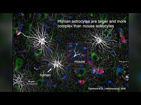






Astrocytes, which constitute approximately 90% of brain volume, function as critical modulators of brain activity through their release of signaling molecules like adenosine. This astrocyte-derived adenosine regulates sleep-wake states by acting on neuronal A1 receptors to promote slow wave activity during sleep, and its dysfunction contributes to various brain disorders including depression, epilepsy, and Alzheimer's disease. The discovery that astrocytes release neurotransmitters and modulate synaptic transmission has fundamentally changed our understanding of brain function, revealing that glial cells are not merely passive support cells but active participants in neural circuit regulation.
Astrocytes and Sleep: How Glial Cells Control Brain Function
Added:Basic neuroanatomy, specifically distinguishing between neurons and various types of glial cells (astrocytes, microglia, and oligodendrocytes).
The fundamentals of synaptic transmission, including how neurotransmitters are released, received, and cleared from the synaptic cleft.
The two-process model of sleep regulation, particularly the concept of homeostatic sleep pressure (Process S).
The biochemical role of adenosine as a byproduct of cellular metabolism (ATP consumption) and its general function in signaling drowsiness.
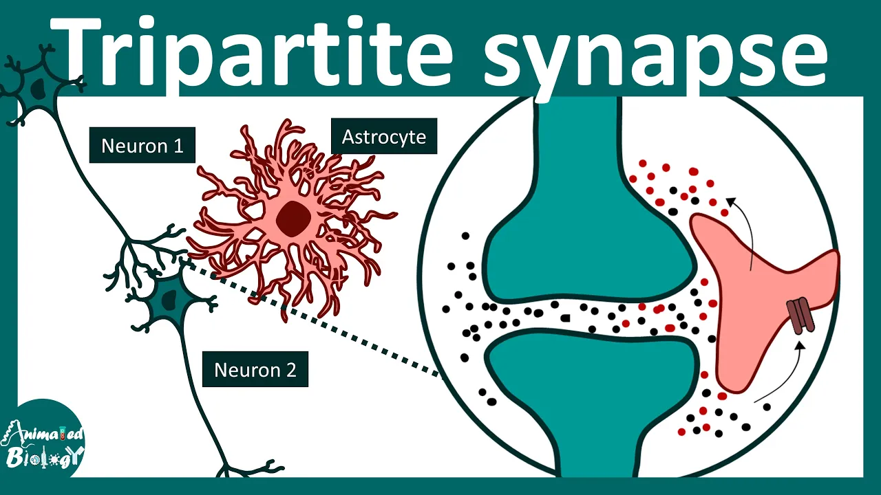
The 'tripartite synapse' concept, exploring how astrocytes actively participate in synaptic integration and information processing.
The glymphatic system, focusing on how astrocytes facilitate cerebrospinal fluid flow to clear metabolic waste during sleep.
The role of glial pathology in neurological and psychiatric disorders, such as epilepsy, major depressive disorder, and neurodegenerative diseases.
Advanced neuropharmacology targeting astrocyte-specific receptors and adenosine signaling to develop novel sleep aids and cognitive enhancers.
473 views5likes1:00:09Original Release: 2013-04-04
Today I have the pleasure of introducing Phil Hayden who's coming from across the street the department of neuroscience where he is the Anata and Gustaf Gryard professor of neuroscience and he's been here at TUS since 2008 or so and before that you started off in England where he got a PhD and um his bachelor's degree in physiology right and then headed to the Midwest in Iowa where he worked as a postocck and did some research there and on his way back to Boston stopped in Pennsylvania where he was in the zoology department and did some research there but we're lucky to have him here to talk about what he does on astro sites which are form gle cells and what they do and why we should care. Okay, thank you.
Thanks very much. It's lovely to be here. I was a little concerned about the introduction because I was told that, you know, I got all the information on you from the internet and when you hear that you get but that's the other Phil Hayden.
Um, so what I'd like to do is tell you a 20-year story. It ted started totally as an accidental observation. We were off on one path in science and there was an observation we could not explain. And over the course of two or three months, we figured out what was going on. And I just changed the direction of research in the lab 180 degrees and said, "It's time to take a risk." And so I will give you a 20-year story. It's actually now a 21-year story, hopefully a little fast in a little bit of a time-lapse pace. Bottom line is I want to tell you that 90% of your brain has been ignored in understanding how the brain functions. And that to understand first how the brain behaves as you're sitting here today we need to understand how the supportive cells ga operate and second almost all disorders of the brain have dysfunction in ga and I'd like to raise the potential with you of thinking of targets within gle cells really as new ways to treat disorders of the brain before going forward I have to disclose that I'm the founder of glycure I'll let you guess what glycure is trying to uh although the work today I will not be talking about the company's uh sciences will be really the academic aspects but from that science I will point you towards opportunities that might be available so the 1900s this is how the brain was thought to operate neurons talk to neurons through synapses and as a result of this this is what how we function today and it's true neurons are really important without neurons we would have a real problem. First, locomotion would be really slow.
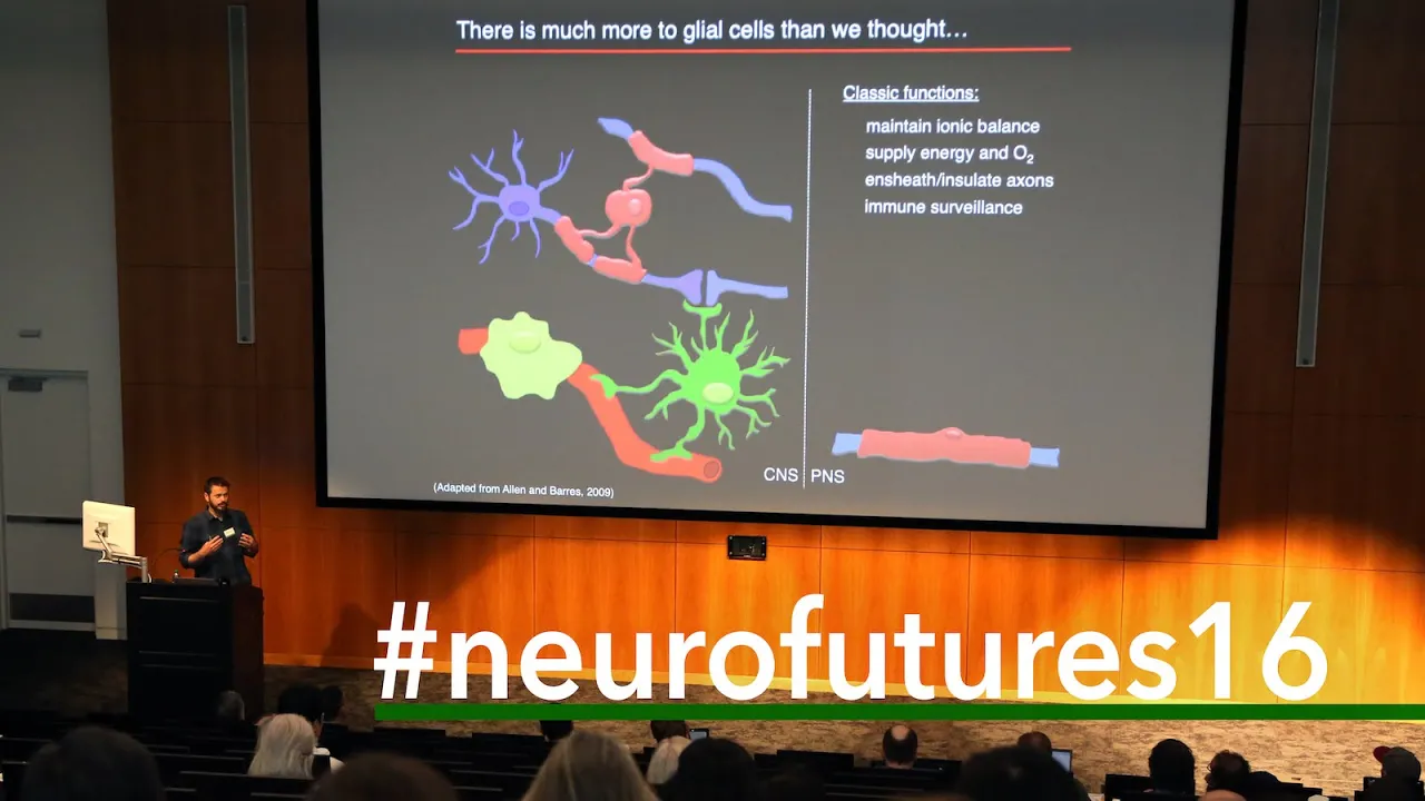
Reflexes would be incredibly slow. But also, neurons are the fundamental element of computation in the brain. However, they only represent 10% of brain volume. One want would one would like to think that the other 90% of the cells in the brain do something important. And these cell types uh do actually do many things and it's worth just taking a moment to discuss them. So here's a presinaptic neuron making synaptic connection onto the dendrite of a post synaptic neuron and the green cell is an illegodendraite and the ilodendrite wraps a milein around the axon which is important for regulating conduction of the action potential. Um in addition to the ilodendrites there are microgle cells.
Microg GA um release cytoines. They also interact with synapses. And more recently, it's been demonstrated that during development as synapses form in an overabundant manner, the microglea use a complement mechanism to actually prune back synapses to refine the connectivity in your brain during the development uh postnatal developmental process. These are also an extremely important cell type that are now heav he heavily being investigated in disorders such as Alzheimer's disease where the pro-inflammatory cytoines um released from these cells and also monocytes invade the brain may be important. I can talk a little bit about our work here later in the discussion time if you wish but I want today to really talk about the discovery made on astroytes which are the largest percentage of gle cell in the brain.
These aststerytes are really intriguing little characters. Very different than neurons. They operate on at least six orders of magnitude slower than neurons, but they do some critical things. And I will discuss those critical things. But for much of the 1990s, we only studied neurons. And it wasn't because we didn't want to study the other cells. We just didn't have the technologies. And this is often the case in science. We have the questions but we don't have the technology that allows us to probe the question. So the 1900s were really dominating by uh by studies of the neurons because we had electrical recording and stimulation techniques. So this allowed us to talk to neurons and listen to their activity and start to decode how they work. But the gal cells are electrically inexcitable. So they're mute and we're unable to understand what they did. And that is why up until about 1990, much of the work in the brain uh studied the neurons and was unable to reveal major things that were occurring in these gal cells. That being said, in the late 1800s, Santiago Roman Cahal did make a statement about his view of what ga may be doing. So a former posttock fellow in my lab, Alfonso Iraq is a faculty member at the Cahal Institute and I so when I visited him, we went to the cow museum.
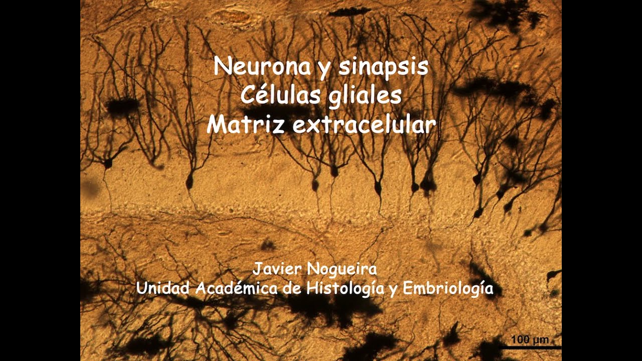
We're actually able to put on the gloves and go through all of the hand pictures that Cahal had drawn. And this is one of them which is taken from the cortex of a man. And here Cahal has drawn the cell types he saw. So these three cells here are neurons. These are the cells we recognize. But this character in the center here is a gio cell. And in fact this is an astrite.
And the structure of the astroite allows you to make some predictions about what it might per uh functions it may perform. So first off its processes touch touch this v vascule contacts the vasculature and contacts the neuron. So the idea was rapidly developed of the notion that the astroite would provide metabolic support to the neuron. Maybe it could provide nutrients, maybe it could take out some of the garbage.
We now know that during synaptic activity the synaptic activity drives biochemical signals within the aststerytes which then talk to the vascule to cause vasoddilation to increase the blood flow during heightened activity. So when you see on discovery channel the beautiful images of functional MRI with changes in blood flow it's actually the aststerytes that are talking to the vascule uh as this intermediate.
Additionally, the astroite makes exuberant contacts and makes them with many neurons. And one idea that's now emerging is that they can coordinate synaptic activity in little volumes in the brain. Now, what Cahel looked at this and in a way that Cahal manages to do everything. He predicted that the astroitic processes may extend and retract and as a consequence regulate sleep and wake. And today I'm going to talk to you about sleep and wake with molecular genetics and how the astroite can influence this. So they could um they extend between neurons act as circuit breakers to facilitate sleep.
But when retracted allows circuits to communicate facilitating wakefulness. So I'm not going to say about extension or retraction but I'll show you if you stay up late tonight and feel drowsy you have to blame your astroite. Your astroite is trying to drive you to sleep. The aststerite releases a compound called adenosine. And a denosine receptor antagonist that make wakes us up is caffeine. So in the morning when you drink caffeine, thank your astroites for allowing you this addiction. So during from 1900ish to uh there was the initial studies on respiration by Lord Adrian where we record respiratory barrage of action potentials in I think 1930. We came through Hodkin Hutsley cats and so on and we understood how neurons work. The revolution technological revolution that occurred that allowed us to gain insight into how these glyos cells uh operate was provided in two manners. First, Roger Chen who later got a Nobel prize on green fluorescent protein developed chemically synthesized fluorescent indicators of calcium that you could introduce into cells. This allowed a readout of the biochemical activity of the calcium signal. The second technological advance was relatively high-speed high sensitivity cameras would that have been developed for the military were declassified and could now be actually used in biological studies.
So in I remember that I remember the day this paper was published. It had this is one of those papers that has had a massive impact on your GA and it was from Steve Smith's lab and what he did he took astraytes put them into cell culture image calcium and added the major excitatory transmitter of the brain glutamate to the dish and he saw calcium signals oscillate. We had no idea what it meant, but this showed that not only can a neuron talk to a neuron, but it has the potential to talk to the astroite. But what does the calcium signal do? We really didn't know. This now comes the accidental discovery. So I was working with a student Vlad Para and we're collaborating with a colleague at the Iowa State Vet School who studied pain and he had dorsal root ganglin expplants in culture. let the axons grow and we take the superfuse and measure glutamate with HLC. So he was able to provide compounds like bradicinine capsain because it was studying paint and he would find that glutamate would be released into the dish. I said well why don't we cut out the expplan and let all the axons die.
So we did this and we thought we had an empty culture dish but the culture dish still released glutamate. So we looked in the microscope and there were these cells. I didn't even know what the cells were. So we later figured out they were migrating out of the dorsal root ganglia and proliferating. So then we went on and did later studies and purified aststerytes. And here is a purified bed of aststerytes. Apply bradicaninine mobilizes calcium. Glutamate is released and this release of calcium sorry this release of glutamate. Calcium is both necessary and sufficient for the release of glutamate.
It's just like a nerve terminal. Calcium is necessary and sufficient. The time scale is different here. This is occurring on a time scale of seconds and minutes as opposed to milliseconds and tens of milliseconds. So astroytes can do something similar. They just do it slower. They can release glutamate. They can also release ATP which will be a major transmitter I discuss today. This is my former postto mentor Ben Kedar and his postto Peter Guthrie uh did a lovely experiment. So when you have aststerytes in culture if you get a calcium elevation in one it will propagate as a calcium wave and the first experiment he did he just cut made a scratch between aststerytes the wave would come to the edge then there was a delay and suddenly the wave would start across this non-neuronal boundary so there must have been a diffusable signal so he then took a pipet and he just sucked the saline above cells that had a calcium elevation and then applied it to cells at a And when he applied that saline that had been collected from around cells that were had excitation, it was able to promote excitation and through methods that you can all anticipate antagonists and luciferin lucis bioassay. He demonstrated that the calcium signal releases ATP. ATP then acts on purinergic receptors to mobilize calcium that stimulates more ATP release and you get this regenerative wave of calcium.
So several transmitters in culture have been identified that can be released from aststerytes. And the challenge was to begin to understand what happens as we get to more intact nervous systems, brain slices, circuits, behavior. And that is the story I'd like you to take you through. By 1998, so we've we just done four year four years in five minutes. So we're on schedule. By 1998, we had discovered that if you now have neurons in contact with aststerytes that the astroite releasing chemicals can modulate synaptic transmission and so we developed the concept of a tripartite synapse whereby the pre there's the presinaptic terminal post synaptic element and they talk to one another.
But the third element is the aststerite and it is very important for modulating how that synapse performs. And in this diagram we're showing a few of the important steps. First off, with presinaptic activity, you get an accumulation of extracellular potassium.
And as every one of you who knows the ner equation, if you accumulate extracellular potassium, you deolarize neurons and you're going to end up with epileptform activity. The aststerytes have a very high resting potassium conductance. So when neurons release potassium, it can be buffered into the astroite. So that's one important uh function. Another is you want to clear away chemical transmitter. For example, if you release chemical transmitter into the cleft and if it stays there, the postsaptic receptors will desensitize and so subsequent action potentials will not have no effect. Astroytes express a high density of transporters that then bind avidly the the transmitter internalize into the astroite and then supply it back to the nerve terminal as renewable transmitter.
But in the early 90s, the breakthrough discovery was they contain receptors.
And the list of receptors probably goes from the top of this building down into the basement. Just as neurons have a plethora of receptors, astroytes have a plethora of receptors. They have metabotropic receptors, ionotropic receptors. But what happens is you don't call a voltage change and an action potential. You call biochemical signaling that's slower.
So from this tripartite syninnapse how can we go from cell culture to insitue to invivo behavior disorders of the brain we needed cell specific molecular genetics because we can pharmacologically manipulate but we can't target a pharmacological agent just to an astroite as opposed to a neuron. So the way we perform this uh I'm going to talk only about ATP and denosine today and mo mostly on adenosine and I'll show you a uh an approach we've taken which can impair the accumulation of adenosine impact sleepostasis and is intriguing in relation to um mediating anti-depressive effects of sleep uh sleep deprivation. So I'm I'm going to give you four little nuggets. Astroytes and sleepasis. We can measure adenosine now in freely behaving mice and make the molecular genetic changes in those mice to see whether it comes from the astroite. We'll do another astroitic manipulation that reinforces that what we have identified in one and two is in fact correct. And then we'll talk about uh anti-depressive like behaviors in mice. And then at the end we can wander into many directions. For example, we're now doing all these studies in aging mice in mice with trans genes to make them have an Alzheimer's like phenotype and we see very clear phenotypic changes. So, how do we perturb the system? I'd like to say we're smart, but we're not. We're copycats. We just like to use the literature to our advantage.
And in the field of synaptic transmission, it's known that exocytosis, this is just the corner of a vicle, uh requires a protein complex called the snare complex consisting of three proteins and four snare domains that form a complex that's necessary to allow a visical diff to fuse. The blue protein also named vamp 2. Uh we expressed this, this is Chiang's thesis.
He expressed the snare domain in the absence of the vesicular tether and by doing that he was able to show that if I just he just accepts this expresses this domain it competes with the indogenous proteins and prevents the complex forming. If the complex doesn't form you don't get regulated exocytosis. That was his thesis. Don't tell him I summarized it in one slide.
So with that knowledge we then went on and said we need to make a mouse in which we can express just this snare domain only in aststerytes and we make it conditional. So we work with Ken McCarthy's group and the results are summar because this is published I'll just give you the summary. So we use the tetracyc system the tet off mouse and we first need to get cell selectivity and second conditional expression. So we used the human GFAP promoter to drive a transactivator TTA. We then made mice with a TET operator that's responsive to the transactivator to drive the snare domain and a reporter gene EGFP.
So we take one mouse containing the G GFAT TTA gene. The others carrying this mate them and then offspring that carry all of these genes in aststerytes because this is an astroitic promoter.
We will get transgene expression but so that it's conditional we include doxycyc in the food at the time that we bring the mother and father together.
Doxycycline binds to the transactivator and prevents it from inducing gene expression. So at the time that we put the appearance together in the cage, doxycyc in the food and we keep them on a diet of doxycyc until the mice are beyond weaning. So we're then we remove doxycyc over two weeks trans genes are expressed. So we're only studying these effects in adult mice. So is it expressed in aststerytes?
So these sort of green bushes here are the incredibly fine processes of the astroite. We have never found this to be we've counted thousands of cells. We have never found it to be expressed in neurons NG2 ga aligodendrites nor in microglea only in astroytes. So here in pink is the parameal uh neurons aver1 no transgene expression. So what does it do? So our next accident we actually when we made this mouse we were trying to do something else. Everything that's good in my lab is an accident. Uh actually I believe when people find things that we don't predict that's when I really believe the experiments. So I'll summarize what we found. It's known in the people have taken brain slices for decades now and it's known there's a basil level of adenosine that acts on nerve terminals to reduce transmitter release. And what we found was when this trans gene is expressed in the aststerite, this adenosine dependent inhibition of transmission was removed. So let me show you mechanistically how we think this occurs. ATP is released from the astroite and this ATP release is prevented by snare expression. ATP there's ecucleotidases that hydrayze ATP to adenosine. That adenosine then acts on neuronal receptors to inhibit transmission. So when we express the trans gene we remove extracellular adenosine. Now as I mentioned earlier caffeine promotes wakefulness and so we knew that caffeine is an antagonist of adenosine receptor.
So we asked well maybe the astroite influences sleep and wakefulness in some manner. So we went ahead and re took these mice and started to study sleep. So this is a sleeping mouse. We have cortical EEGs and EMGs and it's and this is a tethered approach. You'll also see in the slides I put up here either a picture of a mouse or a picture of a brain slice. So if I forget to tell you whether it's in vivo or in site you'll know which type of experiment we're doing. So we collaborated with Marcus Frank at pen and um learned about EEGs and I because I learned it I'm going to try and explain it to you.
So, EEGs I used to think were squiggly lines, but there's a lot of information that I had no idea was present. Here are the three vigilant states that mice exhibit. Humans exhibit many additional states. During wakefulness, you have high frequency low amplitude EEG activity because it's relatively desynchronized. But when an animal goes to sleep and it switches into nonrapid eye movement sleep, non-REM sleep, you now get synchronous activity at low frequency of neurons because and so you get these higher amplitude low frequency oscillations or slow wave activity. If you then switch from non-REM to REM sleep, you now get more desynchronization and higher frequency low amplitude events similar to wake.
But you can discriminate between wake and sleepfulness because the EMG is in a postural muscle. So when the animal's awake, you see the EMG activity, but when it's asleep, you don't. If you take if you um perform fast fura transform, you can now actually plot the frequency and power at different frequencies. So for example, here you have this theta activity in the mouse. So when it's awake, it switches to non-rem. get now significant representation of slowwave activity in non-REM sleep. Now what's known is the drive for you to go to sleep is proportional to this power of slowwave activity. So if I was to sleep deprive you tonight as soon as you switch into non-REM sleep you would find that your power would be elevated. So what I want to do is first let's check that that is true. Then second, let's make molecular manipulations to the astroite. So let's imagine we've been at work all day. So we've had a baseline day and then you go to sleep at your normal time. So we're I'm going to be showing you zeitgeber times. So this zeitgeber zero is just remember zeggeber zero.
That's essentially when you go to sleep for a mouse. It's when lights turn on because they're nocturnal. But this is the onset of sleep. So we have an elevated slowwave activity because there's some pressure to sleep. Then as the animals sleep that pressure declines. So if you're awoken for example here you've got little pressure to drive you back to sleep. But on the next day we sleep deprive the mice for six six hours and then when they go to sleep you can see there's a significant enhancement in the power of the slowwave activity. And this just this this isn't something we've demonstrated. This is just something that others have shown. So the amount of slow wave activity is proportional to the drive to sleep. So actually I think uh you could argue that we should have got an IRB protocol. We got the cook okay for the students sleep depriving the mice. But maybe we should have had a IRB protocol because students had to come to the lab for a 6 a.m. experiment which some say is cruel and unusual punishment. All right. So let's now perturb the astroite.
first baseline then with sleep deprivation. So the white um white circles represent a wild type littermate mouse and black represents when the snare trans units only in the astroite.
So on a baseline day animals go to sleep slow wave activity is very small. Sleep deprive them and there's a significant attenuation of the increase in slowwave activity or the drive to go to sleep. And we remember we're measuring the electrical activity in the brain, but we're perturbing the astroite. So the astroite is influencing this circuit. This is all well and good and honestly I didn't want to believe it.
But what I did believe was the next experiment is where you monitor the compensatory increase in sleep time with sleep deprivation. So if you were to stay up late tonight, you know what's going to happen tomorrow? You're going to sleep a little longer.
So let's look at that experiment. This is a wild type mouse. On a baseline day, it sleeps about 40% of the time. But then with sleep deprivation, it gets a compensatory increase in sleep time. Express snare in the astroite, they don't compensate. You can sleep deprived. They have the same amount of sleep. It's almost like a party gene.
You can stay up late at night and you can get up and go to work the next morning. There um this is also shown here. This is the amount of compensatory increase in sleep in a wild type versus snare. Is it mediated by adenosine receptors? So we now do the same experiment but we introduce um we have osmotic mini pumps and canula intra cerebro ventricularly and uh either with vehicle or an A1 antagonist and here let's look on the sleep deprivation day the slowwave activity is elevated with vehicle but with A1 antagonist it's not and if you look at the compensatory increase in sleep time here we get 12% increase in vehicle but significant attenuation with an A1 antagonist So this was the work really Mike Alassa who was a former graduate student in the laboratory and Mike and uh Marcus Frank should really be taking the credit I merely talk about it. So let me tell you what our current working model is. So the aststerite releases ATP in a snare sensitive manner to give rise to adenosine. That adenosine acts on neuronal A1 receptors. So we for example we've done conditional A1 knockouts just in neurons and find similar phenotypes.
I showed you that the A1 antagonist has a similar phenotype to the snare. The last enzyme in the hydraysis has now been shown if that that enzyme is knocked out it has the same phenotype.
Interestingly in humans with polymorphisms in adenosine diaminise which reduce their ability to metabolize adenosine you that leads to greater adenosine and greater drive to go to sleep. So both human and mice data are in agreement with one another. But you're all being very forgiving because we've not actually measured a denosine. So can we measure a denosine?
So we've taken uh two strategies. First we'll talk about brain slices then I'll talk invivo. So in the brain slices what we we did we said well if we isolate brain slices at different times of day would there be a different level of denosine relatively simple experiment you might say so again now we're in the hippocample slice and we'll just come to this summary graph here so remember zeitgeber zero this is the end of wakefulness zeggeber 4 the animals have been asleep And here we've then performed a sleep deprivation for this 4hour period. So they've stayed awake. So a wild type animal adenosine tone and this is by monitoring synaptic transmission and the relief of the A1 inhibition is elevated but significantly less in a snare animal. If you let the animal sleep before you cut the brain slices, adenosine tone declines and there's no difference with a snare animal. And I want to point to back to this in a moment. But if you sleepdeprive the animals for this 4-hour period, nice adenazine tone that's astroite and snare dependent. So for all the students and postocs in the audience, I want to point something out very important. If you only ever did your experiment at this time of day, you would say the astroite has no contribution. Whereas if you ca if you were an early bird, you'd say they do have a contribution.
And so actually one thing I want to promote to many people now is do experiments at more than one time of day. Most of the experiments we do in rodents are during the light phase which is when they should be sleeping. And we do learning and memory tasks during the light phase. Can you imagine me waking you up at 3:00 in the morning and say take the SATs. And so this is where I think we have to start thinking reverse light cycle. Do some studies in light phase.
do some others in dark phase and you can then really begin to understand what's going on. The other important thing is the brain slice has a memory of the time of day that we isolated the brain slice. Presumably the intrinsic biochemistry is different. Well, this is not a direct assay of adenosine per se. So we wanted to now because we're using um synaptic transmission and adenosine antagonist to do this. So we want now to measure adenosine using bio sensors. So these bio sensors are really fantastic devices and what it is is we'd like to measure adenosine and use amprometric detection but adenosine is not good for amprometric detection. So instead you coat the electrode with enzymes that metabolize adenosine to a product that can be amperometrically detected. So this enzyme uh sorry this electrode is coated with adenosine diaminase nucleioide phosphorase and zanthine oxidase which allows adenosine to be metabolized to hydrogen peroxide and we can then detect an electron uh when we polarize the electrode to plus 600 molts. Hydrogen peroxide donates this electron. So we can detect a current but when we detect that current we don't know is it because a denosine was present or maybe inosine or maybe hopanthine. So we have to use two electrodes. One electrode with all three enzymes and one in which the first enzyme is missing. Calibrate both electrodes then subtract the signals to give us a pure adenosine measurement. So Ian Schmidt has done this in the laboratory. And here's an example. This is in a brain slice. So here he's got the current from an a the three enzyme bioensor. We'll call it a denosine bio sensor. the two enzyme biosensor innocine bioensors and then with subtraction. So he places an adenosine bioensor gets a current brings in the inosine bio sensor then we subtract those signals here then he removes the innocine bio sensor. So now this adenosine bio sensor is measuring all of the metabytes in the dish. And what we do is we measure the difference between this signal with both bio sensors as opposed to when both bio sensors are out of the brain slice followed by a calibration. So let's do this in in site you zeitge zero animals have just have been awake. Here's a different wild type the paired bioensor signal subtracted compared to out of the brain slice. So this is our basil adenosine. If the animals have been allowed to sleep, we take them at zeitgeber six, there's less adenosine. But if we sleep deprive them from 0 to six before cut and slice, there's elevated adenosine. Sneer animal, lower adenosine, lower denosine, lower adenosine. So this is what you see here.
The animals been awake. Elevated adenosine that is astroite and sneer dependent. By zabber 6, there's no difference between the two. The aststery was providing the adenosine here. But if we sleep deprive, we now have continued elevated adenosine that's astroite dependent. So that's nice. Let's go on and do this now in vivo. And this was been a technical challenge. And this is Tamar Bloodstein and Ian Schmidt. And I remember they're not here, I don't think. So I can actually make this comment. I remember sitting at the table in my office saying you have to do this in vivo. You have to have two bio sensors in vivo and EEG in EMG. and they said it would be impossible. I said, "Leave the room and come back when you figured it out." And give them their credit. They figured it out. And these experiments were working absolutely beautiful. So this is a mouse. It's got two bio sensors into his hippocampus. It's got the EEG and EMG electrodes. And this is showing the placement of a bio sensor into area C1 of the hippocampus. So let me show you RO choices.
EEG, EMG, the adenosine, the three enzyme bio sensor and the two enzyme bio sensor. And here the animal is just waking and sleeping at will and then we wake it ourselves. Remember during non-REM sleep you have the high amplitude event. Okay, so this is nonREM and there's low activity in the postural muscle. We wake an animal up during sleep deprivation. more activity in the EMG, the lower amplitude wakefulness signal and these bio sensors change. All right, so let's now score this for the different vigilant states, calibrate, subtract, and see what we find. So here's an animal spontaneously switching between vigilance states and then we wake it up. Here, for example, the animal wakes up on its own, adenosine rises, goes back to sleep, it declines.
We wake it, adenosine rises.
We wake it again. A denosine rises. This is quite incredible. The mouse is just moving around. It's in its cage. And you see denosine going up and down, up and down depending on whether it's awake or going to sleep. So now let's take that sleep deprivation period and average across all animals where time zero represents the synchronization of wake u enforced wakefulness. So the animals were in non-RAM sleep. Adenosine was declining.
We wake them up, it increases, then they go back to sleep. Is this regulated in vivo by an astroite snare mediated process? Of course, you know it's going to be right because I asked the question. Yes, this is awesome. We're This is I can't tell you how tough these experiments are. And uh so the hypothes so what we believe is happening is that ATP is released and we're ATP is much a much more difficult cookie to measure because of the high activity of the enzymes out here. Uh hydrayzeed to adenosine the axon A1 receptors promote slow wave activity and now we've measured it express snare when the animal wakes up. If you express snare adenosine doesn't rise. All right.
So I haven't really provided you any evidence that hydrarolysis is needed. So let's now take another mouse. Let's take a mouse where CD73 is knocked out. The last enzyme required for hydraulysis of ammp to adenosine. Way cool. These results came in last week. So it's my brain is really enjoying them.
So what that says is not only is it a snare sensitive release that gives rise to adenosine but hydrarolysis of the higher energy precursors is required to give rise to that denosine. This is enforced wakefulness. Spontaneous wakefulness works as well. So here again we synchronize the timing at time zero with a transition to a spontaneous wakefulness. Adenazine declines, the animals wake and we get about 100 nanomolar increase in adenazine that is snare sensitive. The the results with CD73 knockouts are still being analyzed.
It takes a long time to go through this data. But this is you might guess I'm a little excited.
We wake we have a snare sensitive release of ATP. We get a hydraulic ecucleidase dependent accumulation of denosine that regulates slow wave activity. In humans there's adenosine diaminise polymorphisms regulated adenosine and slowwave activity. There's also experiments done in mice with adenosine kynise manipulations. And if you increase adenosine kynise you phosphorate adenosine and you reduce extracellular adenosine reduces sleep pressure. If you do the opposite it increases denosine increases sleep pressure. There's always the possibility that things are going on we don't understand. So we wanted to find an alternative way of attacking the same problem and ask truly is the astroite involved. So one of my um colleagues Mike and Netagar published years ago that conexins can regulate ATP release from aststerittes.
So we thought what if we can knock out conxin 43 would we see a similar phenotype? Now conexin 43 is expressed in the heart and in the brain it's expressed only in aststerytes. So if you have a conexin 43 knockout it's a lethal mouse because your heart doesn't work. So we've now used a mouse in which we cross again the astroitic promoter with crebinise to flux conexin 43. When you do this you lose a substantial amount of the conexin 43 in the brain.
You're all experts now at interpreting all these sleep diagrams and hopefully these sleep diagrams are not raising your adenosine levels too much. So here is a baseline period increased slow wave activity a little pressure to sleep as you sleep it dissipates conexin 43 knockout flap. Is that only conexin 43 or manipulating all other connections are not being changed and conexin 43 is sufficient to generate the calcium?
Yeah. The question is is it only conexin 43 or other conexins changing? So for example there's conexin 30 in astroytes and in this particular line conxin 43 is only changed and there's no compensatory change in connection in 30 and there was a public there was a a um presentation by Christian Jean a week ago and what he showed that was that he could take brain slices and look at these slow oscillations that underly slow wave activity and he found that if he manipulated connects in 43 he could change those through an identity dependent pathway. But if he directly manipulated connection 30, there was no effect. So with sleep deprivation, we see the similar effect. So this is good.
It's supportive. The astroite might be doing something important. You now know how to make adenosine biosensor measurements. So here's our adenosine signal. Then both electrodes out of the slice, but with the connection knocked out, there's less adenosine, which is shown here. And note this is at zygraber zero after the animals have been awake when we know there's the increase in adenosine. So these two animals seem similar but as you look further there are actually differences and here is the hourby hour percent wakefulness nonrem sleep and REM sleep. So from zgabber zero through to 23. There's no difference between a snare and a wild type but there is in the connection 43.
For example, in the conexin 43, they are awake less in the dark phase. There are some differences. Bottom line is in terms of the sleep homeostatic phenotypes, they're identical showing the astroytes important, but the conexin 43 has more phenotypes and there's more down into the basement. What we think is happening is conex the idea is kexin 43 actually forms a nexus that organizes several other proteins in the astroite and by perturbing kexin 43 we perturb the snare pathway but also other pathways which gives rise to additional phenotypes but importantly this says the aststerite contributes to the control of adenosine and sleep homeostasis.
Well, rather than going on and telling you more about the intricacies of the molecular pathways and receptor trafficking, I thought I would now bring this to an interesting area. We've been we've now been using these types of mice in studying several different translationally relevant phenotypes. And I want to talk to you about depression. So, first, how do you study depression in a mouse? You can ask it if it's depressed, but if it answers, you go see a psychiatrist.
It's extremely hard to study depression in mass. You can't study depression. But there are certain behaviors that are have comp core phenotypes related to human depression. So I'm just going to try and say depressive like and I will leave it there. What is known is that um about 60 to 70% of the of depressed patients can respond to a night of sleep deprivation with an elevation of mood. So what we've done is we've asked well does sleep deprivation change the parameters we're measuring in depressive like phenotypes and if it does does that require the astroite and we've used three different um measurements. Initially for a fast screen we use the proud swim task and the second the tail suspension and then later we use sucro's consumption to model anonia. I consider this task to be much like if I was to fall off my sailboat in mid-Atlantic. If I was depressed, how long would I swim before I came be gave up became immobile. So we look at amount of immobility. So this is a um a t like a top view through a time series of images of a mouse swimming in a tank. So if you imagine collapsing all of those images into one frame. And so we're now looking in pseudo color at the amount of time in those frames that the mouse was in one location. So if it was in one location a long time, it's a warm color showing immobility. So this is a wild type mouse which was allowed to sleep as much as it wanted. If we sleep deprived the mouse for a full night in advance of the task, you can see it looks like there's less immobility.
We only ever swim the mice once because the act of swimming in this task will change their behavior. So we look at the histograms. Sleep deprivation significantly reduces immobility. If we express snare within the astroite, sleep deprivation is no longer has a significant effect on immobility. Now as an important control which I didn't discuss earlier but was present in those experiments we then take mice where we keep them on a diet of doxycyc so the trans genes aren't expressed. So this controls for insertional effects of the transgene if doxycyc is present sleep deprivation is still effective. Okay. So trans gene on sleep deprivation is ineffective. Trans gene not expressed is ineffective.
Tail suspension test sleep deprivation reduces immobility. Snare expression in the astroite prevents it. Interestingly, we did chronic treatment of mice with aramine for 14 days. And when we use do a mipramine and follow with a force swim pass, there's a reduction in immobility. But this is not sensitive to snare expression in the astroite either suggesting sleep deprivation acts through a different pathway or that the aipramine compon sensitive component is upstream of the snare pathway. So does this asteric ATP adenosine contribute? So we've gone to adenosine one receptor knockout mice. Hopefully from this amino chemistry you can see the A1 receptor is missing. uh and in addition A1 antagonist delivered into the brain. A1 receptors are present throughout the body. So we can't control here for any systemic effects of A1 knockout. So we deliver our antagonist to the brain. And we ask does do we see similar effects? So here is our control. Sleep deprivation reduces immobility. But in an A1 knockout, sleep deprivation is ineffective. And with intracererebra ventricular delivery of an A1 antagonist, sleep deprivation is ineffective. Tail suspension test, we get the same results. Sleep deprivation action immobility is sensitive. It requires the A1 receptor both pharmacologically and with the knockout mouse.
So is it that the mouse needs to be awake or is it that by staying awake adenosine is elevated to act on A1 receptors? Is there a way we can dissociate these two? So what we decided to do was say let's add an A1 receptor agonist to stimulate the downstream effect and let the mice go to sleep and when they wake up what would we find? So we did intra cerebral ventricular administration of an A1 agonist. We used EEG to confirm the agonist was working and then we did the swimming experiments. So with vehicle delivery this was in the for swim task the immobility time. If we provided the A1 agonist through the nighttime but the mice slept when they woke immobility was reduced just as if we had sleepd deprived them. Then in other mice, we didn't deliver the agonist any longer. We assessed them 24 hours later and there was still significant reduction in immobility and this recovered over um several days. Importantly, when we measure the EEG, you can see the EEG, there's changes in uh in in uh slowwave activity in the EEG which is recovering on a similar time course. So the actual I'll call it the structure of the EG and the circuit behavior is changing in addition to this immobility. We then looked at sucrose consumption. So it's a very different task and delivering this agonist also it changed sucrossse consumption suggesting on those very different behavioral tasks that when we bring this together we like to conclude that the effects of sleep deprivation known in humans we can model to a degree in mice and the anti-depressive like effects of sleep deprivation in mice require the astroite and the adenosine pathway now if I was to give you some elicit it drugs, you would become more mobile. So, it's important in all of these experiments that we ask whether this is specific to these tasks or whether we're just promoting hyperactivity. So, I've not shown you all the controls, but if we look at locomotivity, for example, there's no hyperactivity by modifying doing sleep deprivation, by making transgenic manipulations, knockouts, or pharmacology. It's very specific for the tasks which is why we're concluding that they're affecting this mouse uh component of depressive light behavior.
So you're probably sick of this diagram by now, but you know what they say. Um long-term learning requires multiple trials. So hopefully by now you're starting to get the idea that what we think is happening when an animal wakes up, we get a receptor-driven release of ATP that gives rise to adenosine. That adenosine signals on neurons and work going on indicates that that regulates um NMDA receptor trafficking and and learning processes. 70 million people in the United States have a chronic sleep disturbance of some sort. And sleep disturbance is a comorbid with many disorders of the brain. And we have a continuous chicken and egg problem. How much is the sleep disturbance a consequence of a primary pathology? And how much does a sleep disturbance contribute to the pathology?
And this is a problem that's emerging. One area we're very interested in is epilepsy with kids. when they go on vacation, they stay up late night, late at night, and they often can have a breakthrough seizure even though they're normally controlled by their um pharmacological agents. So, sleep deprivation is known to reduce the threshold for seizures. So, we're taking these same mice and Jiron Clazodonte is doing continuous video EEG monitoring of epileptic mice for 5 months and is finding that this pathway has significant modulation of seizures.
Sally Macccyver is looking into alcohol phenotypes. This is the favorite project in the lab. Um, for example, one of the major indicators of relapse in a recovering alcoholic is sleep uh a lack of sleep consolidation. And so Sally is finding that the sleep homeostatic pathway which regulates adenosine and the ability of alcohol which inhibits the uptake of adenosine that there's a convergence in the sleep homeostat. I've talked about depression and now we're working in aged mice and in Alzheimer's mouse models looking at this sleep homeostthenic pathway.
There's major problems with sleep in in Alzheimer's patients and Parkinson's patients. And so our goal is by understanding the basic biology of how astroytes work with synaptic networks.
We can be understand how the brain works and then begin to gain insights into disorders of the brain. But before you think that I think that it's all about GLEA, this is how I really think the brain works. The brain is a team and the brain requires a fast car and a driver to get to the finish line. Those are your neurons. But the difference between coming first and second can be way how well your engine is tuned, whether or not you get gas on time, whether or not you get your tires changed. And the pit crew are the gal cells. and they are there monitoring and responding and tuning this high-speed network so it can have optimum performance. And if the car doesn't work properly, if the driver doesn't work properly, and if the pit crew doesn't work properly, you don't have a good race. And so what we're now doing is really looking at ways to optimize the pit crew to facilitate neuronal function. The people who've done the work are Ian Schmidt and Tamar Blutstein on all the bio sensors. Dustin Hines did the depressive like studies.
Mike Halasa was the original one who got us into sleep and Jerome and Dustin have been doing the connection 43 work and these are our collaborators Nick Dale for bio sensors, Ted Ael for some learning and memory task and Marcus Frank for EEG, Elelliana Skeis for the connection 43 and this is the NIH that funds us. Thank you very much.
Thank you very much. Um, we're open for questions. Alan, that was a wonderful talk. Thank you. And um, so I I I don't know anything about this field. So, me neither. Excuse the question. But am I correct that I saw that the change in the amount of sleep that you started with when you start to monitor that venison tone was about 10%. Is that correct? So, after all this action is done, we're talking about 10%.
So um I don't so the adenosine doesn't change in the snare animal it doesn't change the amount of time awake so but it's the pressure to go to sleep and so something I haven't shown you is the latency for example from lights on until the animal's first going to sleep so that's like you're laying on your pillow at night and with the astroite very important for let pushing you to sleep right then the total amount of sleep time under a baseline condition has not changed when you sleep deprived mouse you go from about 40% uh recovery sleep in baseline to about 50% recovery sleep uh after sleep temperature and that differential is near is that the differential you're talking about yes I was just wondering it seems like um so when you look at all the molecules that change that are engaged and you're measuring what seems to me to be a small parameters like 10% difference. So another way that you rationalize it in terms of 25%. Yeah, exactly. And and and how how easy can you sort of specifically identify particular processes when you only have a measure that's changing little percent. It's a 25% in the change in the total amount of uh sleep that they're having. But it's only 10% in the day.
And it's very easy to measure extremely easy, very reliable. Yeah. And these EEG measures, I've been surprised how reliable they are in the measurements.
And there's a lot more going on. If we have another three hours, I'll tell you the other things going on as well. But what's good about this? The idea now is if we activate an acidic receptor cause a change in denine within minutes, you see an increase in symmetric NMDA receptors. So one of the ideas um is that when you wake up the adune pushes MBA receptors into the synaptic cleft so that you are perfectly primed for synapse specific learning but then one of the downsides is A1 receptors inhibit the production of cyclic AMP which is needed for memory consolidation. So when you go to sleep tenin drops which will now allow cyclic AMP to go and consolidate memories. So we have memory consolidation phenotypes as well that I didn't do as well as uh LCP and learning phenotypes.
I'm wondering if you can talk a little bit about the flexibility of this system you've described in terms of aging. I think you you talked most of the first part of the your talk the experiments were done in younger animals. How does that how would that be? How would that look if they were older? So I'll tell you the results of studies that came in last week where it's not yet ready for publication but it's intriguing and what we're doing is we're using animals. So these were mainly like 8 to 12 weeks of age. So we've been using animals at 5 months and 10 months of age that are littermates of animals that carry a uh they're called 5x FAD mice that carry some of the human genes for Alzheimer's disease. They lay down amaloid plaques and so on. And what we see is preliminarily at five months of age the wild type animal phenotype is fine. By 10 months of age they're starting to get more sleep fragmentation and the ability of the mice to respond to sleep deprivation is impaired. So there's like and so one thing we want to do now is well how is the control of adenosine different? If we introduce the Alzheimer's gene, what you see at five months of age, they have an accelerated age phenotype that's normally age dependent. So at five months, they're not responsive to sleep deprivation.
They have more of the sleep transplantation. So these are things we want to continue bringing forward. And what is the cause? I don't know, but maybe I'll give you a I love long- winded answers. In the epileptic brain, in human temporal lobe epilepsy, there's an enzyme glutamine synthetase that disappears. and glutamine synthetase uh glutamate is taken up converted to glutamine and then it can cross the extracellular space without activating receptors and it turns out inhibitory transmission requires glutamine for a renewable source of GABA. So in the epileptic brain this precursor of GABA is gone. So we just perturbed the brain in a mouse even just by introducing a virus astroytes lose glutamine synthetase. Now gabaurgic uh system is compromised and in the region of the brain you have massive epileptform like activity. The aststerytes are very plastic. You get a slight change in their environment or an injury to them and their whole biochemical processing goes. So what we're anticipated the hypothesis is that with age that what we're going to do is the enzymes that regulate this adenosine pathway are going to become disregulated. And I I'll have another example if we run out of questions if you remind me.
So you mentioned earlier that some of the gal cells are associated with the vascule some of those images. Is there any role for how how some of these adaptations take place that sort of appear for example. So most of these aststerittes are polarized and have one process that goes to the vascule and the other process is wrap synapses. So probably all of the astroytes are in contact with the vascule and whether there's becoming more of an awareness as I'm sure you're aware that changes in the brain lead to changes that you can detect in the blood and vice versa. And the relative role of microglea versus aststerytes for regulating cytoine release and what comes into the blood is a hot topic of debate at the moment or dis investigation. Yeah.
Thanks for the talk. Um I'm wondering about the other side of the coin from the depressive like behavior if there are any tools to behaviorally look at episodes of mania and mice or if anyone has considered the role of astrocated events in signaling. Now mania is an interesting one because it's some people some people say uh that the final common pathway of mania is sleep deprivation.
You sleep deprive someone can trigger mania then they don't sleep. It's just a vicious circle. But I don't know of any work going on in uh in modeling mania in mice. Sometimes acrob I feel a little like that.
Um this past you believe it's a sight specific role or global.
So I I anticipate that different regions of the brain the astroite operates differently in that I mean you you start off and you think let's think of a dopamineergic rich brain region as opposed to a glutamate rich it's going to be different you can envision that there are some regions of the brain that are responsive to sleep deprivation and others are not and in those that are not I wouldn't anticipate this and a student uh Jennif is studying another brain region where she sees there's different signaling events going on. So I think we're going to have very high degree site specificity even within the hypoc campus. For example, in area C3 where there's a moss fiber terminals the astroytes interact the whole synapse. In area CA1 you just get a finger of an astroite associated with 50% of the terminals. So you can imagine in one area that will probably take up glutamate there'll be no spillover. In another region they have the opportunity for spillover. So I think every synapse, every brain, even sub regions has the potential to be different. And it's just such a we're sort of where the neurosciences were in 1930. We got a lot of low hanging fruit. We got to figure out which ones to pick first. Let's thank again our speaker [Applause]
Up Next

5:27
Microglia: Structure and Function in CNS Inflammation
@khanacademymedicine
123K views•2013-09-24

21:55
Deciphering Neural Circuits Controlling Anorexia in Mice | Richard Palmiter
@AllenInstitute
2.3K views•2011-10-10

4:10
Vagus Nerve (CN X): Anatomy, Nuclei & Functions Explained
@Alilamedicalmedia
305.2K views•2022-10-31

13:03
How Exercise Benefits Your Brain: Science Explained
@TED
11.4M views•2018-03-21
Related Study Plans & Knowledge Roadmaps
Structured learning paths in Neuroscience
neuroscience
Appetite Regulation: Brain Circuits & Hormones
01Foundations of Endocrinology and Brain Anatomy
02Introduction to Energy Homeostasis
03Peripheral Signals: Leptin, Ghrelin, and Insulin
+ 2 more learning modules
14h5 modules
Explore Roadmap
neuroscience
Neuroeconomics: Value, Risk & Brain Mechanisms
01Foundations of Neuroscience and Brain Anatomy
02Introduction to Neuroeconomics and Subjective Value
03Dopaminergic Prediction Errors & Reinforcement Learning
+ 3 more learning modules
18h6 modules
Explore Roadmap
neuroscience
Spatial Navigation: Cells, Grids & Brain Maps
01Foundations of Brain Anatomy and Memory
02Place Cells and Head-Direction Cells
03Grid Cells: The Brain's Coordinate System
+ 2 more learning modules
12h5 modules
Explore Roadmap
neuroscience
BCIs: Neural Decoding & Prosthetic Control
01Neuroscience Foundations: How the Brain Signals Intent
02Signal Acquisition: Recording the Brain's Activity
03Neural Decoding: Translating Brain Waves into Data
+ 2 more learning modules
25h5 modules
Explore Roadmap