Brain Aging: Healthy vs Pathological Changes
Learning Goal: Analyze the cognitive and neurological changes associated with healthy versus pathological aging, and formulate a lifestyle-based cognitive training and social engagement plan to build cognitive reserve in older adults.
- Prerequisites: None (Introductory neurobiology concepts will be covered in Module 1).
- Estimated Total Study Time: 12 Hours (Includes video lectures, analysis exercises, and plan formulation).
Module 1: Neurobiology Basics: How the Brain Works
Module Overview
To understand how the brain ages, we must first understand how it functions under baseline, healthy conditions. This module introduces you to macroscopic brain anatomy (with 3D visualization), the microscopic pathways of neural communication across synapses, and the specific functions of the prefrontal cortex (PFC) and hippocampus—the two regions most vulnerable to both healthy aging and pathological cognitive decline.
Recommended Videos
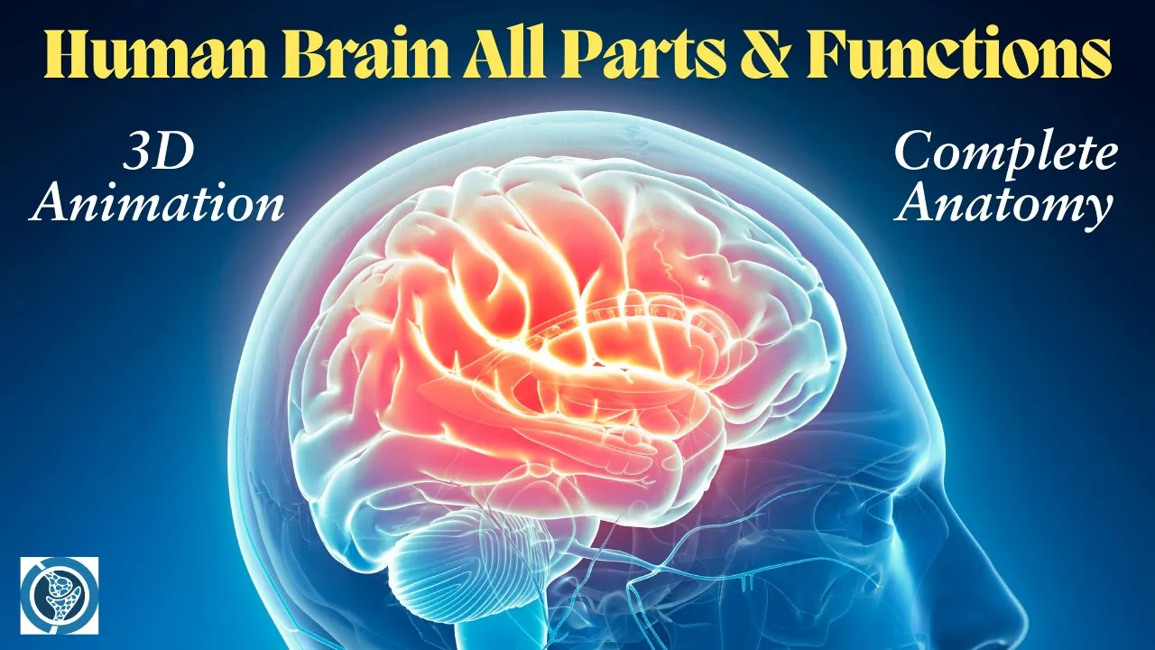
- Why this video: This video uses 3D animations to bridge the gap between abstract anatomy and spatial reality. It provides a visual walk-through of the cerebrum, frontal lobe, and temporal structures, laying the structural groundwork needed to locate key cognitive regions.

- Why this video: Understanding cognitive decline requires a cellular-level perspective. This video details synaptic transmission, illustrating how electrical action potentials trigger the chemical release of neurotransmitters across the synaptic cleft, and how this process governs neural circuit processing.

- Why this video: Dr. Wendy Suzuki delivers a highly focused breakdown of the two crucial brain regions involved in aging: the prefrontal cortex (which governs executive functions, focus, and personality) and the hippocampus (which converts short-term memory to long-term memory).
Knowledge Checkpoint
- Label the four primary lobes of the cerebrum and locate the prefrontal cortex and temporal lobes on a diagram.
- Explain the step-by-step chemical and electrical process that occurs when a presynaptic neuron transmits a signal to a postsynaptic neuron.
- Contrast the primary functional responsibilities of the prefrontal cortex (executive control) with those of the hippocampus (consolidation of declarative memory).
Module 2: Healthy Aging vs. Dementia and Pathology
Module Overview
As the brain ages, structural and chemical changes naturally occur, leading to benign cognitive slowing. However, pathological processes deviate sharply from this trajectory. This module draws a clear clinical line between normal, age-related cognitive slowing (e.g., minor multitasking delays, occasional word-finding blocks) and neurodegenerative pathologies such as Alzheimer’s Disease and Vascular Dementia.
Curriculum Note: While clinical resources separating subtle early-stage pathology from healthy slowing can be scarce, this module relies on comparative expert diagnoses and neuropathological criteria to clarify these distinctions.
Recommended Videos

- Why this video: This clinical video provides clear criteria for distinguishing benign forgetfulness (e.g., misplacing keys temporarily) from pathological cognitive impairments that alter everyday function, helping learners avoid false positives when evaluating age-related changes.

- Why this video: Dr. Najeeb outlines the cellular neuropathology of Alzheimer's disease. He breaks down the formation of extracellular beta-amyloid senile plaques, intracellular neurofibrillary tangles (hyperphosphorylated tau), and the downstream consequence: progressive synaptic loss and cerebral atrophy.

- Why this video: Renowned dementia care expert Teepa Snow maps brain performance changes over time. She demonstrates how healthy aging exhibits predictable, marginal declines (starting after age 25 at a rate of roughly 0.01% to 0.1% annually) compared to the rapid, functional step-downs associated with progressive dementias.
Knowledge Checkpoint
- Differentiate between normal age-related cognitive changes (e.g., occasional word-finding difficulties with intact wisdom/intellect) and pathological symptoms (e.g., progressive disorientation, loss of executive autonomy).
- Describe the structural abnormalities that characterize Alzheimer's disease at the cellular level: beta-amyloid plaques and hyperphosphorylated tau tangles.
- Explain how vascular changes and cumulative mini-strokes contribute to the step-wise cognitive decline seen in vascular dementia, as opposed to the gradual decline of Alzheimer's.
Module 3: The Science of Cognitive Reserve
Module Overview
Why do some individuals possess advanced Alzheimer's pathology (amyloid plaques and tau tangles) upon autopsy, yet showed zero symptoms of cognitive decline during their lives? This phenomenon is explained by Cognitive Reserve and Brain Reserve. This module explores the theoretical models of neuroprotection pioneered by Dr. Yaakov Stern, distinguishing between passive structural hardware (brain reserve) and active software-based optimization (cognitive reserve).
Recommended Videos

- Why this video: This is a comprehensive, deep-dive lecture by Dr. Yaakov Stern himself. He explains how cognitive reserve allows the brain to tolerate pathology without clinical expression, discussing the lifetime epidemiological factors (education, occupational complexity, cognitive engagement) that build this resilient buffer.

- Why this video: A clear conceptual resource that defines the difference between the passive model (Brain Reserve—e.g., absolute brain volume, number of neurons, synaptic density) and the active model (Cognitive Reserve—e.g., efficiency, flexibility of neural networks, recruitment of alternative pathways).

- Why this video: This concise summary reinforces Dr. Stern's landmark 2002 framework, focusing specifically on how individuals utilize functional compensation to bypass physical brain damage.
Knowledge Checkpoint
- Define the passive model of Brain Reserve (structural capacity, neuron count, synaptic density) and contrast it with the active model of Cognitive Reserve (functional efficiency, alternate network recruitment).
- Summarize Dr. Yaakov Stern's core hypothesis regarding why individuals with high cognitive reserve can cope with more neuropathological damage before showing clinical symptoms.
- Identify three major lifetime factors that act as clinical proxies or builders of cognitive reserve.
Module 4: Lifestyle Medicine: Exercise, Nutrition & Sleep
Module Overview
If cognitive reserve is the brain's defense system, lifestyle medicine is the toolkit used to build it. This module covers the physiological mechanisms of three main lifestyle factors: high-intensity aerobic exercise (which triggers Brain-Derived Neurotrophic Factor/BDNF and neurogenesis), deep non-REM sleep (which activates the glymphatic clearance system to wash away amyloid-beta), and the MIND diet (which lowers inflammation and protects the blood-brain barrier).
Recommended Videos
- Why this video: Dr. Wendy Suzuki explains the chemical cascade of exercise. Movement triggers the release of neurotransmitters (dopamine, serotonin, noradrenaline) and, crucially, Brain-Derived Neurotrophic Factor (BDNF), which acts as a "fertilizer" to stimulate neurogenesis in the hippocampus.

- Why this video: Dr. Matthew Walker explains the glymphatic system. He describes how, during deep non-REM sleep, glial cells shrink, allowing cerebrospinal fluid to wash through the interstitial space and clear toxic proteins like beta-amyloid before they aggregate into plaques.
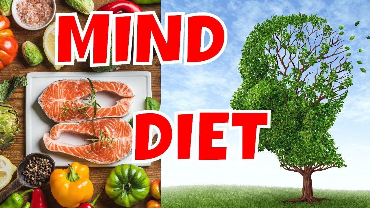
- Why this video: This video details the MIND Diet (Mediterranean-DASH Intervention for Neurodegenerative Delay), outlining the specific foods (leafy greens, berries, nuts, olive oil, fatty fish) and nutritional mechanics that lower inflammation and support cerebral blood flow.
Knowledge Checkpoint
- Explain how aerobic exercise induces the release of BDNF, and outline the pathway through which BDNF promotes neurogenesis and synaptic plasticity in the hippocampus.
- Describe the function of the glymphatic system during deep sleep, explaining how astrocyte structural changes allow CSF to flush metabolic waste out of the brain.
- List the primary dietary components of the MIND diet and explain how they help maintain vascular health and reduce neuroinflammation.
Module 5: Cognitive Training, Neuroplasticity & The Social Brain
Module Overview
Can you train your brain like a muscle? This module analyzes the science of neuroplasticity in older age and evaluates the efficacy of computerized "brain games" versus real-world skill acquisition. Crucially, this module addresses a key gap in standard longevity models by focusing on The Social Brain. We will examine how social isolation acts as a toxic stressor that accelerates brain atrophy, and why social connection is one of the most powerful protective factors against cognitive decline.
Recommended Videos

- Why this video: This video offers a balanced, science-based critique of brain training software. It explains the concept of "near transfer" vs "far transfer"—showing that while computerized games make you better at the game itself, they rarely translate into improved daily cognitive functions.

- Why this video: This video challenges ageist assumptions about learning capacity. It shows how the brain retains neuroplastic potential throughout life, demonstrating that learning complex new motor and cognitive skills remains highly achievable in older age.

- Why this video: This video presents findings from a UK study of 462,000 participants, showing that social isolation increases Alzheimer's risk by 14% and vascular dementia risk by 17%—comparable to the cardiovascular damage caused by smoking.

- Why this video: Professor Barbara Sahakian reviews neuroimaging data linking social isolation to structural brain changes, including reduced gray matter volume in temporal regions and a 26% overall increase in dementia risk.
Knowledge Checkpoint
- Define the terms "near transfer" and "far transfer" in the context of cognitive training, and explain why learning a new language or musical instrument offers superior protective benefits compared to playing digital brain games.
- Describe how social isolation alters brain structures (such as temporal lobe volume) and quantify its impact on dementia risk based on recent epidemiological studies.
- Outline how to design an effective cognitive training and social engagement plan for an older adult that combines physical, cognitive, and social components.
Course Map
Below is the recommended learning flow and module dependencies:
Key People Index
- Dr. Yaakov Stern (Columbia University) — Pioneer of the Cognitive Reserve model. His epidemiological and neuroimaging research explains why stimulating mental activities build resilience against brain pathology.
- Dr. Wendy Suzuki (New York University) — Renowned neuroscientist specializing in neuroplasticity. Her work focuses on how aerobic exercise stimulates hippocampal neurogenesis through BDNF release.
- Dr. Matthew Walker (University of California, Berkeley) — Author and leading sleep scientist. His research highlights how deep sleep activates the glymphatic system to clear beta-amyloid and tau proteins.
- Professor Barbara Sahakian (University of Cambridge) — Clinical neuropsychologist whose work links social isolation to structural brain changes and increased dementia risk in older adults.
- Teepa Snow — Leading dementia care specialist who translates neurodegenerative changes into practical, empathetic caregiving strategies.
Final Self-Assessment
Complete this comprehensive self-assessment to verify your mastery of healthy versus pathological brain aging:
- I can label a diagram of the brain with the prefrontal cortex, temporal lobe, and hippocampus, and state their cognitive functions.
- I can explain synaptic transmission, including how neurotransmitters move from the presynaptic terminal to postsynaptic receptors.
- I can list three key differences between normal cognitive slowing (e.g., slower processing speed, word recovery) and pathological cognitive decline (e.g., loss of daily functional independence, visual-spatial disorientation).
- I can describe the microscopic hallmarks of Alzheimer’s disease: beta-amyloid plaques (extracellular) and neurofibrillary tangles (intracellular hyperphosphorylated tau).
- I can define the difference between Brain Reserve (physical, structural assets) and Cognitive Reserve (active functional and network adaptability).
- I can identify the biochemical pathway through which physical exercise triggers BDNF production to promote neurogenesis in the dentate gyrus of the hippocampus.
- I can describe how the glymphatic system clears metabolic waste during deep non-REM sleep, and explain why chronic sleep deprivation increases neurodegenerative risk.
- I can list five key foods recommended by the MIND diet and describe their protective cardiovascular and anti-inflammatory properties.
- I can explain why digital "brain games" usually fail to produce "far transfer" effects, and contrast them with the benefits of learning complex, real-world skills.
- I can summarize the statistical and physiological consequences of chronic social isolation on temporal lobe volume and overall dementia risk.
- I can write a personalized lifestyle plan for an older adult that combines physical movement, cognitive challenges, and social engagement to actively build cognitive reserve.