Glial Cells: Astrocytes, Myelin & Microglia
Learning Goal: Analyze the active roles of glial cells in brain function, focusing on how astrocytes modulate synaptic transmission, microglia prune connections, and oligodendrocytes facilitate signal propagation via myelination.
- Prerequisites: Basic biology (familiarity with cells and membranes). No prior neuroscience training is required.
- Estimated Total Study Time: 9 hours
Module 1: Foundations of Neurobiology: Neurons & Synapses
Before diving into the complex world of glial cells, we must master the primary communication network they support. This module covers how biological neurons generate electrical changes (action potentials) and cross-communicate using chemical neurotransmission at the synaptic gap.
Recommended Videos
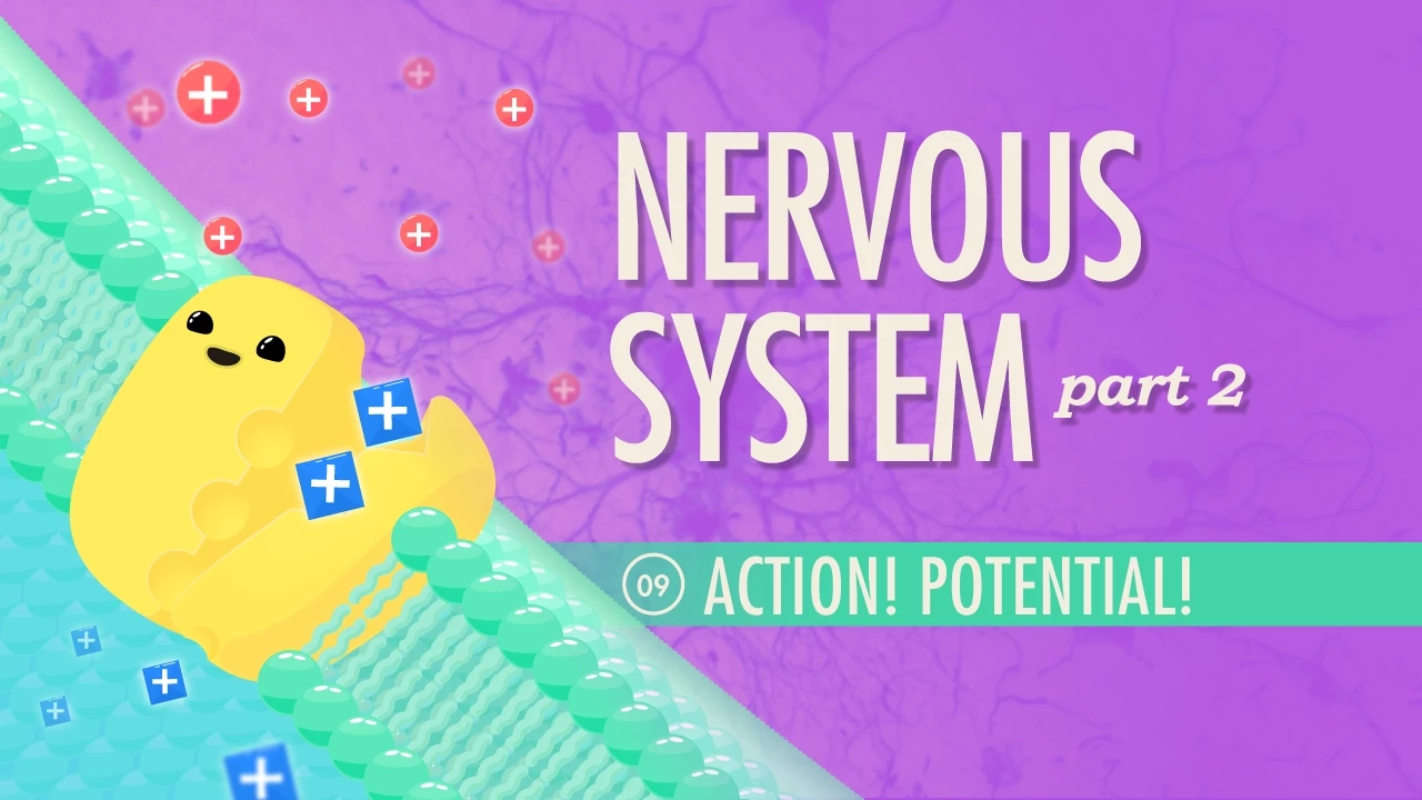
Why this video: This fast-paced, high-yield animation provides the essential background on resting potentials, membrane polarity, and the cascade of events that generates an electrical action potential down the neuronal axon.
Knowledge Checkpoint:
- Define the specific threshold voltage (typically ) required to trigger an action potential.
- Contrast the roles of voltage-gated sodium () channels during depolarization and voltage-gated potassium () channels during repolarization.
- Explain the "all-or-none" law of nerve impulses.
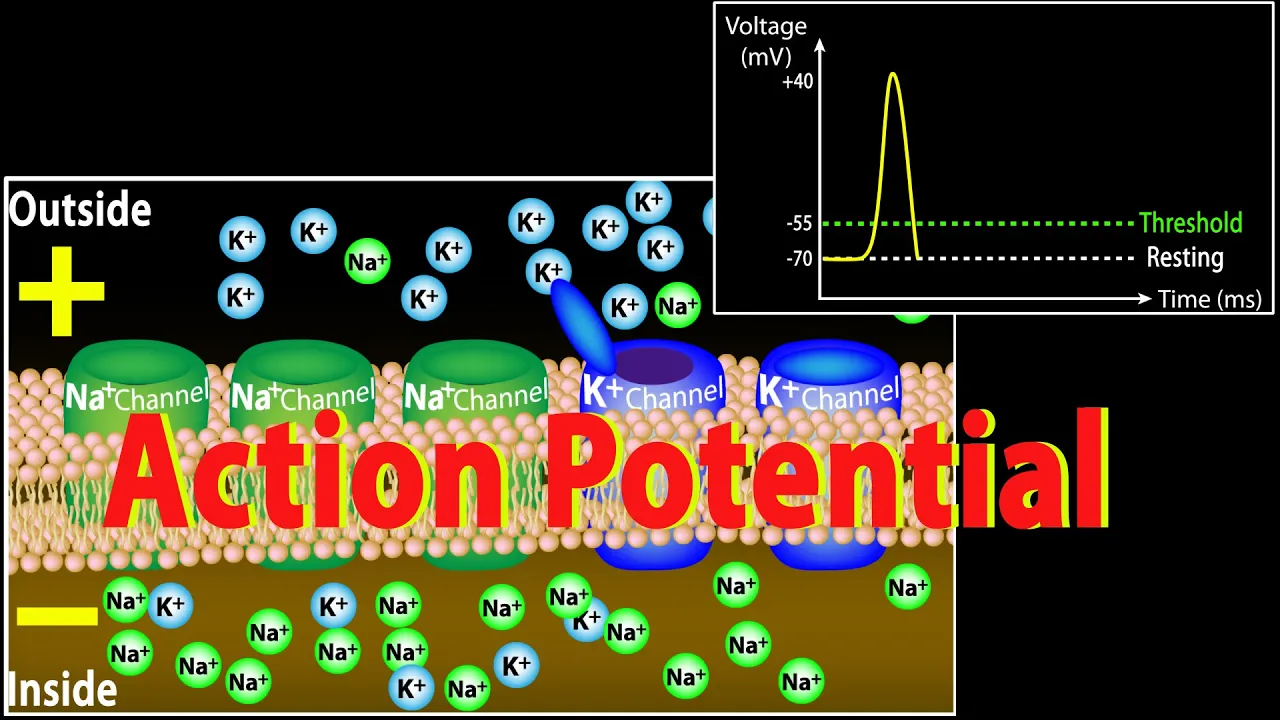
Why this video: A clear, step-by-step visual dissection of ionic shifts. It maps out membrane voltage changes on a classic coordinate graph, visually reinforcing how ions physically travel through the cell membrane.
Knowledge Checkpoint:
- Identify where the hyperpolarization (undershoot) phase occurs on an action potential graph and why it happens.
- Describe the function of the Sodium-Potassium Pump ( ATPase) in restoring resting concentrations after an action potential.
Why this video: Dr. Neela Bakore uses highly pedagogical chalkboard-style illustrations to break down chemical synapse architecture. It clarifies how an electrical impulse translates into chemical vesicular release.
Knowledge Checkpoint:
- Draw or describe a chemical synapse, explicitly labeling the presynaptic terminal, synaptic cleft, and postsynaptic membrane.
- Explain the key role that calcium () influx plays in initiating the exocytosis of neurotransmitter-filled vesicles at the terminal.
- Explain the functional difference between excitatory and inhibitory receptor activation on the postsynaptic side.
Module 2: Introduction to Glia: The Brain's Hidden Half
For over a century, glial cells were dismissed as simple structural filler ("glia" is Greek for glue). This module challenges that dogma by introducing the major classifications of glial cells within the central nervous system (CNS) and their general roles in keeping the brain functional, healthy, and organized.
Recommended Videos
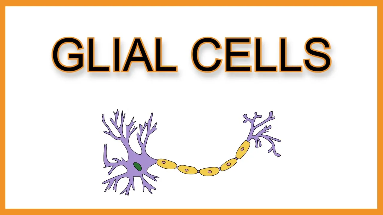
Why this video: An exceptionally clear, memory-friendly overview of glial taxonomy. It maps out the five primary glial cell lines, systematically organizing them by location (CNS vs. PNS) and core functions.
Knowledge Checkpoint:
- Differentiate between the glial types of the Central Nervous System (astrocytes, microglia, oligodendrocytes, ependymal cells) and the Peripheral Nervous System (Schwann cells, satellite cells).
- State the primary function of each main glial cell type.
- Distinguish the myelin-producing capacity of a single oligodendrocyte from that of a single Schwann cell.

Why this video: This lecture dives into the physiology of neuroglia, discussing their relative abundance, developmental origins, and how they physically associate with neurons to sustain metabolic homeostatic states.
Knowledge Checkpoint:
- Explain how neuroglial cells differ fundamentally from biological neurons in terms of electrical excitability and mitotic capability.
- Explain how glial cell dysregulation can disrupt the brain's extracellular microenvironment.
Module 3: Oligodendrocytes and Myelination
Biological electrical signals must travel rapidly across long distances. This module details how oligodendrocytes act as the insulation specialists of the CNS, wrapping axons in lipid-dense layers of myelin to enable saltatory conduction.
Recommended Videos

Why this video: This video explains how the presence of myelin impacts cable properties, lowering electrical capacitance and increasing axial resistance to drive signal velocity up to 120 meters per second.
Knowledge Checkpoint:
- Define "saltatory conduction" and explain how the signal "jumps" down a myelinated pathway.
- Describe the structural architecture of the Nodes of Ranvier and identify which voltage-gated ion channels are highly concentrated there.
- Explain how myelin decreases membrane capacitance and prevents lateral ion leakage.

Why this video: This concise guide focuses specifically on the unique cellular anatomy of oligodendrocytes, showing how they extend multiple long, flat processes to wrap around different axons.
Knowledge Checkpoint:
- Explain the anatomical origin of the word "oligodendrocyte" and how its morphology reflects its function.
- Contrast how myelination happens in the brain (via oligodendrocytes) with how it occurs in the limbs (via Schwann cells).

Why this video: Focuses on the physical and biochemical properties of myelin. This video illustrates how a glial cell membrane wraps up to 100 times around a single axonal segment, leaving a highly structured lipid sheath.
Knowledge Checkpoint:
- Explain why myelin is primarily composed of lipids (fats) and how this biochemical makeup serves as an electrical insulator.
- Discuss the physical consequences to signal propagation when these lipid layers degrade, as seen in demyelinating conditions like Multiple Sclerosis.
Curriculum Gap Alert: The physical wrapping mechanics of myelin are highly dynamic. While the videos above explain the concepts well, they lack high-definition 3D rendering of the membrane spiraling process.
Independent Search Recommendation: If you need a stronger visual mental model of this wrapping process, search YouTube for: "Myelination and saltatory conduction 3D animation" or "How oligodendrocytes wrap myelin sheath physiology".
Module 4: Astrocytes & The Tripartite Synapse
This module introduces the star of synaptic regulation: the astrocyte. Astrocytes do not merely support neurons structurally; they listen to synaptic communication, clear out excess chemical messengers, buffer ions, and actively respond via internal calcium waves.
Recommended Videos

Why this video: An academic video that defines the tripartite synapse. It explains how astrocytic processes physically wrap around chemical synapses to monitor and modulate neurotransmission.
Knowledge Checkpoint:
- Define the three structural parts of the "tripartite synapse."
- Explain how astrocytes clear glutamate from the synaptic cleft and describe the astrocyte-specific pathways used to recycle it back to the neuron safely.
- Describe the process of astrocytic "potassium buffering" during intense neuronal activity.

Why this video: While neurons communicate using electrical impulses, astrocytes communicate chemically. This video breaks down the mechanism of astrocytic calcium signaling, showing how intracellular calcium surges enable coordinated communication across glial networks.
Knowledge Checkpoint:
- Contrast neuronal electrical signals with astrocytic chemical signaling (calcium waves).
- Explain how a localized signal at one astrocytic process can generate a calcium wave that travels to neighboring astrocytes.

Why this video: Dr. Phil Haydon, a pioneer in the tripartite synapse field, reviews the evidence that astrocytes release their own chemical signals (gliotransmitters) to actively modulate synaptic transmission.
Knowledge Checkpoint:
- Define "gliotransmitters" and list common examples (e.g., D-serine, ATP, glutamate).
- Describe how astrocytic signaling can feedback to strengthen or weaken synaptic communication.
Curriculum Gap Alert: Astrocyte calcium signaling is a complex process. The available video resources are mostly dense academic presentations rather than simplified step-by-step visual animations.
Independent Search Recommendation: To complement these lectures, search YouTube for: "Astrocytes tripartite synapse calcium signaling explained" or "Astrocyte calcium waves animation" for shorter, high-yield educational visualizations.
Module 5: Microglia: Immune Defense & Synaptic Pruning
In this final module, we look at microglia: the brain's resident immune cells. Originally seen as simple defenders, we now know microglia act as active sculptors of neural networks during development and learning, physically eating away weak or inactive synapses through phagocytosis.
Recommended Videos
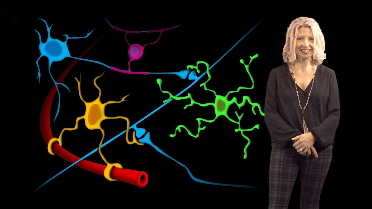
Why this video: Dr. Beth Stevens, a world leader in microglia research, shares her work on how these cells transition between highly dynamic "surveying" states and "active" defense states.
Knowledge Checkpoint:
- Identify where microglia originate developmentally and why they differ from other brain cells.
- Contrast the physical shape of a "surveying" (resting) microglial cell with that of an "activated" (phagocytic) microglial cell.
- Explain how microglia continuously inspect their local environment in healthy brain tissue.

Why this video: Features live in vivo time-lapse imaging that captures microglia in action as they move through the brain, monitor active synapses, and physically prune away weak neural connections.
Knowledge Checkpoint:
- Describe the physical, real-time dynamics of microglia processes as they interact with synaptic connections.
- Explain how activity-dependent synaptic pruning contributes to normal brain development and plastic changes.

Why this video: A brief, clear look at the physical process of phagocytosis ("cell eating"). It illustrates how microglia surround, engulf, and break down debris to keep the brain clean.
Knowledge Checkpoint:
- Define "phagocytosis" and explain how microglia use it to engulf damaged cells, foreign invaders, or unwanted synapses.
- Explain why keeping the brain clear of cellular debris is critical for protecting nearby neurons from damage.
Curriculum Gap Alert: Dr. Beth Stevens' lecture provides excellent scientific context, and Dr. Ruthazer's video shows real live-imaging data, but there is a lack of simplified, step-by-step 3D animations of the molecular signals involved in synaptic pruning (such as the complement cascade).
Independent Search Recommendation: To fill this visual gap, search YouTube for: "Microglia synaptic pruning live imaging animation" or "Complement system synaptic pruning neuroscience animation".
Course Map
This flowchart shows the recommended pathway through the curriculum, highlighting how the modules build on each other.
Key People Index
These researchers have helped transform our understanding of glial cells from simple filler tissue to active partners in brain function:
- Dr. Phil Haydon (Tufts University): A pioneer in studying astrocytic communication. His research helped establish the "tripartite synapse" model, demonstrating that astrocytes release chemical messengers (gliotransmitters) to actively modulate synaptic transmission.
- Dr. Beth Stevens (Harvard/Boston Children's Hospital): A leading researcher in microglial biology. Her work revealed that microglia use immune system proteins (the complement cascade) to identify, target, and prune away weak synapses during brain development.
- Dr. Ed Ruthazer (McGill University): An expert in high-resolution in vivo imaging. His work uses live time-lapse microscopy to show the real-time physical interactions between microglia, astrocytes, and developing neural pathways.
- Dr. Ben Barres (Late Professor at Stanford University): A legendary neurobiologist who spent his career championing glial research. He developed innovative methods to isolate and culture glial cells, proving they are essential for synapse formation, health, and survival.
Final Self-Assessment
Test your understanding of the active role of glial cells across the entire curriculum with this comprehensive checklist:
- Action Potential Mechanics: Explain how an action potential is generated, specifically identifying the roles of voltage-gated and channels during depolarization and repolarization.
- Chemical Synapse Physiology: Describe how calcium influx at the presynaptic terminal triggers neurotransmitter release into the synaptic cleft.
- Glial Taxonomy: Name the five primary types of glial cells in the CNS and PNS, and state their general physiological functions.
- Myelination Structure: Describe how an oligodendrocyte wraps its cell membrane around an axon, and list the major lipids that make up the myelin sheath.
- Saltatory Conduction Physics: Explain how myelin decreases membrane capacitance and increases lateral insulation to allow electrical signals to "jump" down an axon.
- Tripartite Synapse Dynamics: Identify the three components of a tripartite synapse, and describe how astrocytes physically participate in this structure.
- Glutamate Clearance: Detail how astrocytes clear glutamate from the synaptic cleft and convert it into glutamine to prevent toxic buildup (excitotoxicity).
- Astrocyte Calcium Signaling: Explain how astrocytes communicate chemically using intracellular calcium () waves instead of electrical action potentials.
- Microglial Activation States: Contrast the physical shape and behavior of "surveying" (resting) microglia with "activated" (phagocytic) microglia.
- Synaptic Pruning Mechanics: Describe how microglia use phagocytosis to target and eat away weak or inactive synapses during development and learning.