Pain Pathways: Nociception to Chronic Pain
Learning Goal: Analyze the neural pathways of pain perception and modulation, detailing how nociceptive signals travel from peripheral receptors to the cortex, and how descending pathways and endogenous opioids gate pain transmission and contribute to chronic pain states.
- Prerequisites: Basic knowledge of cellular biology and introductory human anatomy.
- Estimated Total Study Time: 12 Hours
Module 1: Foundations of Neurobiology and Signal Transmission
This module establishes the foundational neurobiology necessary to understand signal transmission. You will explore how the nervous system is organized, how neurons generate electrical impulses (action potentials) to carry signals over distances, and how these electrical messages are translated into chemical signals across synaptic junctions.
Recommended Videos
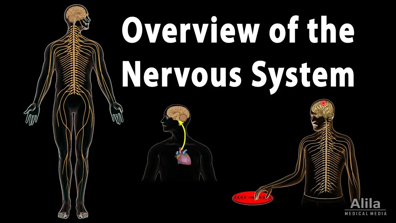
Why this video: This concise, visually clear animation outlines the macro-architecture of the nervous system, showing how the peripheral nervous system (PNS) collects environmental sensory information and transmits it to the central nervous system (CNS) for integration. This structural framework is critical for placing pain pathways in anatomical context.
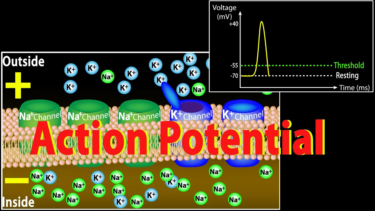
Why this video: Understanding pain signaling requires a firm grasp of the action potential. This video details the ionic fluxes (sodium influx and potassium efflux) that dictate resting membrane potential, depolarization, repolarization, and the refractory periods of excitable neuronal membranes.
Why this video: This video introduces the mechanics of synaptic transmission, explaining how voltage-gated calcium channels trigger neurotransmitter exocytosis to bridge the synaptic cleft, illustrating how a physical action potential becomes a chemical message.
Knowledge Checkpoint
- Describe the structural and functional divisions between the central nervous system (CNS) and the peripheral nervous system (PNS).
- Explain the electrochemical gradient shifts of and ions across each phase of an action potential (depolarization, repolarization, hyperpolarization).
- Detail the cellular cascade starting from action potential arrival at the presynaptic terminal to vesicle docking and neurotransmitter release.
Module 2: Peripheral Nociception: How the Body Detects Pain
This module covers the specialized sensory receptors (nociceptors) that detect noxious mechanical, thermal, and chemical stimuli. You will examine the physiologic differences between fast-conducting A-delta () fibers and slow-conducting C fibers, and investigate the biochemical cascades that drive peripheral sensitization.
Recommended Videos
Why this video: This video introduces free nerve endings (nociceptors) and explains how environmental noxious energy is converted into electrical potentials. It specifically touches on how Transient Receptor Potential (TRP) channels open to generate excitatory graded potentials.

Why this video: This segment offers a direct physiological comparison between myelinated fibers and unmyelinated C fibers. It connects fiber diameter and myelination status to the clinical presentation of "first pain" (sharp, localized) versus "second pain" (dull, aching, diffuse).
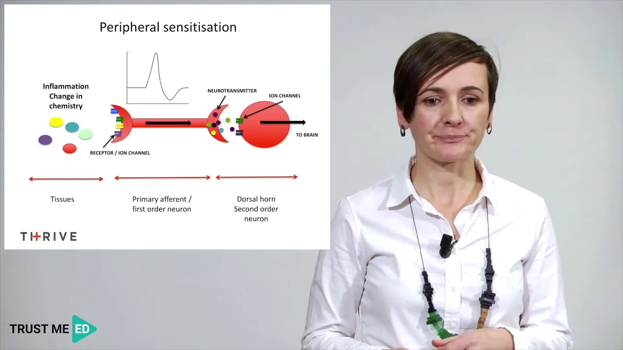
Why this video: This video unpacks the physiological changes in tissue chemistry that follow an injury, demonstrating how inflammatory mediators alter nociceptor threshold responses.
Independent Study Gap Focus
While the provided videos introduce peripheral sensitization and receptor activation, they do not deeply analyze specific molecular transduction channels. To bridge this gap:
- Search Target: “TRPV1 thermal transduction mechanics” and “NaV1.7 sodium channel pain signaling mutation”.
- Key Concept to Research: Focus on how the TRPV1 channel responds to noxious heat () and protons, and how voltage-gated sodium channels like , , and act as threshold generators for action potentials in nociceptive fibers.
Knowledge Checkpoint
- Differentiate between and C fibers in terms of myelination, diameter, conduction velocity, and clinical sensory quality.
- Explain how Transient Receptor Potential (TRP) channels convert physical heat or chemical irritants into membrane depolarization.
- Detail the constituents of the "inflammatory soup" (prostaglandins, bradykinin, histamines) and how they modulate nociceptors to produce hyperalgesia.
Module 3: The Ascending Pain Pathways to the Brain
This module traces pain signals from the periphery into the central nervous system. You will follow the primary afferent fibers as they enter the spinal cord dorsal horn, synapse within the substantia gelatinosa, cross the midline (decussate), and ascend through the spinothalamic tract of the anterolateral system to reach the thalamus and primary somatosensory cortex.
Recommended Videos

Why this video: Dr. Najeeb delivers a detailed, step-by-step lecture on the anatomy of the spinothalamic tract, tracing first-, second-, and third-order neurons from the peripheral receptors to the cerebral cortex. This is a definitive guide to the classical anterolateral system.
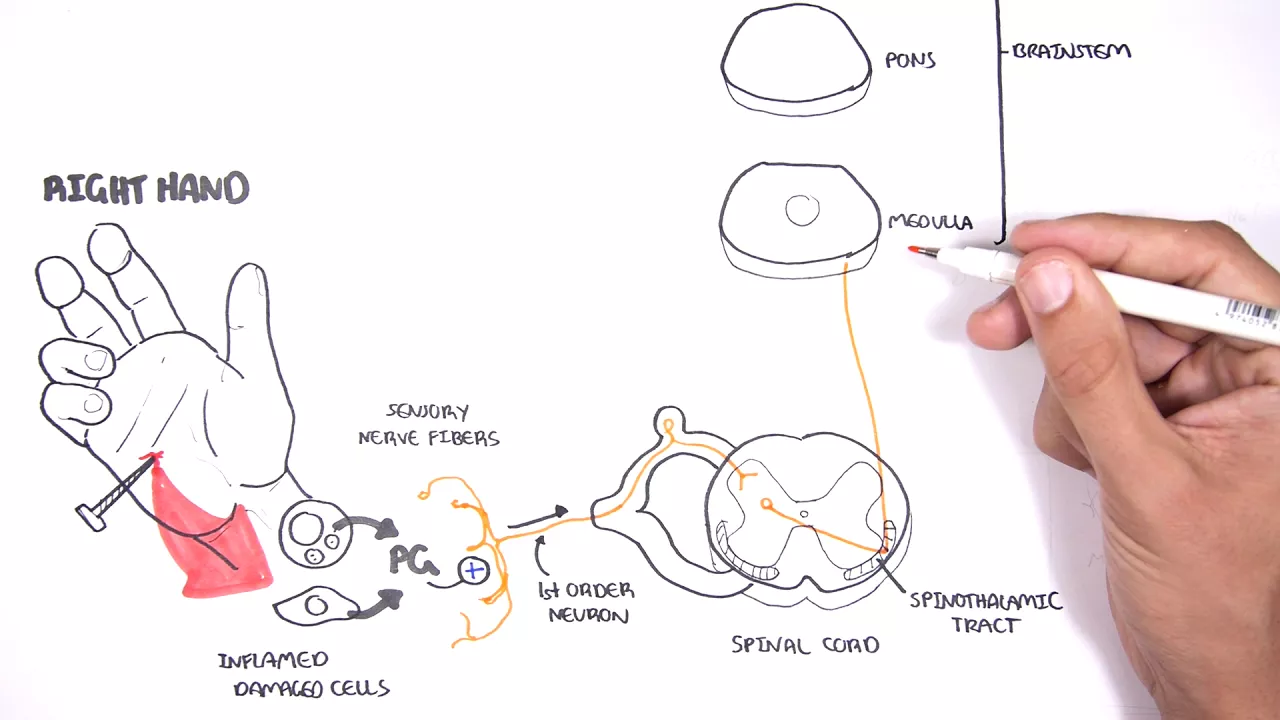
Why this video: This video uses hand-drawn illustrations to clearly map pain transduction, transmission, and modulation. It explicitly illustrates the micro-anatomy of the spinal cord's dorsal horn (including the substantia gelatinosa) and shows where decussation occurs.
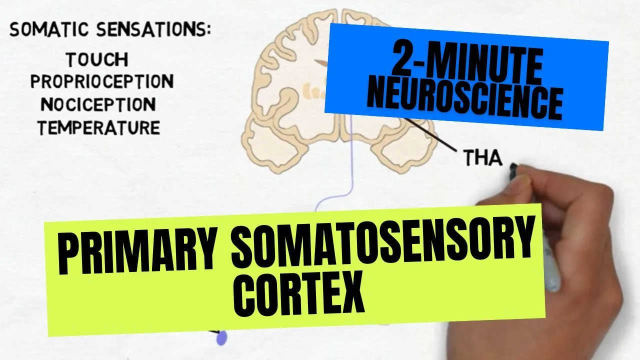
Why this video: This high-yield video covers the primary somatosensory cortex (postcentral gyrus). It describes the spatial arrangement of the cortical homunculus, which allows the brain to map and localize pain to specific body regions.
Knowledge Checkpoint
- Trace a pain signal from a pinprick on the right index finger to the left postcentral gyrus, naming all intermediate synapses, locations of decussation, and tracts.
- Identify the anatomical laminae of the dorsal horn where and C fibers preferentially terminate.
- Define the primary role of the thalamus in pain transmission and identify which specific thalamic nuclei project to the primary somatosensory cortex.
Module 4: Descending Modulation and Gate Control Theory
Pain perception is not a simple, passive recording of injury; it is actively modulated by both local spinal cord circuits and descending pathways from the brainstem. This module covers the classic Gate Control Theory of Melzack and Wall, the descending monoaminergic inhibitory tracts, and the cellular mechanisms of endogenous opioids.
Recommended Videos
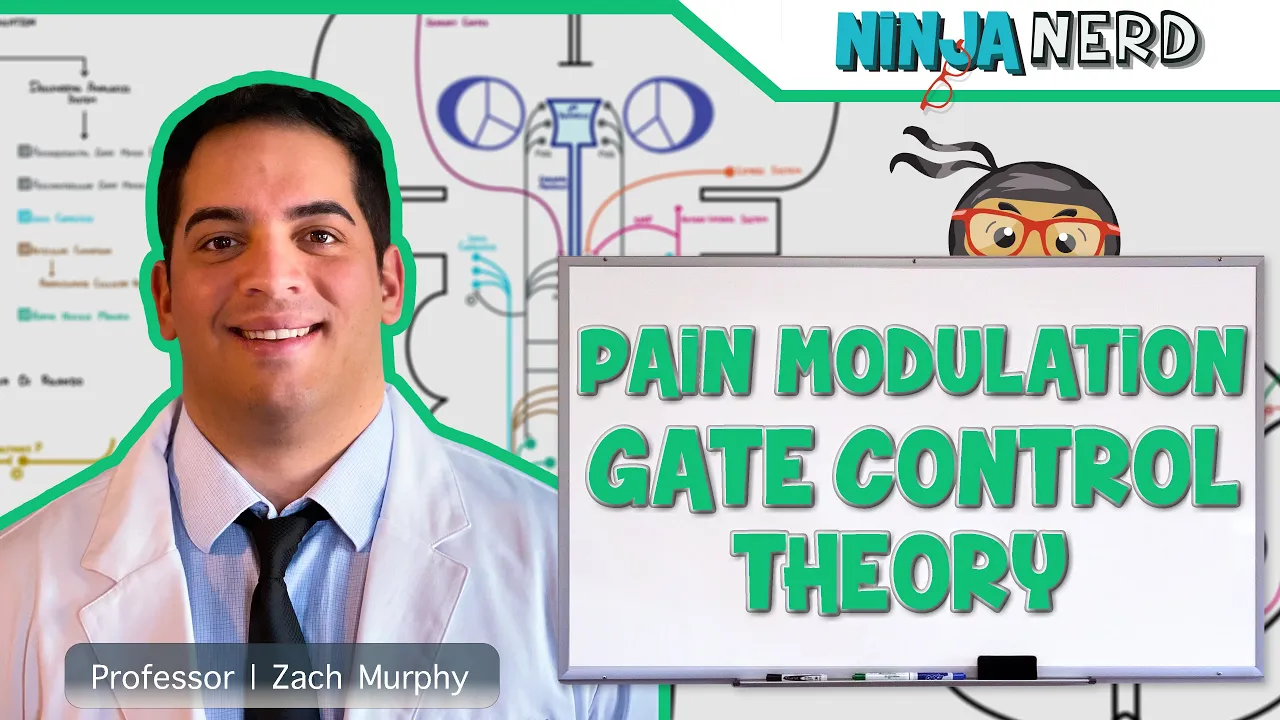
Why this video: Ninja Nerd delivers an exceptional, comprehensive lecture on the mechanical gating of pain. The video illustrates how stimulating large-diameter tactile fibers () activates inhibitory interneurons in the dorsal horn, turning down the volume of nociceptive signals.

Why this video: This structural and biochemical video focuses on GPCR-mediated opioid receptor activation. It explains how endogenous opioids (endorphins, enkephalins) bind to mu, kappa, and delta receptors to inhibit pain signaling at both presynaptic and postsynaptic levels.

Why this video: This video introduces the descending inhibitory pathway. It outlines how higher brain centers engage the Periaqueductal Gray (PAG) and the Rostral Ventromedial Medulla (RVM) to suppress nociceptive transmission at the level of the spinal cord.
Independent Study Gap Focus
To fully master the cellular mechanisms of endogenous opioids at the synaptic level:
-
Search Target: “Presynaptic inhibition G-protein beta-gamma calcium channels” and “Postsynaptic hyperpolarization GIRK channels enkephalin”.
-
Key Concept to Research: Focus on how opioid receptor activation inhibits presynaptic voltage-gated calcium channels (preventing release of glutamate and Substance P) and activates postsynaptic inwardly rectifying potassium channels (causing hyperpolarization of the spinothalamic tract neuron).
 Presynaptic Terminal (Primary Afferent)
       [ Ca2+ Channel ] <-- (Inhibited by Opioid GPCR βγ subunit)
              |
             [X] No Glutamate / Substance P release
              |
-----------------v----------------- Synaptic Cleft | Postsynaptic Dendrite (Spinothalamic Neuron) [ GIRK K+ Channel ] <-- (Activated: K+ efflux) | Hyperpolarization
Knowledge Checkpoint
- Draw a diagram of the Gate Control Theory, illustrating the interactions between C fibers, fibers, inhibitory interneurons, and projection neurons.
- Map the anatomical connections of the descending pain inhibitory pathway, starting from the cortical regions down to the PAG, RVM, and dorsal horn.
- Explain the cellular effects of opioid receptor activation on presynaptic calcium channels and postsynaptic potassium channels.
Module 5: The Neurobiology of Chronic Pain, Sensitization, and Glial Activation
In chronic pathological states, neuroplastic changes and sustained neuroinflammation transform pain from a protective symptom into a disease of the nervous system itself. This module explores central sensitization, the structural rewiring of dorsal horn synapses, and the role of non-neuronal glial cells (microglia and astrocytes) in driving chronic pain.
Recommended Videos
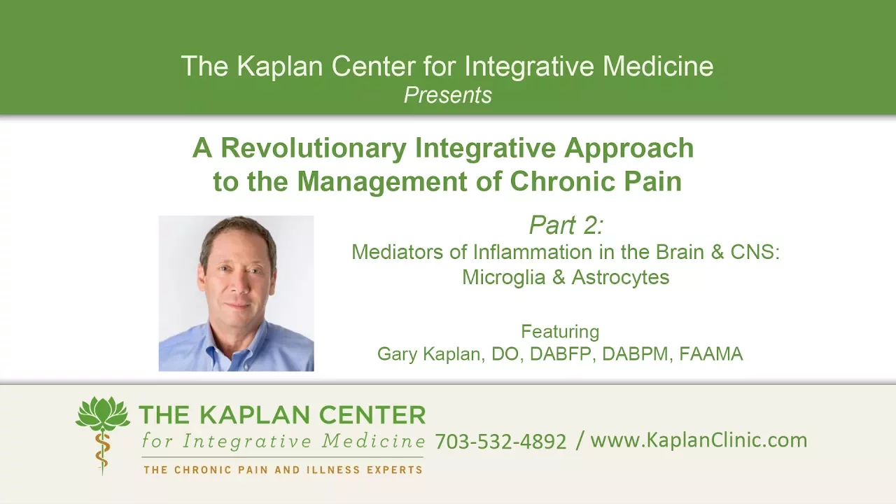
Why this video: This lecture explores neuroinflammation, highlighting how microglia (the CNS's innate immune cells) and astrocytes switch from supportive roles to highly reactive states, releasing proinflammatory mediators that sustain chronic pain.

Why this video: This video explores the differences between acute and chronic pain, defining central sensitization as an abnormal "volume control" setting in the central nervous system. It examines how persistent injury leads to prolonged hyperexcitablity of spinal and cortical pain circuits.

Why this video: Pediatric anesthesiologist Elliot Krane explores how glial cells (microglia) release signaling molecules that act as a "three-dimensional cloud of neurotransmitters," spill out from the synapse, and amplify baseline pain signaling.
Independent Study Gap Focus
While the provided videos cover glial activation, they do not address the specific signaling cascades of microglia-mediated synaptic modulation. To close this gap:
- Search Target: “Microglia BDNF TrkB chloride transporter KCC2 chronic pain”.
- Key Concept to Research: Read about how activated microglia in the dorsal horn release Brain-Derived Neurotrophic Factor (BDNF). BDNF binds to TrkB receptors on second-order projection neurons, causing a downregulation of the potassium-chloride cotransporter KCC2. This alters the intracellular chloride gradient, turning the normally inhibitory actions of GABA and glycine into excitatory depolarizations.
Knowledge Checkpoint
- Define hyperalgesia and allodynia, distinguishing between them in terms of sensory thresholds and underlying physiology.
- Describe the molecular changes that occur in central sensitization, focusing on NMDA receptor activation and the removal of the magnesium block.
- Explain how reactive microglia and astrocytes contribute to central sensitization and neuroinflammation.
Course Map
Below is a flowchart mapping the modules, key concepts, and structural progression of the pain pathway curriculum.
Key People Index
- Ronald Melzack & Patrick Wall: Proposed the Gate Control Theory of Pain in 1965, introducing the concept that non-painful sensory inputs ( fibers) can close a neural "gate" in the spinal cord, preventing pain signals ( and C fibers) from traveling to the brain.
- Dr. Daniel J. Clauw: A leading clinical researcher in fibromyalgia and central sensitization, known for his work in defining chronic overlapping pain conditions (COPCs) as disorders of central nervous system volume control.
- Dr. Elliot Krane: A pioneer in pediatric pain management who helped establish the modern model of chronic pain as a neuroglial pathology rather than simply persistent nociception.
Final Self-Assessment
Complete this comprehensive self-assessment to verify your mastery of the material:
- Action Potential Ionics: Can you draw an action potential curve from memory and label where channels open/inactivate and where channels open/close?
- Synaptic Signaling: Can you explain the roles of calcium, synaptotagmin, SNARE proteins, and ligand-gated ion channels in transmission?
- Fiber Differentiation: Can you explain why fibers produce sharp, well-localized "first pain," while unmyelinated C fibers produce slow, diffuse "second pain"?
- Transduction Channels: Can you describe how the TRPV1 channel changes conformation in response to heat or capsaicin to allow calcium and sodium influx?
- Anatomical Mapping: Can you trace the spinothalamic pathway, identifying the locations of the first-order, second-order, and third-order cell bodies?
- Anatomical Decussation: Do you know exactly where spinothalamic second-order neurons cross the midline, and how this relates to sensory deficits in spinal cord injuries (such as Brown-Séquard syndrome)?
- Gate Control Mechanics: Can you explain how rubbing a bumped knee decreases pain perception using the synaptic connections of mechanoreceptors, C fibers, and inhibitory interneurons?
- Descending Inhibition: Can you describe how the Periaqueductal Gray (PAG) communicates with the Rostral Ventromedial Medulla (RVM) to suppress spinal cord dorsal horn transmission?
- Opioid Pharmacology: Can you outline how G-protein coupled opioid receptors block presynaptic neurotransmitter release and hyperpolarize postsynaptic membranes?
- Glial Neuroinflammation: Can you explain how activated microglia release BDNF to downregulate KCC2, and why this shifts GABA/glycine signaling from inhibitory to excitatory?
- Wind-up & Central Sensitization: Can you describe the roles of substance P, glutamate, and NMDA receptor activation in driving the hyperexcitability of spinal projection neurons?
- Clinical Allodynia: Can you explain the physiological difference between hyperalgesia (increased sensitivity to pain) and allodynia (pain caused by non-painful stimuli)?