Visual Perception: Retina, Cortex & Streams
Learning Goal: Deconstruct the neurobiology of visual perception, tracing sensory signal transduction from the retina through the lateral geniculate nucleus to the primary visual cortex, and exploring the functional divergence of the dorsal and ventral streams in processing motion and object recognition.
- Prerequisites: Basic knowledge of cellular neuroscience (action potentials, synaptic transmission, and GPCR signaling cascades).
- Estimated Total Study Time: 12 Hours
Module 1: Anatomy of the Eye and Light Detection
Module Overview
This module establishes the anatomical and structural framework of visual reception. Before signals can be processed by the brain, light must be focused and refracted by the cornea and lens, projecting an image onto the retina. Rather than treating the retina as a simple light-sensitive sheet, this module unpacks its complex microscopic architecture, examining its ten distinct histological layers. This high-level neuroanatomical mapping provides the crucial context needed to understand subsequent signal processing steps.
Recommended Videos

- Why this video: This exhaustive lecture provides a highly technical, rigorous overview of eye anatomy. It covers the three ocular tunics (fibrous, vascular, and sensory), detailing the role of the cornea and lens in refraction, the ciliary body in accommodation, and the gross spatial arrangement of the retina. This provides the macroeconomic foundation for understanding how light is captured before cellular transduction begins.
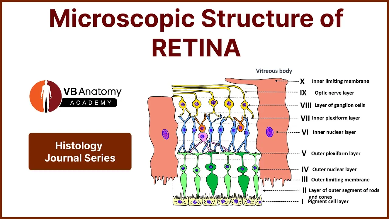
- Why this video: Direct addressing a key gap in standard textbooks, this video details the ten distinct layers of the retina under microscopic examination. It walks through the pigment epithelium, photoreceptor outer segments, external limiting membrane, outer nuclear layer, outer plexiform layer, inner nuclear layer, inner plexiform layer, ganglion cell layer, nerve fiber layer, and internal limiting membrane. Mastering this structural hierarchy is essential before exploring functional synaptic signaling.
Knowledge Checkpoint
- Understand the boundaries and functional roles of the fibrous (sclera/cornea), vascular (choroid/ciliary body/iris), and nervous (retina) tunics of the eye.
- Differentiate between the ten histological layers of the retina and describe which cell bodies and synaptic connections reside in each (e.g., the outer nuclear layer containing photoreceptor cell bodies vs. the inner nuclear layer containing bipolar, horizontal, and amacrine cell bodies).
- Explain how the structural properties of the fovea (displacement of inner layers) minimize light scattering to maximize visual acuity.
Module 2: Retinal Processing and Phototransduction
Module Overview
This module explores the molecular and cellular mechanisms of phototransduction and local retinal computation. You will trace the G-protein coupled receptor (GPCR) pathway that allows photons to hyperpolarize photoreceptors—a counterintuitive process where light reduces the release of the neurotransmitter glutamate. From there, you will study how horizontal and bipolar cells shape receptive fields, establishing lateral inhibition and the classic "center-surround" architecture that allows the visual system to detect contrast rather than absolute luminance.
Recommended Videos
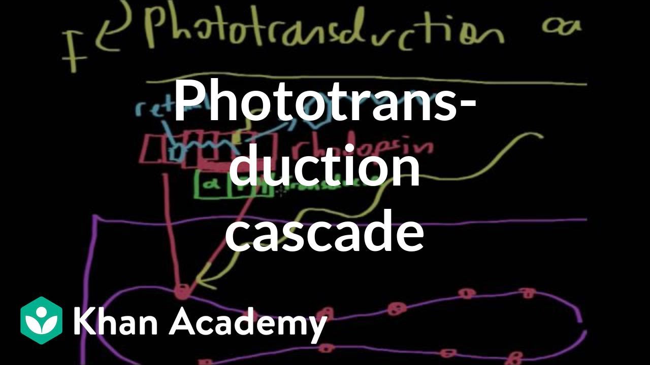
- Why this video: This video offers an exceptionally clear, step-by-step breakdown of the biochemical cascade of phototransduction. It details how the absorption of a photon converts 11-cis retinal to all-trans retinal, conformational changes in rhodopsin, activation of the G-protein transducin, activation of cyclic GMP (cGMP) phosphodiesterase, the resulting decrease in intracellular cGMP, closure of cyclic nucleotide-gated (CNG) sodium/calcium channels, and final membrane hyperpolarization.

- Why this video: This video focuses on the neural circuitry of the outer plexiform layer. It explains how horizontal cells provide inhibitory feedback to neighboring photoreceptors. This lateral pathway is the physiological basis of lateral inhibition and is crucial for creating the contrast-detecting antagonistic receptive fields of downstream retinal ganglion cells.

- Why this video: This animation visually demonstrates the center-surround receptive field organization of retinal ganglion cells (RGCs). It clearly shows how ON-center/OFF-surround and OFF-center/ON-surround systems respond to varying configurations of light and dark, highlighting why these cells are tuned for edge and contrast detection rather than diffuse, ambient illumination.
Knowledge Checkpoint
- Diagram the molecular phototransduction cascade, identifying the roles of 11-cis retinal, rhodopsin, transducin, phosphodiesterase (PDE), cGMP, and cyclic nucleotide-gated (CNG) channels.
- Explain why photoreceptors are depolarized in the dark (the "dark current") and hyperpolarized in the light, specifying the ionic movements involved.
- Describe how glutamate acts on metabotropic receptors (mGluR6) on ON-bipolar cells versus ionotropic receptors (AMPA/Kainate) on OFF-bipolar cells to create parallel pathways.
- Contrast the response of an ON-center retinal ganglion cell when light falls on its center versus when light falls on its surround, explaining the synaptic role of horizontal cells in this process.
Module 3: Visual Pathways: Optic Nerve to the LGN
Module Overview
Once visual signals leave the retina via the axons of retinal ganglion cells, they travel along the optic nerve, undergo partial decussation at the optic chiasm, and project to the Lateral Geniculate Nucleus (LGN) of the thalamus. This module traces this pathway, focusing on visual field mapping and clinical lesion deficits. Crucially, we dive deep into the laminar organization of the LGN, analyzing how it acts as a gatekeeper that segregates visual information into parallel Magnocellular (M), Parvocellular (P), and Koniocellular (K) processing pathways.
   [Left Visual Field]       [Right Visual Field]
         \   /                     \   /
          \ /                       \ /
        [Left Eye]               [Right Eye]
         /     \                   /     \
Temporal/       \Nasal       Nasal/       \Temporal
       /         \               /         \
      |           \             /           |
 [Left Optic]      \           /      [Right Optic]
   Nerve            \         /          Nerve
      |              \       /              |
      |               \     /               |
      |               X Chiasm              |
      |              /       \              |
      \             /         \             /
     [Left Optic Tract]     [Right Optic Tract]
             |                       |
         [Left LGN]             [Right LGN]
       (Layers 1-6)            (Layers 1-6)
Recommended Videos

- Why this video: This lecture segment provides a clear, high-yield explanation of the functional and structural anatomy of the LGN. It explicitly details the division between the Magnocellular pathway (layers 1 and 2, which process motion and low-spatial-frequency contrast) and the Parvocellular pathway (layers 3 through 6, which process color, fine details, and high-spatial-frequency shapes), filling a critical gap in standard online visual pathway overviews.

- Why this video: This comprehensive lecture details the spatial mapping and parallel streams from the retina to the LGN. It covers retinotopic organization (how spatial relationships in the retina are preserved in the thalamic architecture) and explains how nasal fibers cross at the chiasm while temporal fibers remain ipsilateral, ensuring that each side of the brain processes the contralateral visual hemifield.

- Why this video: This video walks through the clinical representation of the visual fields and teaches you how to map lesions in the visual pathway to specific visual deficits. By mapping damage at the level of the optic nerve, optic chiasm, optic tract, optic radiations, and visual cortex, you will learn to identify hemianopias, quadrantanopias, and macular sparing.
Knowledge Checkpoint
- Map the flow of visual information from the left and right visual hemifields to the contralateral hemisphere's optic tract.
- Detail the laminar organization of the LGN: identify which layers are Magnocellular (layers 1-2) vs. Parvocellular (layers 3-6) and explain the functional differences between these pathways.
- Define the Koniocellular pathway, identifying where these cells are located (interlaminar regions) and their primary functional role.
- Predict the visual field deficit resulting from:
- A complete lesion of the right optic nerve (ipsilateral blindness).
- A sagittal transection of the optic chiasm (bitemporal hemianopia).
- A lesion of the left optic tract (right homonymous hemianopia).
Module 4: Primary Visual Cortex (V1) and Feature Detection
Module Overview
Primary Visual Cortex (V1, Brodmann Area 17, or Striate Cortex) is where basic visual features are first extracted by cortical circuits. In this module, you will analyze how inputs from the LGN terminate in specific sublayers of cortical layer 4, and how those signals are integrated to build receptive fields for orientation-selective neurons. You will study David Hubel and Torsten Wiesel's Nobel Prize-winning work on simple and complex cells and examine the columnar architecture of V1, culminating in the concept of the cortical "hypercolumn."
           +-----------------------------------+
           |             HYPERCOLUMN           |
           |  (Processes 1 point in visual space) |
           +-----------------+-----------------+
           | Left Eye Input  | Right Eye Input |  <-- Ocular Dominance
           |  [0°][45°][90°] |  [0°][45°][90°] |  <-- Orientation Columns
           +-----------------+-----------------+
           |   COLOURED BLOB |   COLOURED BLOB |  <-- Wavelength/Color
           +-----------------+-----------------+
Recommended Videos

- Why this video: This academic video dives into the micro-anatomy of V1. It details the six major neocortical layers, showing exactly where LGN inputs terminate (layer 4C, further divided into 4C for magnocellular and 4C for parvocellular streams). It then explains the functional hierarchy of simple, complex, and hypercomplex (end-stopped) cells within these layers.

- Why this video: This historical footage shows Hubel and Wiesel’s actual physiological experiments recording from the cat visual cortex. Hearing the crackle of action potentials as bars of light are projected on a screen provides an unforgettable demonstration of how V1 neurons ignore uniform illumination and instead fire selectively to edges, lines, and movement of precise orientations.

- Why this video: This video tackles the complex structural architecture of V1 by breaking down the "hypercolumn." It explains how ocular dominance columns (representing left vs. right eye input) and orientation columns (representing 180 degrees of orientation preferences) are physically grouped alongside mitochondrial-rich "blobs" (which process color) to form a self-contained modular processing unit for a single point in the visual field.
Knowledge Checkpoint
- Describe the laminar flow of information into and through V1: specify where Magnocellular and Parvocellular LGN axons synapse in layer 4C.
- Contrast the receptive fields of simple cells with those of complex cells, explaining how a complex cell's receptive field can be constructed from the inputs of multiple simple cells.
- Define the term "end-stopped" (or hypercomplex) cell and explain what visual features these cells detect.
- Sketch or explain a cortical hypercolumn, detail its size (approx. 1 mm x 1 mm), and identify its constituent parts: ocular dominance columns, orientation columns, and cytochrome oxidase blobs.
Module 5: The Dual Stream Hypothesis: Dorsal and Ventral Pathways
Module Overview
Once primary features are extracted in V1, visual processing splits into two parallel association pathways. This module evaluates the Dual-Stream Hypothesis. The Dorsal Stream ("Where/How" pathway) projects dorsally to the parietal lobe and handles spatial awareness, motion processing, and visually guided action. The Ventral Stream ("What" pathway) projects ventrally to the temporal lobe, processing color, shape, and complex object/face recognition. You will analyze these streams through clinical and comparative neurology, examining deficits like optic ataxia and visual agnosia.
                                [Dorsal Stream] ---> [Parietal Lobe]
                               / (Where / How)        - Motion (MT/V5)
                              /                       - Spatial awareness
                             /                        - Optic Ataxia (deficit)
[Retina] ---> [LGN] ---> [V1 / Striate]
\ [Ventral Stream] ---> [Temporal Lobe]
(What) - Form/Color (V4)
- Face recognition (FFA)
- Visual Agnosia (deficit)
Recommended Videos

- Why this video: This video offers a clear, structured introduction to the dual-stream hypothesis first proposed by Ungerleider and Mishkin and later expanded by Goodale and Milner. It traces the anatomical pathways from V1 into the parietal and temporal cortices, laying out the fundamental functional differences of these systems.
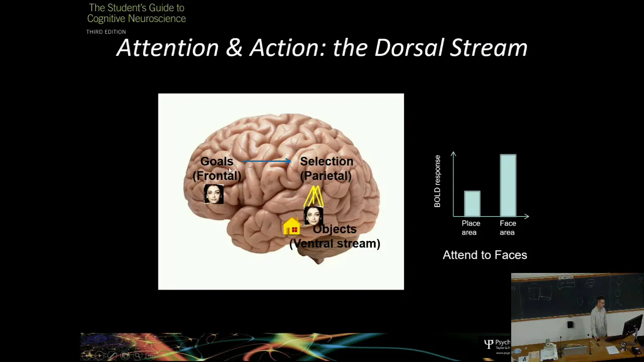
- Why this video: This academic summary addresses a critical learning gap by directly comparing the clinical manifestations of dorsal and ventral stream damage. It contrasts visual agnosia (ventral damage), where patients cannot perceptually identify objects but can accurately reach out and grasp them, with optic ataxia (dorsal damage), where patients can identify objects but fail to coordinate visually guided hand movements to interact with them.

- Why this video: In this archival interview, legendary neurologist Dr. Oliver Sacks discusses visual agnosia, famously illustrated in his book The Man Who Mistook His Wife for a Hat. Sacks describes the clinical presentation of visual agnosia, explaining how patients see visual features clearly but cannot synthesize them into recognition, illustrating the profound specialization of the ventral temporal stream.
Knowledge Checkpoint
- Contrast the anatomical trajectories and endpoints of the dorsal and ventral streams.
- Explain the distinction between the "Where" (Ungerleider & Mishkin) and "How" (Goodale & Milner) interpretations of the dorsal stream.
- Compare the symptoms of optic ataxia and visual agnosia, and map each deficit to its corresponding anatomical pathway.
- Identify the role of specialized cortical regions within these streams, specifically area MT/V5 (motion processing) in the dorsal stream and V4 (color processing) and the Fusiform Face Area (FFA - face recognition) in the ventral stream.
Course Map
This flowchart maps the logical and chronological flow of visual information from the external environment to high-level association cortices, highlighting the parallel processing lines.
Key People Index
- David Hubel & Torsten Wiesel
- Context: Awarded the Nobel Prize in Physiology or Medicine in 1981 for their discoveries concerning information processing in the visual system. They mapped V1's functional architecture, discovered simple and complex cells, and showed how cortical orientation selectivity and ocular dominance columns develop.
- Oliver Sacks
- Context: A British neurologist, naturalist, and author who wrote influential clinical case studies. His work bringing conditions like visual agnosia and prosopagnosia to the public's attention highlighted the modular, parallel nature of visual processing in human clinical cases.
- Mortimer Mishkin & Leslie Ungerleider
- Context: Neuroscientists who first proposed the "Two-Streams Hypothesis" in 1982, describing the separation of visual processing into a dorsal "where" pathway and a ventral "what" pathway based on lesion studies in non-human primates.
- Melvyn Goodale & David Milner
- Context: Re-conceptualized the dual-stream hypothesis in 1992, arguing that the dorsal stream is better understood as a "how" (action-guided) system rather than just a "where" (spatial-location) system, pointing to patients with visual agnosia who retained the ability to guide actions toward objects they could not consciously identify.
Final Self-Assessment
To verify your mastery of the material, write out detailed answers to the following questions:
- Can you trace a photon of light from the cornea through the eye's refractive media, listing every retinal layer it passes through before it strikes the photopigment rhodopsin?
- Can you write out the biochemical cascade of phototransduction from light activation to hyperpolarization, identifying every secondary messenger and channel state?
- Do you understand why photoreceptors release more neurotransmitter (glutamate) in the dark than in the light, and can you explain how this glutamate release differentially affects ON-center and OFF-center bipolar cells?
- Can you explain how lateral inhibition, mediated by horizontal cells, allows retinal ganglion cells to act as contrast and edge detectors rather than absolute light detectors?
- If a patient has lost vision in their entire left visual field in both eyes (left homonymous hemianopia), where in the visual pathway is the lesion located?
- Can you explain the structural and functional differences between the Magnocellular and Parvocellular layers of the LGN, including which layers they are and what visual information they carry?
- Can you explain how the receptive fields of V1 simple cells are constructed from center-surround LGN inputs, and how complex cells build upon simple cell inputs?
- Can you describe the functional components of a cortical hypercolumn, explaining how ocular dominance columns, orientation columns, and blobs process a single region of visual space?
- What is the difference between visual agnosia and optic ataxia, and how do these clinical conditions support the "how" versus "what" division of the dual-stream hypothesis?
- Which specific areas of the visual association cortex would show increased metabolic activity when a subject is watching a fast-moving object versus when they are trying to identify a stationary face?