Bacteriophages: Life Cycles & Phage Therapy
Learning Goal: Analyze the replication cycles and therapeutic applications of bacteriophages, focusing on the lysogenic-lytic switch and phage therapy for antibiotic-resistant bacteria.
- Prerequisites: Basic college-level microbiology, molecular biology concepts (DNA replication, transcription, and translation), and general genetics.
- Estimated Total Study Time: 9 Hours
Module 1: Introduction to Bacteriophages
This module establishes the foundational biology of bacteriophages (phages)—the most abundant biological entities on Earth. You will study their discovery, unique anatomical structures, genetic composition, and highly specific molecular mechanisms for targeting and attaching to bacterial hosts.
Recommended Videos

Why this video: Presented by world-renowned virologist Dr. Graham Hatfull, this academic lecture outlines the historical discovery of phages by Felix d'Herelle and Frederick Twort. It introduces the plaque assay—the foundational method used to visualize phage activity—and establishes the scale, variety, and ecological significance of these bacterial predators.

Why this video: This high-definition 3D animation visualizes the intricate anatomical components of the Myoviridae family (exemplified by T4). It shows the exact structural sequence of infection: long tail fiber alignment, baseplate conformational shifts, short tail fiber locking, and tail sheath contraction.

Why this video: This short, scientifically accurate molecular animation showcases the physical process of DNA translocation. It visualizes how the tail sheath of a T4 phage acts as a microscopic syringe, puncturing the outer membrane of Escherichia coli and violently injecting its genetic material into the host cytoplasm.
Knowledge Checkpoint
- Summarize the contributions of Frederick Twort and Felix d'Herelle to the discovery of bacteriophages.
- Diagram a tailed bacteriophage, labeling the icosahedral capsid, tail sheath, baseplate, long tail fibers, and short tail fibers.
- Describe how long tail fibers achieve host specificity by interacting with bacterial surface receptors (e.g., LPS or OmpC).
- Explain the thermodynamic and mechanical forces that drive DNA ejection from the high-pressure phage capsid into the host cell.
Module 2: The Lytic and Lysogenic Replication Cycles
This module examines the two main reproductive pathways used by bacteriophages to replicate within bacterial hosts: the lytic cycle (rapid, destructive replication) and the lysogenic cycle (silent, integrated replication).
Recommended Videos
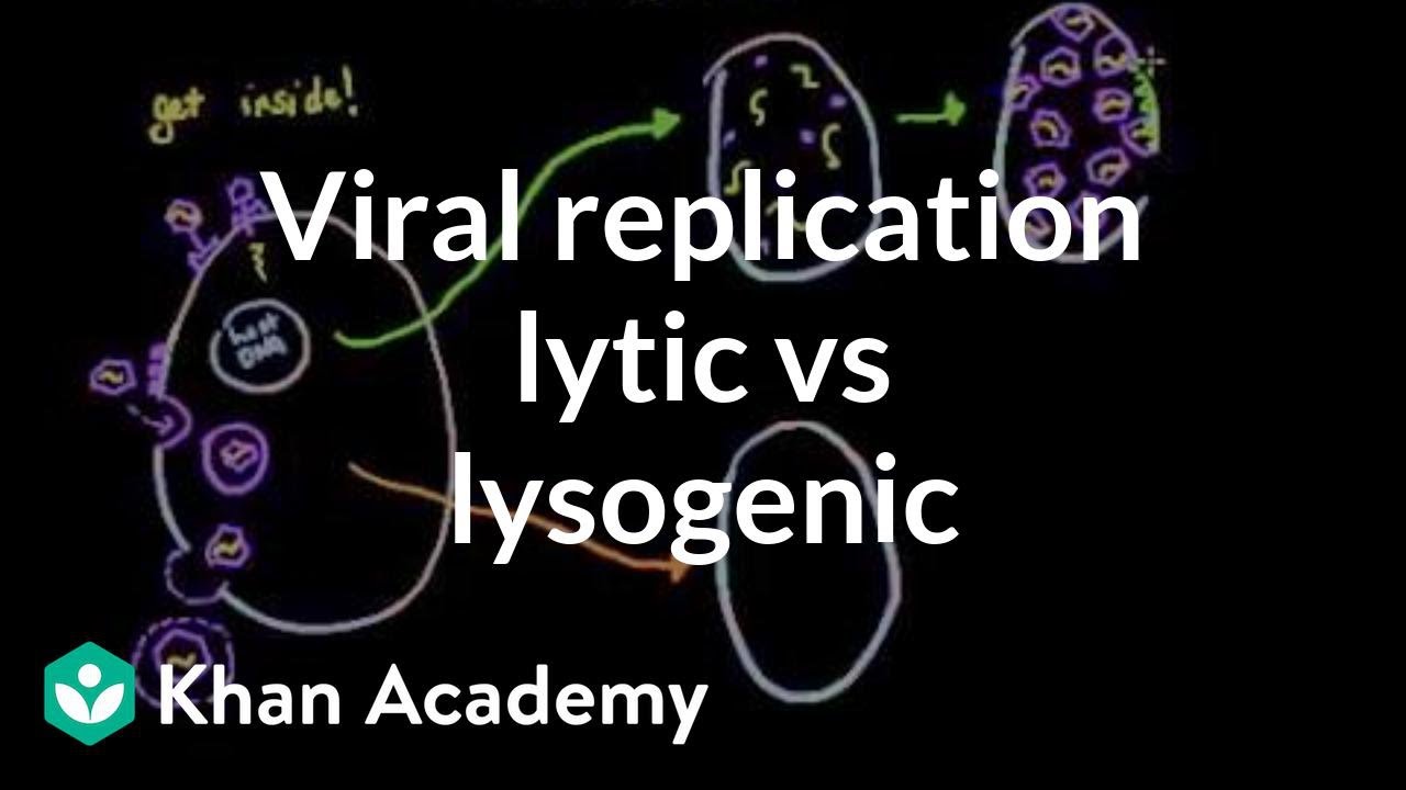
Why this video: This MCAT-focused lecture provides a clear conceptual comparison of the two lifecycles. It explains how virulent phages cause host cell lysis, and how temperate phages integrate into the host chromosome to replicate as a "prophage" without killing the host.
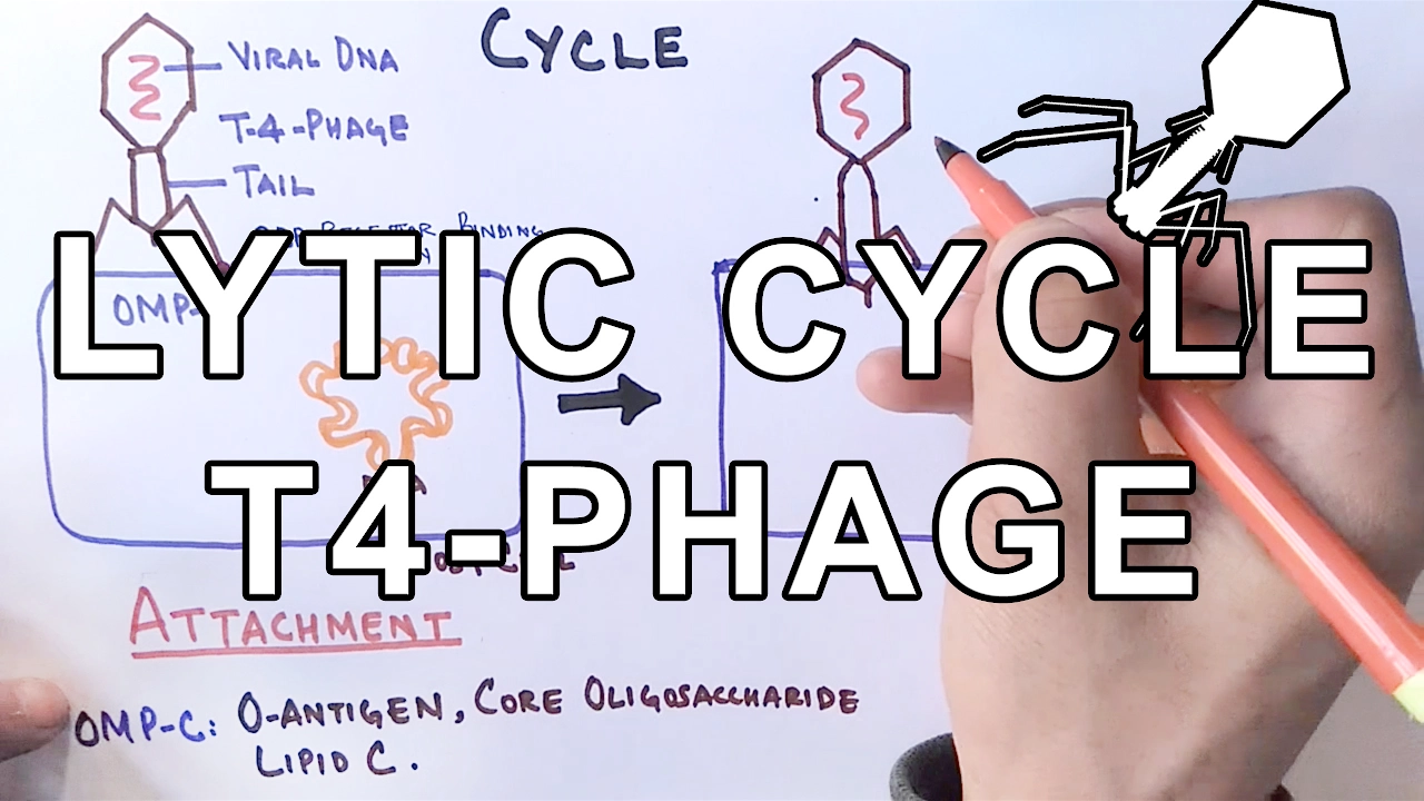
Why this video: This biochemically focused video details the molecular steps of the lytic cycle. It highlights host chromosome degradation, transcriptional hijacking, phage protein synthesis, capsid packaging, and the coordinated actions of endolysins and holins during host cell lysis.
Why this video: This video provides a step-by-step breakdown of lysogeny. It explains how phage DNA is integrated into the bacterial chromosome via integrase, how it remains dormant as a prophage during host binary fission, and what environmental factors prompt it to enter the lytic cycle.
Knowledge Checkpoint
- Compare the host cell's fate in the lytic cycle versus the lysogenic cycle.
- Define "prophage" and explain how lysogenic conversion can alter the phenotype of a host bacterium (e.g., toxin production).
- Detail the functions of holins (forming membrane pores) and endolysins (degrading peptidoglycan) in facilitating host cell lysis.
- Distinguish between virulent phages (restricted to lytic growth) and temperate phages (capable of both cycles).
Module 3: The Lysogenic-Lytic Switch
This module explores the genetic regulatory mechanisms that control the transition of a temperate bacteriophage (specifically Lambda phage) from lysogenic dormancy to active lytic replication, driven by host SOS responses to stress.
Recommended Videos
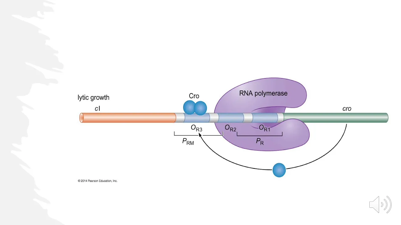
Why this video: This comprehensive video explains the competitive transcriptional circuit that governs the lysogenic-to-lytic decision. It covers the roles of the CI repressor and Cro proteins, their binding dynamics at operator sites, and how environmental inputs influence this molecular switch.

Why this video: This video focuses on the molecular mechanics of the CI and Cro genetic switch. It explains how these proteins interact with operator regions (, , and ) to repress or promote the promoters and , providing a clear look at transcriptional regulation.
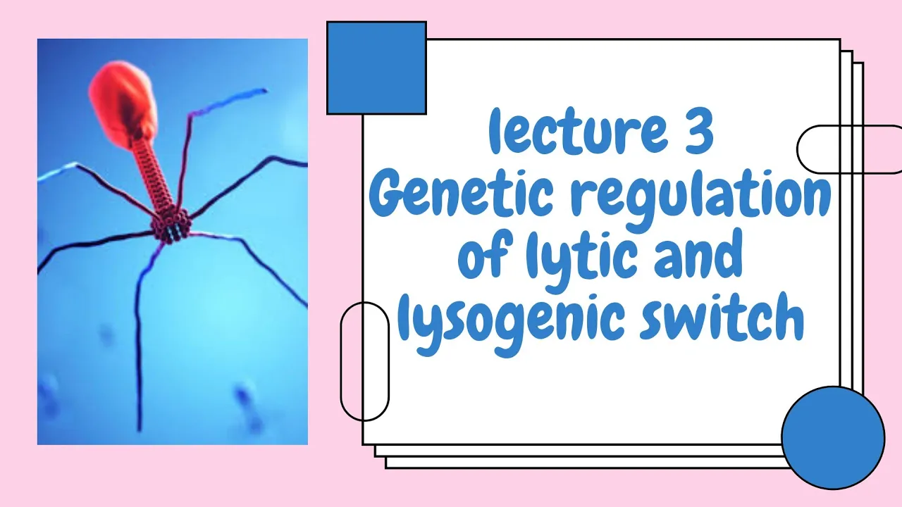
Why this video: This detailed academic lecture explores the thermodynamics of operator binding. It covers the helper proteins CII and CIII, the role of host proteases (like FtsH) under varying nutritional conditions, and how DNA damage triggers the RecA-mediated cleavage of the CI repressor during the SOS response.
Knowledge Checkpoint
- Explain how the competitive binding of CI and Cro to operator sites and controls transcription from and .
- Describe how the host protease FtsH senses nutritional levels and targets the CII protein to influence the initial lysis/lysogeny decision.
- Detail the step-by-step pathway of prophage induction: DNA damage RecA activation autocatalytic cleavage of CI transcription of lytic genes.
- Explain the role of the CIII protein in protecting CII from proteolytic degradation.
Module 4: The Antibiotic Resistance Crisis
This module analyzes the global health threat of multi-drug resistant bacterial pathogens. It reviews how small-molecule antibiotics function, how bacteria evolve defenses against them, and why alternative treatments like phage therapy are needed.
Recommended Videos

Why this video: This PBS FRONTLINE documentary offers a detailed look at the clinical reality of the antibiotic resistance crisis. Through patient case studies and interviews with public health officials, it illustrates the impact of pan-resistant "superbugs" and the economic challenges of antibiotic development.

Why this video: This animated lecture explains the molecular mechanisms of antibiotic resistance. It illustrates the four main bacterial defenses: enzymatic drug inactivation (e.g., beta-lactamases), efflux pumps, target modification (e.g., ribosomal protection), and outer membrane permeability changes.
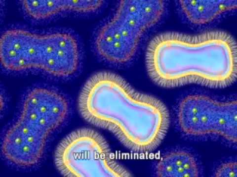
Why this video: Produced by the US Food and Drug Administration (FDA), this video explains how selective pressure drives the expansion of resistant subpopulations. It also details the three routes of horizontal gene transfer: conjugation (plasmid transfer), transformation (naked DNA uptake), and transduction (phage-mediated transfer).
Knowledge Checkpoint
- Describe the four primary biochemical mechanisms bacteria use to resist traditional small-molecule antibiotics.
- Differentiate between conjugation, transformation, and transduction as mechanisms of horizontal gene transfer (HGT).
- Explain how sublethal exposure to antibiotics accelerates the selection and spread of resistance genes within a clinical population.
- Summarize why the traditional pharmaceutical pipeline has struggled to produce novel classes of small-molecule antibiotics.
Module 5: Phage Therapy and Clinical Applications
This final module covers the practical application of bacteriophages as personalized therapeutics. You will study phage isolation protocols, therapeutic design (phage cocktails), the evolutionary dynamics of phage resistance, and the regulatory pathways for experimental clinical use.
⚠️ Independent Study Focus: Regulatory & Manufacturing Gaps
While public educational videos provide excellent coverage of clinical history and basic isolation protocols, they often lack deep technical details on Good Manufacturing Practice (GMP), personalized clinical trial designs, and formal regulatory pathways (such as the FDA IND process).
To supplement this module, you are encouraged to independently read peer-reviewed literature on:
- FDA Investigational New Drug (IND) pathways for emergency, single-patient phage therapy.
- GMP production standards for clearing host cell proteins, DNA, and endotoxins (lipopolysaccharides) from therapeutic phage preparations.
- The evolutionary "arms race"—how bacteria mutate surface receptors (such as outer membrane proteins or lipopolysaccharides) to resist phages, and how phages counter-evolve to recognize modified receptors.
Recommended Videos
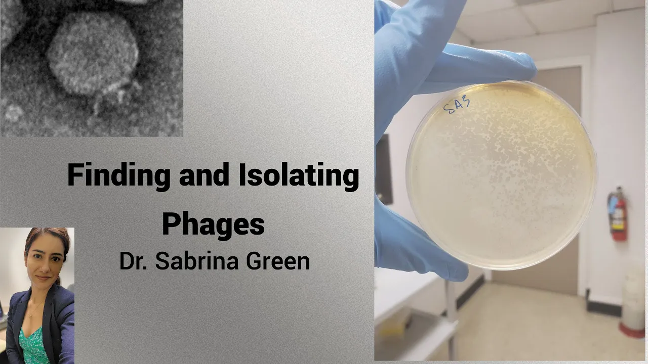
Why this video: Produced by a clinical phage research facility (TAILOR Labs at Baylor College of Medicine), this video demonstrates how phages are sourced from environmental reservoirs like bird feces and wastewater. It covers sample homogenization, centrifugation, and 0.22-micrometer membrane filtration to isolate cell-free viral filtrates.

Why this video: This wet-lab protocol video demonstrates the direct isolation of bacteriophages from soil samples. It explains how to dislodge phages from soil particles using phage buffer, perform low-speed centrifugation, filter the supernatant, and set up plaque assays to confirm the presence of viable phages.
Why this video: This academic presentation covers the formulation challenges of phage therapeutics. It details the complexity of designing "phage cocktails" to target multiple bacterial strains, phage-antibiotic synergy, stability testing, and the challenges of high-volume manufacturing.
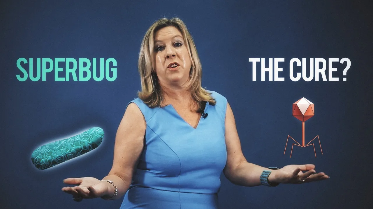
Why this video: This video documents the landmark clinical case of Tom Patterson, who was cured of a multi-drug resistant Acinetobacter baumannii infection. It highlights the collaboration between research labs, the FDA, and clinical teams that catalyzed the modern revival of clinical phage therapy in the West.
Knowledge Checkpoint
- Describe the wet-lab steps required to isolate, filter, and purify bacteriophages from environmental sources like wastewater or soil.
- Explain the benefits of using a multi-phage "cocktail" compared to single-phage monotherapy in clinical settings.
- Explain how bacteria can develop resistance to phages (e.g., receptor mutation) and how this resistance can sometimes make the bacteria more sensitive to traditional antibiotics (receptor trade-off).
- Explain why endotoxins must be removed from phage preparations before they can be administered intravenously to patients.
Course Map
This flowchart illustrates the learning progression and dependencies of the modules in this curriculum:
Key People Index
The following historical and contemporary scientists played key roles in developing the concepts covered in this curriculum:
- Frederick Twort (1877–1950) & Felix d'Herelle (1873–1949): Independent co-discoverers of bacteriophages. d'Herelle coined the term "bacteriophage" (bacteria-eater) and pioneered early, pre-antibiotic clinical applications.
- Esther Lederberg (1922–2006): Discoverer of Lambda phage in 1951, which became the primary model organism for studying lysogeny and genetic regulation.
- Alexander Fleming (1881–1955): Discovered penicillin in 1928, initiating the modern antibiotic era that temporarily eclipsed western development of phage therapy.
- Dr. Graham Hatfull: Professor at the University of Pittsburgh and HHMI investigator. He leads the SEA-PHAGES program, cataloging mycobacteriophage diversity and developing personalized phage therapeutics.
- Dr. Stephanie Strathdee: Epidemiologist and advocate whose efforts to secure emergency FDA approval for her husband, Tom Patterson, led to a successful phage therapy case that helped revitalize Western clinical interest.
Final Self-Assessment
Complete this comprehensive self-assessment to verify your mastery of the curriculum's learning goals:
- Identify the structural components of Siphoviridae, Myoviridae, and Podoviridae phage families under electron microscopy.
- Detail the physical and biochemical changes that occur in a T4 baseplate and tail sheath during host cell penetration.
- Diagram the lytic cycle, highlighting the timing of host DNA degradation, phage protein synthesis, genomic packaging, and cell lysis.
- Contrast the genomic integration of Lambda phage via integrase at the / sites with the passive replication of a prophage.
- Draw the Lambda genetic control circuit, showing how CI self-regulates via and to maintain lysogeny.
- Explain how Cro binding to shuts down the promoter, driving the phage into lytic replication.
- Connect the bacterial SOS response (triggered by DNA damage) to the cleavage of the CI repressor by RecA.
- List the four major cellular mechanisms of antibiotic resistance and describe their genetic origin (chromosomal mutation vs. plasmid-borne).
- Explain how transduction acts as both a mechanism of bacterial gene transfer and a potential hazard in clinical phage therapy (via the transfer of resistance genes).
- Outline the process of isolating, filtering, and performing a plaque assay to determine the titer (PFU/mL) of an environmental phage sample.
- Explain the evolutionary trade-off of receptor mutation, detailing how phage resistance can lead to decreased bacterial virulence or restored antibiotic susceptibility.