Electrochemical Aptasensors: Design & Testing
Learning Goal: Design, fabricate, and validate an electrochemical biosensor (aptasensor) for detecting small-molecule environmental toxins. This includes mastering gold electrode surface chemistry, characterizing surface modifications using Cyclic Voltammetry (CV) and Electrochemical Impedance Spectroscopy (EIS), and understanding the biophysical signal transduction pathways of redox-labeled aptamers.
Prerequisites
- Chemistry: General college-level chemistry (balancing redox equations, basic thermodynamics).
- Biology: Fundamentals of nucleic acids (DNA/RNA hybridization, secondary structures).
- Lab Safety: Standard laboratory safety protocols, handling of chemical solutions, and personal protective equipment (PPE).
Estimated Total Study Time: 18 Hours
Module 1: Foundations of Electrochemistry
Module Overview
Before building a biosensor, you must understand the interface where chemistry meets electricity. This module introduces the fundamental physical chemistry behind oxidation-reduction (redox) reactions and details how standard electrochemical measurements are set up. You will explore why analytical electrochemistry relies on a three-electrode system (working, reference, and counter electrodes) and the essential role of supporting electrolytes in shielding electrostatic migration.
Recommended Videos
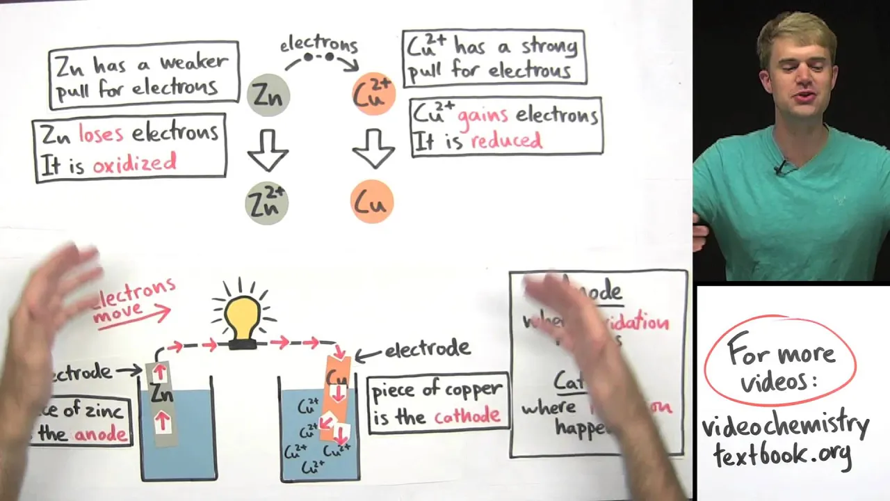
Why this video is valuable:
This video provides an intuitive and visual introduction to the core principles of electrochemistry. It explains how redox reactions operate through the simultaneous transfer of electrons (oxidation as loss, reduction as gain) and lays the conceptual groundwork for understanding electrochemical cells, cell potentials, and the physical separation of half-reactions.
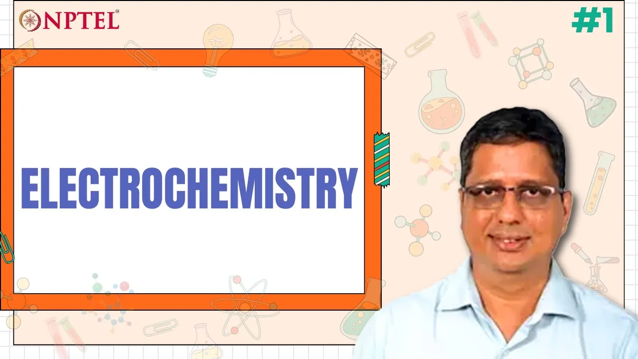
Why this video is valuable:
This advanced academic lecture bridges the gap between basic chemistry and laboratory electrochemical analysis. It explains the physical structure of the electrochemical double layer, the necessity of a three-electrode setup to isolate the potential of the working electrode, and how supporting electrolytes eliminate migration currents so that mass transport is purely diffusion-controlled.

Why this video is valuable:
A brief but practical demonstration of a real-world laboratory setup. It shows the physical arrangement of a three-electrode cell containing a classic ferricyanide () redox probe with potassium chloride () as the supporting electrolyte. This serves as the baseline experiment for validating any electrochemical biosensor.
Knowledge Checkpoint
- Write down the half-reactions for a standard redox couple (e.g., ) and identify which species is oxidized and which is reduced.
- Explain the specific role of the Working Electrode (WE), Reference Electrode (RE), and Counter Electrode (CE) in a three-electrode system. Why is current not passed through the Reference Electrode?
- Define the electrical double layer. How does the addition of a high concentration of supporting electrolyte (such as ) compress this double layer and suppress migration?
Module 2: Introduction to Biosensors & Aptamers
Module Overview
A biosensor translates a biological recognition event into a measurable physical signal. In this module, you will learn the structural classification of biosensors and deep-dive into aptamers—synthetic single-stranded DNA or RNA oligonucleotides engineered to bind specific targets. Unlike antibodies, aptamers can be selectively evolved in vitro using the SELEX process to recognize small-molecule environmental toxins (e.g., heavy metals, mycotoxins, pesticides) with high affinity and reproducibility.
Recommended Videos

Why this video is valuable:
This comprehensive lecture maps out the five key architectural elements of any biosensor: the analyte, the bioreceptor (molecular recognition element), the transducer (converts binding into an electrical/optical signal), the amplifier, and the signal processor. It explains how selecting the correct bioreceptor dictates the selectivity and limit of detection of the assay.
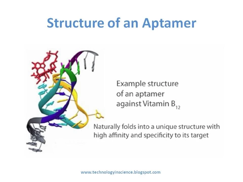
Why this video is valuable:
This tutorial explains the unique biophysical properties of single-stranded DNA/RNA aptamers. It covers how these oligonucleotides fold into precise 3D shapes (hairpins, pseudoknots, G-quadruplexes) to encapsulate target ligands, comparing their stability, production costs, and modification capabilities favorably to classic protein antibodies.

Why this video is valuable:
This video breaks down the SELEX (Systematic Evolution of Ligands by Exponential Enrichment) method. It describes the iterative cycles of partitioning bound and unbound oligonucleotides, elution, and PCR amplification, detailing how to tailor the selection conditions to target small, uncharged organic molecules.
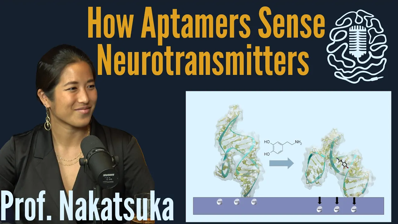
Why this video is valuable:
A short, high-fidelity visual animation showcasing the conformational restructuring that occurs when an aptamer interacts with its target. Understanding this structural transition is essential for designing electrochemical "signal-on" or "signal-off" transduction mechanisms.
Knowledge Checkpoint
- Sketch a block diagram of an electrochemical biosensor and label its five main components.
- Why are aptamers uniquely suited as bioreceptors for small environmental toxins (e.g., Ochratoxin A, Bisphenol A) compared to antibodies? Consider binding pocket geometry, molecular weight, and chemical stability.
- Outline the sequential steps of a SELEX cycle. What is "negative selection" (or counter-SELEX), and why is it crucial to prevent cross-reactivity with structural analogs?
Module 3: Gold Electrode Surface Chemistry
Module Overview
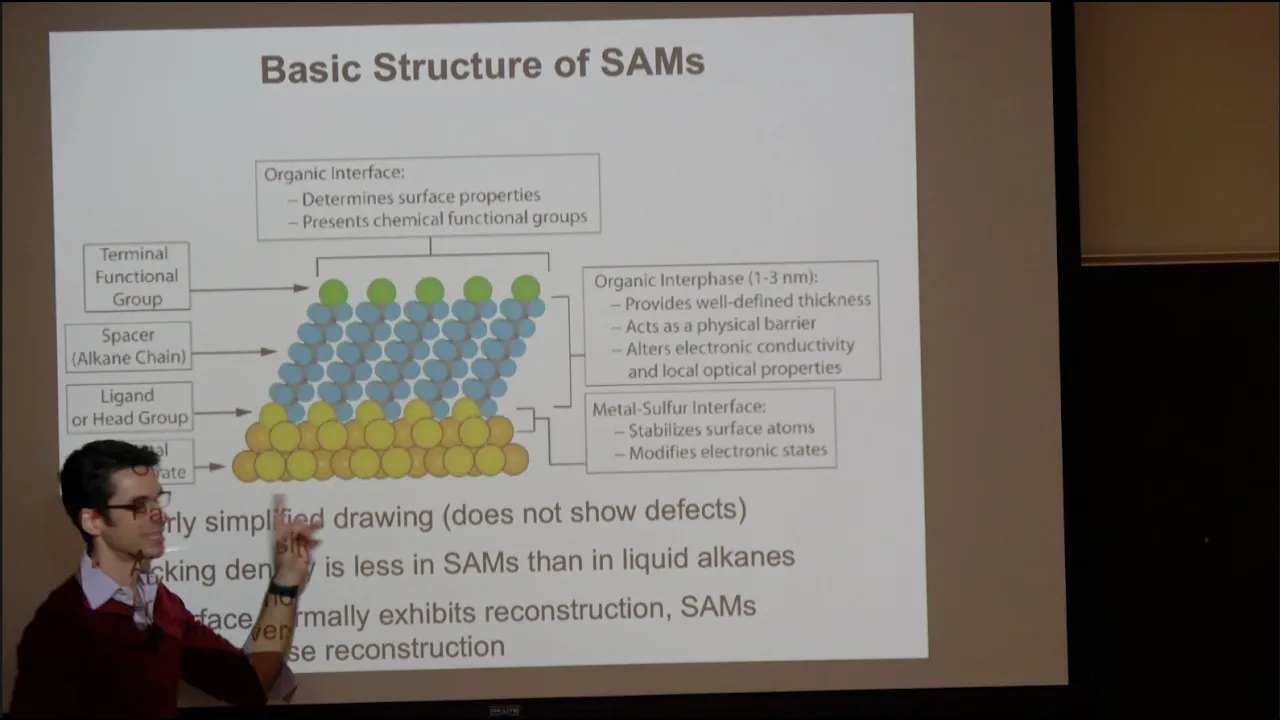
The sensitivity and reproducibility of an electrochemical sensor depend heavily on the quality and cleanliness of the electrode surface. This module covers the rigorous physical and chemical cleaning protocols for gold disk electrodes. You will learn how to prepare a clean gold surface and modify it with thiolated aptamers to form self-assembled monolayers (SAMs) via strong, covalent gold-sulfur () bonds.
   [Aptamer Strand]
          │
      (Spacer)
          │
         [S]  <-- Thiol group
═══════════╧═══════════ <-- Gold Electrode Surface
Recommended Videos

Why this video is valuable:
A highly practical, step-by-step video demonstration showing how to perform physical alumina polishing on a micro-cloth pad in a figure-eight pattern. It also briefly demonstrates the subsequent immobilization of thiolated DNA probes onto the clean gold surface.
Why this video is valuable:
An in-depth academic lecture by Prof. Darren Lipomi explaining the thermodynamics, kinetics, and structural organization of Self-Assembled Monolayers (SAMs). This video provides the theoretical foundation for how thiolated alkanes organize on gold surfaces, covering defect states, packing density, and the role of intermolecular van der Waals forces.

Why this video is valuable:
This webinar excerpt explains the physical chemistry of the thiol-gold bond. While focused on nanoparticles, the core principles of sulfur-gold coordination chemistry, ligand displacement, salt aging, and surface coverage apply directly to functionalizing flat gold disk electrodes.
Critical Laboratory Protocols (Filling the Video Gaps)
Because short videos often bypass the molecular details of electrode preparation, study and master the following two standard protocols:
1. Gold Disk Electrode Polishing and Electrochemical Cleaning Protocol
- Mechanical Polishing: Standard polishing uses an aqueous slurry of alumina () powder on a smooth micro-cloth pad. Polish the electrode surface by moving it firmly in a figure-eight pattern for 2–3 minutes to remove physical passivation layers.
- Sonication: Sonicate the electrode in ultra-pure deionized water () and then in absolute ethanol for 5 minutes each to remove residual alumina particles.
- Electrochemical Cleaning (Acid Cycling):
- Submerge the polished electrode into a three-electrode cell containing degassed .
- Perform cyclic voltammetry (CV) between and vs. Ag/AgCl at a scan rate of .
- Repeat for 20–40 cycles until a stable, highly reproducible gold oxidation peak (at ) and a sharp gold oxide reduction desorption peak (at ) are observed. The integration of this reduction peak can be used to calculate the electrochemically active surface area (ECSA) of your electrode.
2. Thiolated Aptamer Immobilization and Backfilling
- Aptamer Reduction (Deblocking): Thiolated aptamers are often shipped as oxidized disulfide dimers (). Before immobilization, incubate the aptamer with a reducing agent like tris(2-carboxyethyl)phosphine (TCEP) or dithiothreitol (DTT) for 1 hour to yield free active sulfhydryl () groups.
- Self-Assembly: Incubate the freshly cleaned gold electrode in a solution of reduced thiolated aptamer (buffered in a high-salt phosphate-buffered saline, PBS, or Tris-HCl buffer) for 12–16 hours at room temperature. The high salt concentration screens the negative phosphate backbone of the DNA, allowing denser packing.
- Co-adsorption / Backfilling: To prevent non-specific binding of proteins/toxins and to lift up flatly lying aptamer strands, backfill the remaining open gold sites. Incubate the electrode in a aqueous solution of 6-mercapto-1-hexanol (MCH) for 1 hour. This creates a mixed monolayer that ensures the aptamers are oriented vertically and are accessible to target molecules.
Knowledge Checkpoint
- Why is a figure-eight motion preferred over circular motions when hand-polishing a disk electrode?
- During cyclic voltammetry in , what physical process on the gold surface corresponds to the large cathodic peak observed around vs. Ag/AgCl?
- What is the role of 6-mercapto-1-hexanol (MCH) in sensor functionalization? What would happen if you omitted the MCH backfilling step?
Module 4: Electrochemical Analysis Techniques
Module Overview
How do you confirm that your gold surface has been successfully modified with aptamers, and how do you measure binding events? This module focuses on the two primary electroanalytical techniques: Cyclic Voltammetry (CV) and Electrochemical Impedance Spectroscopy (EIS). You will learn to interpret changes in the electron transfer of a redox probe (such as ferri/ferrocyanide) as a direct proxy for the chemical state of the electrode surface.
Recommended Videos

Why this video is valuable:
An outstanding, graduate-level tutorial on the theory and execution of cyclic voltammetry. Emily Penn explains the mathematical and physical origin of duck-shaped CV curves, the peak potential separation (), the Randles-Sevcik equation, and how to distinguish between diffusion-controlled processes and surface-confined redox reactions.
Why this video is valuable:
EIS is a powerful, non-destructive technique for analyzing surface changes. This presentation details the fundamental mathematics of AC impedance, the physical meaning of Nyquist plots (semicircle vs. linear diffusion line), and how to model data using the classic Randles Equivalent Circuit (incorporating solution resistance , double-layer capacitance or constant phase element CPE, charge transfer resistance , and Warburg impedance ).
Why this video is valuable:
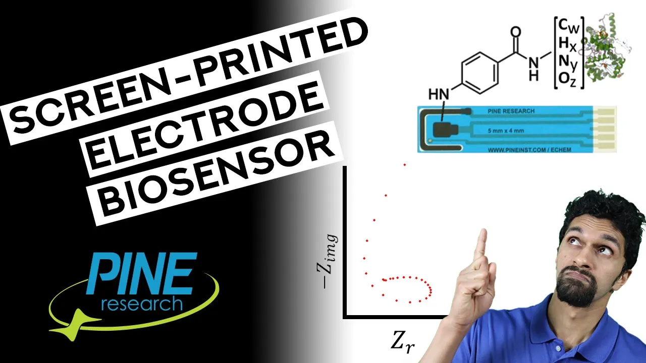
This practical guide links EIS theory directly to biosensor applications. It reviews how to read real-time measurements, analyze impedance responses from screen-printed electrodes, and diagnose instrumentation artifacts.
Biosensor Surface Diagnostics
When characterizing step-by-step surface modifications using a solution-based anionic redox probe (like ), expect the following responses:
A) BARE GOLD B) APTAMER-MODIFIED C) TOXIN-BOUND (SIGNAL-OFF)
Fast Electron Transfer Electrostatic Repulsion Steric & Charge Barrier (Low Rct, High CV) (Higher Rct, Lower CV) (Highest Rct, Lowest CV)
     [Fe(CN)6]³⁻              [Fe(CN)6]³⁻                  [Fe(CN)6]³⁻
         │                         ⤾                            ⤾
         ▼                       ───                          ───
     ─────────                  ┯┯┯┯┯                        ┯┯┯┯┯
      ═══════                  ═══════                      ═══════
- Bare Gold Electrode: Shows a very low charge transfer resistance () on the Nyquist plot (a barely visible semicircle) and sharp, high anodic/cathodic peaks on the CV curve.
- Aptamer/MCH SAM Assembly: Because the phosphate backbone of DNA is highly negatively charged, it electrostatically repels the negatively charged redox probe. This causes the charge transfer resistance () to increase significantly (forming a large semicircle) and decreases the CV peak currents.
- Toxin Binding: When a target environmental toxin binds to the immobilized aptamer, the formation of the bulky aptamer-target complex further passivates the surface. This increases steric hindrance and restricts the redox probe's access to the electrode, resulting in a further increase in (EIS signal-on for resistance change) and a drop in current.
Knowledge Checkpoint
- Draw a standard Randles equivalent circuit. Label each passive element and describe its physical counterpart at the electrode/electrolyte interface.
- Why does peak current scale with the square root of the scan rate () in a diffusion-controlled CV experiment? What does a linear relationship between peak current and scan rate () indicate?
- On a Nyquist plot, which parameter (, , , or ) directly reflects the assembly of non-conductive biomolecules on a conductive gold surface?
Module 5: Aptasensor Design & Environmental Toxin Detection
Module Overview
This final module integrates all previous concepts to design, construct, and evaluate an assay for environmental toxins. You will study how target binding induces conformational changes in the aptamer, and learn how to design "signal-on" and "signal-off" assays using covalently bound redox reporters (such as Methylene Blue or Ferrocene) or solution-phase redox probes.
Recommended Videos
Why this video is valuable:
Prof. Yi Lu, a pioneer in functional nucleic acid sensors, explains how DNA and RNA can be engineered to detect trace amounts of heavy metals (like lead or uranium) and organic small molecules. This video highlights real-world environmental applications and showcases transduction mechanisms.
Why this video is valuable:
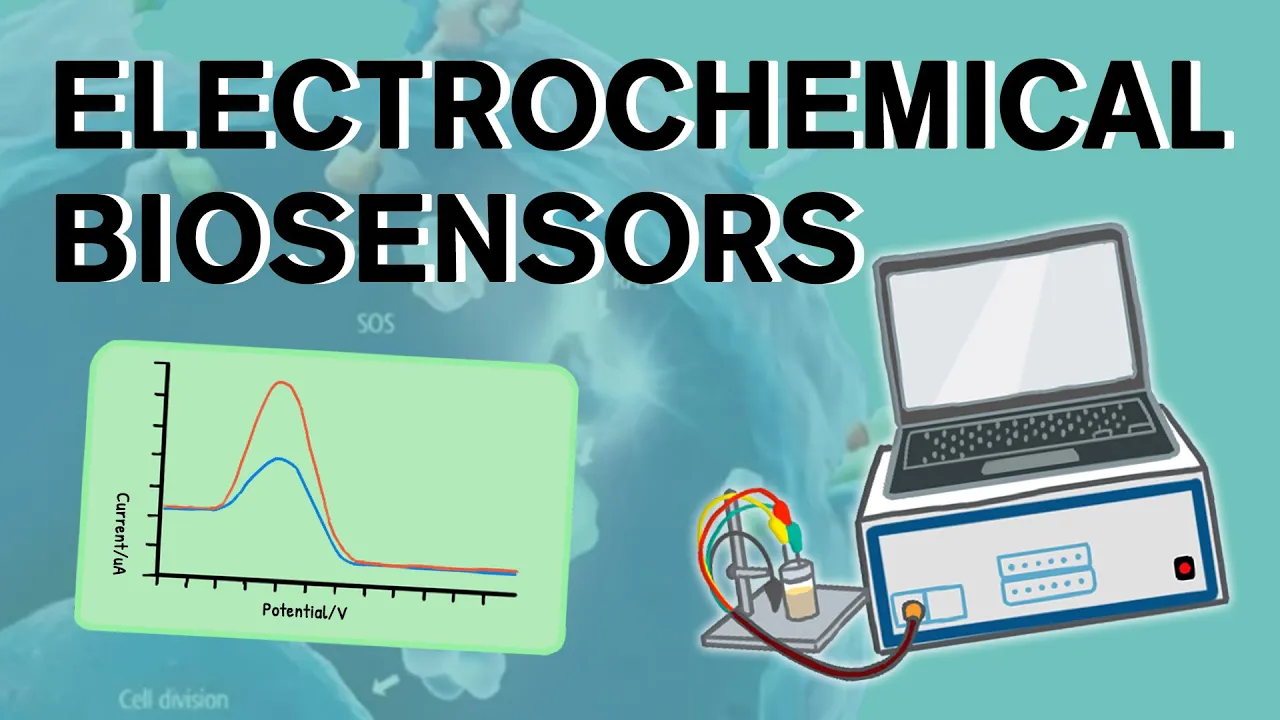
A concise, step-by-step animation showing how affinity-based electrochemical biosensors are fabricated and operated. It reviews antibody/aptamer immobilization and explains how binding changes the local charge density and electron transfer kinetics.
Why this video is valuable:
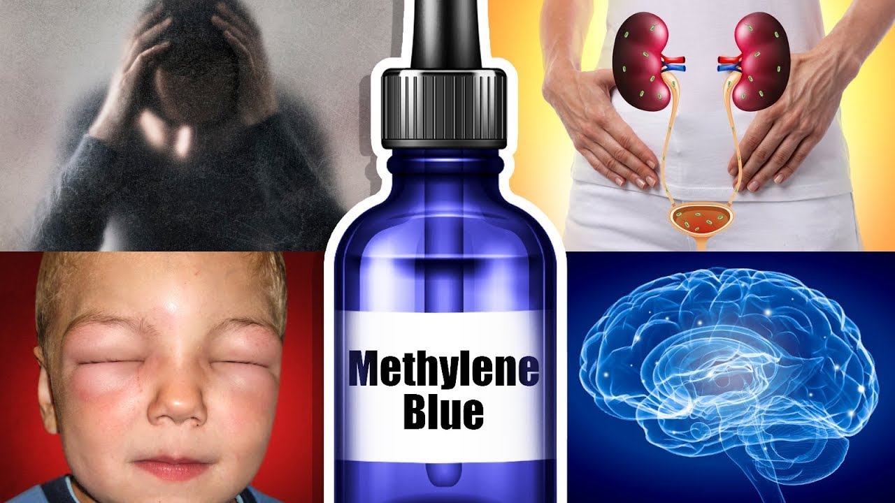
While focusing on biology, this video explains the core physical property of Methylene Blue (MB): its ability to act as an efficient electron shuttle through rapid, reversible redox cycling (interconverting between oxidized Methylene Blue and reduced Leuco-methylene blue). This quick electron transfer makes MB a widely used covalent redox reporter in electrochemical aptasensors.
Detailed Signal Transduction Chemistry (Filling the Video Gaps)
To build a high-performance biosensor for small molecules, you must choose between two primary signal transduction pathways:
A) SIGNAL-OFF MECHANISM (Covalent MB Tag) Target binding pushes the redox tag away from the gold surface, slowing electron transfer.
 [Target] ────┐                   [Target]
              │                      │
   (MB*)      ▼                    (MB*) <-- Pushed away!
     \     /                         │
     [Aptamer]                     [Apt]
      ═══════                     ═══════
    (High Peak)                 (Low Peak)
B) SIGNAL-ON MECHANISM (Covalent MB Tag) Target binding folds a flexible loop, bringing the redox tag closer to the surface.
   (MB*) <-- Far!
     │                           (MB*) <-- Close!
   [Aptamer]                       \ /
      │                          [Apt]
      ▼                            │
    [Target] ───►                  ▼
    ═══════                     ═══════
   (Low Peak)                 (High Peak)
1. Covalently Labeled Aptamers (Conformational Change Model)
In this architecture, the aptamer is synthesized with a thiol group () at the end (for attachment to the gold surface) and a Methylene Blue (MB) or Ferrocene (Fc) tag at the end.
- Signal-Off Pathway: In the buffer solution, the aptamer adopts a flexible conformation where the MB tag is positioned close to the gold surface, allowing fast, direct electron transfer (producing a large peak current during Square Wave Voltammetry). When the target environmental toxin binds, the aptamer folds into a rigid, highly structured complex that forces the MB tag away from the electrode surface. This spatial separation reduces the electron transfer rate, causing a sharp drop in the peak current.
- Signal-On Pathway: The sensor is designed so that the flexible, unbound aptamer keeps the MB tag far from the gold surface (low current). Upon target binding, the aptamer folds into a hairpin or G-quadruplex structure, bringing the MB tag close to the electrode. This structural shift increases the electron transfer rate and yields a higher peak current.
2. Label-Free Solution-Phase Assays
If you use unmodified aptamers, you can monitor target binding using solution-phase redox probes like .
- When the small-molecule target binds, the aptamer folds into a compact structure. This conformational change alters the local charge density and steric hindrance on the electrode surface, modulating the diffusion of to the gold. This change can be quantified as an increase in charge transfer resistance () via EIS.
Knowledge Checkpoint
- Diagram a "signal-off" electrochemical aptasensor that uses a -methylene blue modified aptamer. Show the physical location of the redox tag before and after target binding.
- Why is Square Wave Voltammetry (SWV) or Differential Pulse Voltammetry (DPV) preferred over Cyclic Voltammetry (CV) for quantifying trace concentrations of environmental toxins? (Hint: Consider charging/capacitive current vs. faradaic current).
- You are designing a sensor for a small, uncharged pesticide molecule. Which signal transduction approach would you choose: a label-free EIS-based assay or a covalent redox-labeled (Methylene Blue) SWV-based assay? Defend your choice based on anticipated sensitivity and signal-to-noise ratio.
Course Map
This flowchart shows the recommended learning path and dependencies for each module in the curriculum.
Key People Index
- Prof. Darren Lipomi (UC San Diego): A leading expert in physical organic chemistry and surface science. His research focuses on self-assembled molecular monolayers (SAMs) and mechanical properties of molecular films.
- Prof. Yi Lu (University of Illinois at Urbana-Champaign / UT Austin): A pioneer in the field of inorganic biochemistry and functional nucleic acids. He developed some of the first DNAzymes and aptasensors for detecting heavy metals and organic environmental toxins.
- Dr. Emily Penn (Stanford University / Chueh Group): An electrochemist known for her clear educational work on advanced electroanalytical techniques, cyclic voltammetry theory, and redox thermodynamics.
Final Self-Assessment
Complete this comprehensive self-assessment to verify your mastery of the design, fabrication, and testing of electrochemical aptasensors.
- Fundamentals: I can explain the physical origin of the electrical double layer and explain why a high concentration of supporting electrolyte is required to run a diffusion-controlled electrochemical experiment.
- Three-Electrode Setup: I can set up a three-electrode electrochemical cell, defining the role of the working (gold), reference (Ag/AgCl), and auxiliary (Pt wire) electrodes.
- Aptamer Mechanics: I can explain how the SELEX process selects single-stranded DNA sequences for small-molecule targets and explain how these molecules differ from antibodies.
- Physical Polishing: I can perform physical polishing on a gold disk electrode using alumina slurries in a figure-eight pattern.
- Chemical Cleaning: I can clean a gold electrode electrochemically by cycling it in dilute sulfuric acid () and interpret the resulting voltammogram to confirm a clean gold surface.
- Monolayer Assembly: I can outline the chemistry of the thiol-gold bond and explain why a mixed monolayer using 6-mercapto-1-hexanol (MCH) backfilling is necessary for sensor stability and performance.
- EIS Analysis: I can extract the charge transfer resistance () from a Nyquist plot generated by EIS, and map this parameter to a standard Randles equivalent circuit.
- CV Diagnostics: I can use Cyclic Voltammetry to track each stage of sensor fabrication (bare gold, aptamer immobilization, backfilling, target binding) using a ferri/ferrocyanide probe.
- Signal Transduction: I can explain the biophysical difference between "signal-on" and "signal-off" conformational sensor pathways using covalently bound redox tags like Methylene Blue.
- Analytical Validation: I can design a complete experimental protocol to generate a calibration curve, determine the linear dynamic range, and calculate the limit of detection (LOD) for an environmental toxin in a water sample.