Cellular Respiration: ETC & ATP Synthase
Learning Goal: Deconstruct the biophysical mechanisms of cellular respiration, focusing on the electron transport chain, proton motive force, and ATP synthase rotational catalysis.
- Prerequisites: Introductory biochemistry (understanding of basic chemical thermodynamics, covalent bonding, and enzyme function) and cell biology fundamentals.
- Estimated Total Study Time: 14 Hours
Module 1: Foundations of Cellular Energy and Mitochondrial Anatomy
Module Overview
To understand how cells harness energy, we must first analyze the thermodynamic "currency" of the cell—Adenosine Triphosphate (ATP)—and the highly specialized compartmentalization of the mitochondrion. This module establishes how the structural features of ATP's phosphoanhydride bonds yield high negative Gibbs free energy upon hydrolysis, and how mitochondrial anatomy (specifically the outer membrane, inner membrane, cristae, and matrix) acts as a physical barrier necessary for creating spatial concentration gradients.
Recommended Videos
Why this video
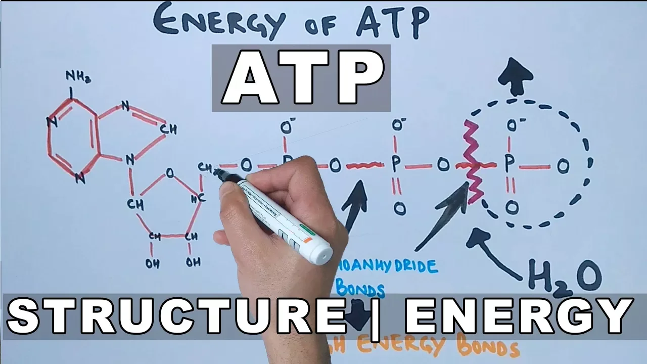
This video breaks down the specific chemical structure of ATP, explaining how the combination of an adenine base, a ribose sugar, and three phosphate groups forms a highly unstable, high-energy molecule. It introduces the crucial role of the phosphoanhydride bonds between the beta and gamma phosphates, which form the mechanical basis for energetic coupling in downstream cellular reactions.

Why this video
Understanding structural biophysics requires knowing where these processes occur. This video details the double-membrane anatomy of mitochondria, illustrating how the inner mitochondrial membrane foldings (cristae) maximize the surface area available to host thousands of electron transport chain proteins, and how the intermembrane space serves as a confined chamber for proton collection.

Why this video
This video provides the thermodynamic foundation for ATP hydrolysis. It explains why the standard Gibbs free energy change (\Delta G^\circ') is highly negative (approximately or ). It highlights the primary biophysical drivers of this exergonic reaction: relieving electrostatic repulsion among negatively charged oxygen atoms, resonance stabilization of the free orthophosphate (), and ionization/hydration of the products.
Knowledge Checkpoint
- Draw the complete chemical structure of ATP, labeling the phosphoanhydride bonds and the location of the terminal nucleophilic attack during hydrolysis.
- Explain how resonance stabilization and electrostatic repulsion contribute to the highly exergonic nature of ATP hydrolysis (\Delta G^\circ' \approx -30.5\text{ kJ/mol}).
- Identify the exact locations of the mitochondrial outer membrane, inner membrane, intermembrane space, cristae, and matrix, and state which compartments contain the high and low concentrations of protons during active respiration.
Module 2: Generating Electron Carriers: Glycolysis to the Citric Acid Cycle
Module Overview
Before mechanical or electrochemical energy can be generated, cells must systematically extract high-energy electrons from carbon fuels. This module traces the oxidation of glucose through glycolysis in the cytoplasm, the transport of pyruvate, and its entry into the Citric Acid Cycle (Krebs cycle) within the mitochondrial matrix. The primary focus is the reduction of coenzymes and to and , which act as mobile, high-energy electron shuttles that feed the Electron Transport Chain.
Recommended Videos
Why this video
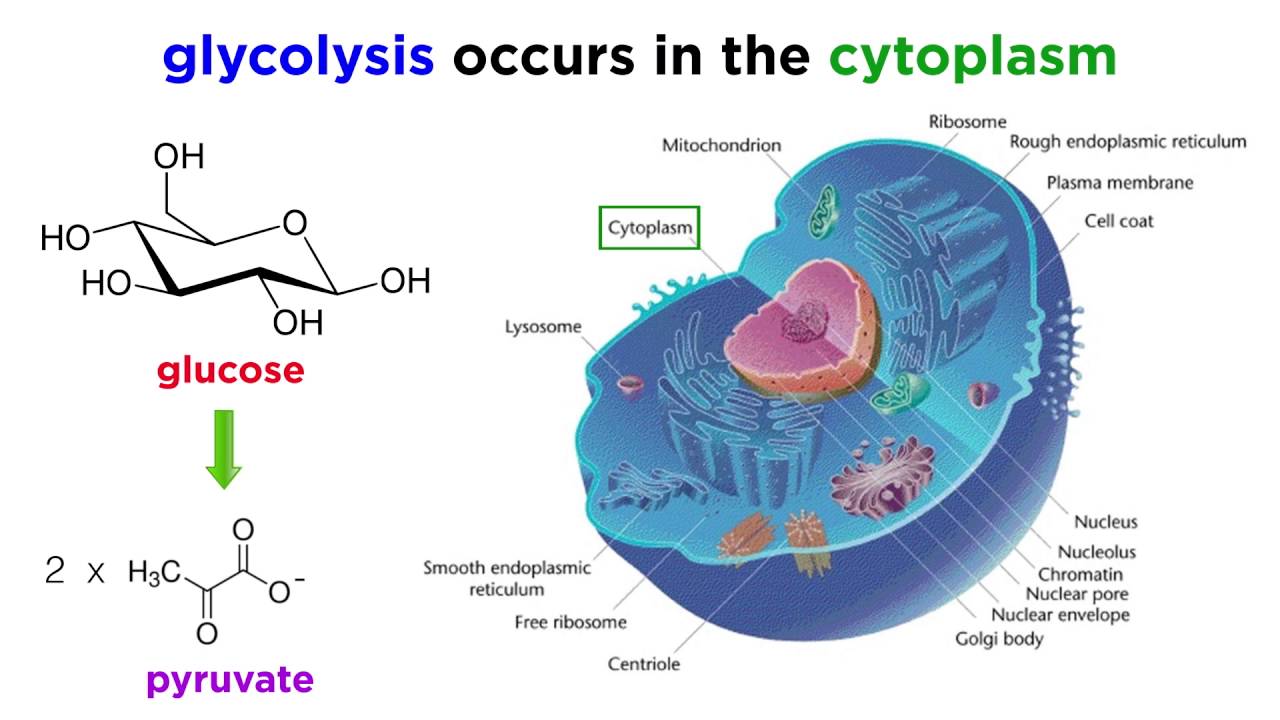
This concise video covers the breakdown of glucose in the cytoplasm. It provides a quick look at how the 6-carbon glucose is enzymatically split into two 3-carbon pyruvates, yielding a net output of and molecules. This is the first critical step in extracting reducing equivalents from external fuel sources.
Why this video
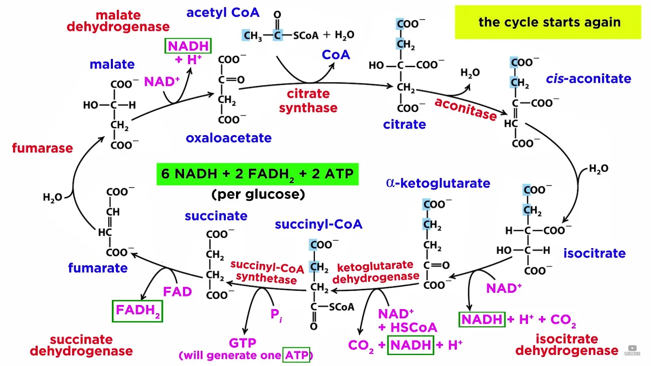
This video explains how pyruvate transitions into the mitochondrial matrix to undergo decarboxylation and enter the Citric Acid Cycle. It breaks down the 8-step loop where acetyl-CoA is fully oxidized, explaining the chemistry behind the reduction steps that generate the crucial pool of and required for the electron transport chain.
Why this video
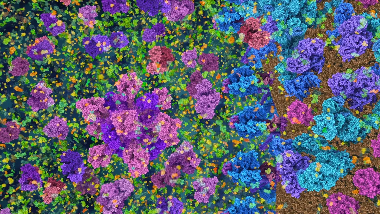
This high-fidelity molecular animation provides a physical, structural view of the enzymes of the Citric Acid Cycle in action within the crowded matrix. It helps you visualize how substrates physically dock within the active sites of these large multi-subunit enzymes, such as pyruvate dehydrogenase and isocitrate dehydrogenase, highlighting the exact moments of carbon decarboxylation and coenzyme reduction.
Knowledge Checkpoint
- Detail the net biochemical yields of glycolysis and the Citric Acid Cycle per single starting molecule of glucose.
- Explain the structural and chemical differences between and , and explain why is considered a high-energy electron carrier.
- Contrast how and are biochemically synthesized, identifying the specific enzymes in the Citric Acid Cycle responsible for their generation.
Module 3: The Electron Transport Chain (ETC) and Redox Reactions
Module Overview
This module explores the inner mitochondrial membrane to examine the mechanical and biophysical properties of the Electron Transport Chain (ETC). You will analyze Complexes I, II, III, and IV, tracing how electrons from and move along a thermodynamic gradient of increasing redox potentials (E^\circ'). You will study how these sequential, exergonic redox reactions are coupled to the physical pumping of protons from the matrix into the intermembrane space, transforming chemical energy into a physical concentration gradient.
Recommended Videos
Why this video
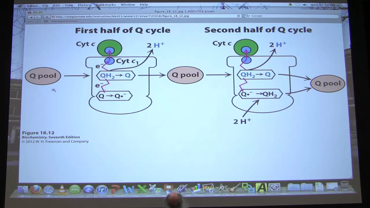
This complete, rigorous university lecture provides an in-depth look at the biochemistry of oxidative phosphorylation. Dr. Ahern goes beyond simple models to detail the specific electron transfer intermediates (such as iron-sulfur clusters, flavin mononucleotides, coenzyme Q, and cytochromes) and explains the thermodynamic changes that drive proton translocation.

Why this video
This video focuses on the specific molecular mechanics of Complexes I, II, III, and IV. It details the exact paths of electrons through each protein structure: Complex I ( Dehydrogenase), Complex II (Succinate Dehydrogenase), Complex III (Cytochrome Complex), and Complex IV (Cytochrome Oxidase). It shows how these complexes pump protons to establish the electrochemical gradient.
Why this video
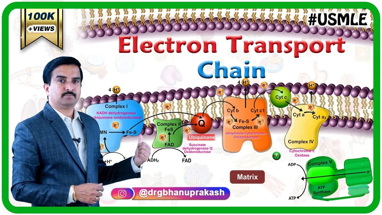
Combining clinical and molecular details, this video uses clear animations to review the movement of electrons from and to the final electron acceptor, oxygen (). It covers the physical movement of mobile electron shuttles like Coenzyme Q (ubiquinone) and Cytochrome , while illustrating the effects of classic respiratory inhibitors (such as rotenone, antimycin A, cyanide, and carbon monoxide) on specific complexes.
Knowledge Checkpoint
- Trace the path of electrons from to , listing each complex, mobile carrier, and cofactor involved, along with the total number of protons pumped across the membrane.
- Trace the path of electrons from to , explaining why entering via Complex II bypasses Complex I and lowers the net amount of proton pumping.
- Describe the structural and physical changes that occur in Complex IV when is reduced to , including the role of copper () and iron () centers.
- Define redox potential (E^\circ') and explain how the sequential increase in E^\circ' from Complex I to Complex IV makes electron flow thermodynamically spontaneous (\Delta G^\circ' < 0).
Module 4: The Proton Motive Force and Chemiosmotic Theory
Module Overview
This module covers the thermodynamics of Chemiosmotic Theory, first proposed by Peter Mitchell. Here, we analyze the physical energy stored within the electrochemical gradient across the inner mitochondrial membrane, known as the Proton Motive Force (PMF).
To satisfy a rigorous biophysical understanding, we must define the quantitative relationship of the PMF. The free energy change () associated with transporting a mole of protons from the matrix (inside, low concentration) to the intermembrane space (outside, high concentration) is expressed as:
Since and , this equation transforms into:
where:
- is the ideal gas constant ()
- is the temperature in Kelvin ()
- is Faraday's constant ()
- is the electrical charge of a proton ()
- is the electrical potential difference (membrane potential) across the membrane (in Volts, )
- (typically negative, as the outside intermembrane space is more acidic than the inside matrix)
Dividing the entire free energy equation by Faraday's constant () yields the electric potential equivalent of the Proton Motive Force ( or PMF, expressed in Volts or millivolts):
At physiological temperature ( or ), the constant factor calculates to approximately or . This gives us the practical biophysical formula:
Because is negative (), subtracting this term results in a positive addition to the total potential energy. In typical mitochondria, the membrane potential () is around to (with the matrix being negative relative to the intermembrane space), and the is approximately to units. This results in a total driving PMF of approximately to , with the membrane potential () contributing roughly 70-80% of the total force, and the concentration difference () contributing the remaining 20-30%.
Recommended Videos

Why this video
This lecture provides the mathematical foundation needed for the biophysics of chemiosmosis. It directly introduces and derives the free energy equation for proton translocation: . It walks through the variables, showing how the concentration term and the electrical potential term combine to form the total thermodynamics of the proton motive force.

Why this video
This video explains the physical chemistry of electrochemical gradients. It details how the chemical gradient (entropy-driven movement of molecules from high to low concentrations) and the electrical gradient (electrostatic forces acting on charged ions) combine to drive ion movement through selective channels.
Why this video
This video reviews the historical context and biological details of Peter Mitchell's Chemiosmotic Hypothesis. It explains how Mitchell's once-controversial idea—that ATP synthesis is coupled to a physical proton gradient rather than a high-energy chemical intermediate—changed our understanding of cellular bioenergetics.
Knowledge Checkpoint
- Write the complete thermodynamic equation for the change in free energy () of proton translocation, defining every variable and its standard unit.
- Calculate the total proton motive force ( in mV) across a mitochondrial inner membrane at given a membrane potential () of (matrix relative to intermembrane space) and a matrix pH that is units higher than the intermembrane space pH.
- Explain why the electrical component () contributes significantly more to the total PMF in mitochondria than the chemical component (), whereas in chloroplasts the reverse is often true.
Module 5: ATP Synthase: Rotational Catalysis and Nanomotor Biophysics
Module Overview
This module explores the mechanics of ATP Synthase (Complex V), a molecular nanomotor that converts the kinetic and electrical energy of returning protons into physical rotation, driving the chemical synthesis of ATP.
Structural Mechanics & Proton Pathway
ATP Synthase consists of two coupled motors:
- The Domain: Embedded in the inner mitochondrial membrane, consisting of a ring of hydrophobic -subunits (the rotor) and a stationary -subunit (which contains two hydrophilic half-channels).
- The Domain: Located in the mitochondrial matrix, consisting of a stationary catalytic headpiece ( hexamer) and an asymmetric central spindle ( and subunits) attached to the rotating -ring.
Protons in the intermembrane space enter the outer half-channel of the -subunit. To complete their path to the matrix, they must bind to a highly conserved, negatively charged aspartic acid residue (Asp61 in E. coli) located on one of the -subunits of the rotor ring. Protonation neutralizes this negative charge, allowing that -subunit to rotate into the hydrophobic core of the lipid bilayer.
As the -ring rotates by one step, a proton on the opposite side of the ring enters the matrix half-channel, where the higher pH (lower proton concentration) causes the proton to dissociate from its aspartate residue and diffuse into the matrix.
   Intermembrane Space (High H+)
         |       |
         |  [a]  |  <-- a-subunit outer half-channel
         V       V
 [c1]-[c2]-[c3(H+)]-[c4]... <-- c-ring rotating through membrane
         ^       ^
         |  [a]  |  <-- a-subunit matrix half-channel
         |       |
           Matrix (Low H+)
Boyer's Binding Change Mechanism
The physical rotation of the -ring rotates the asymmetric -spindle inside the stationary catalytic hexamer. Paul Boyer proposed that each of the three active sites on the -subunits cycles through three distinct conformations as the -shaft rotates :
- Open (O) Conformation: Has very low affinity for nucleotides; newly synthesized ATP dissociates, and empty sites are ready to accept new substrates.
- Loose (L) Conformation: Binds ADP and inorganic phosphate () loosely, trapping them in the active site but preventing them from reacting.
- Tight (T) Conformation: Compresses the bound ADP and close together, driving the spontaneous synthesis of ATP by lowering the activation energy.
One full rotation of the central spindle forces all three -subunits through this conformational cycle, resulting in the synthesis and release of exactly 3 ATP molecules.
Quantitative Stoichiometry & P:O Ratios
The exact energetic cost of synthesizing an ATP molecule depends on the stoichiometry of the -ring:
- Let be the number of -subunits in the -ring (e.g., in mammalian mitochondria, in yeast, in some bacteria).
- Because each -subunit must bind and carry exactly one proton to complete a full rotation, protons must translocate through the domain for every turn.
- Since one full turn generates 3 ATP molecules, the physical cost of synthesis is:
In mammals (), this means are required to synthesize 1 ATP inside the matrix.
However, to make this ATP usable by the rest of the cell, it must be exported to the cytoplasm. This requires the Adenine Nucleotide Translocase (which exports in exchange for , consuming the electrical gradient equivalent to charge) and the Phosphate Carrier (which imports alongside , directly consuming 1 proton from the concentration gradient). Consequently, exporting 1 ATP to the cytoplasm consumes exactly 1 additional proton.
The total physiological cost to produce and export 1 ATP is:
For mammalian cells (), this total cost is .
We can now calculate the P:O ratio (the number of ATP molecules synthesized per pair of electrons transferred to oxygen):
- For NADH: Entering at Complex I, the ETC pumps a total of 10 protons (4 from Complex I, 4 from Complex III, and 2 from Complex IV).
- For }FADH_2\text{: Entering at Complex II, the ETC pumps a total of 6 protons (0 from Complex II, 4 from Complex III, and 2 from Complex IV).
Experimental Proof of Rotation (Noji et al., 1997)
The rotation of ATP Synthase was directly demonstrated in a classic single-molecule biophysics experiment by Masasuke Yoshida, Masahiro Noji, and colleagues. They engineered a recombinant domain () and fixed the hexamer face-down onto a glass slide coverslip coated with nickel-NTA. They then attached a short, fluorescently labeled actin filament to the exposed asymmetric -subunit using a streptavidin-biotin linker.
Upon adding ATP to the buffer solution, the motor ran in reverse (acting as an ATPase). Under a fluorescence microscope, the researchers observed the actin filament rotating continuously in a counter-clockwise direction, providing physical proof that ATP Synthase operates as a rotational motor.
Recommended Videos

Why this video
This video explains Boyer's Binding Change Mechanism in detail. It shows how the asymmetric -subunit acts as a rotating cam within the stationary catalytic headpiece (), forcing the active sites into Open, Loose, and Tight conformations to synthesize ATP.

Why this video
This video focuses on the rotational catalysis and the quantitative calculation of the P:O ratio. It explains the relationship between the number of protons pumped by the ETC and the structural stoichiometry of the -ring, illustrating how the physical rotation of the motor sets the energetic efficiency of respiration.

Why this video
Despite the unrelated title, this physics-oriented lecture clip covers the single-molecule biophysics of rotational motors. It includes a schematic overview of the classic Japanese single-molecule experiment (by Noji et al.) that proved physical rotation by attaching a fluorescent actin filament to the -subunit and recording it under a microscope.
Why this video
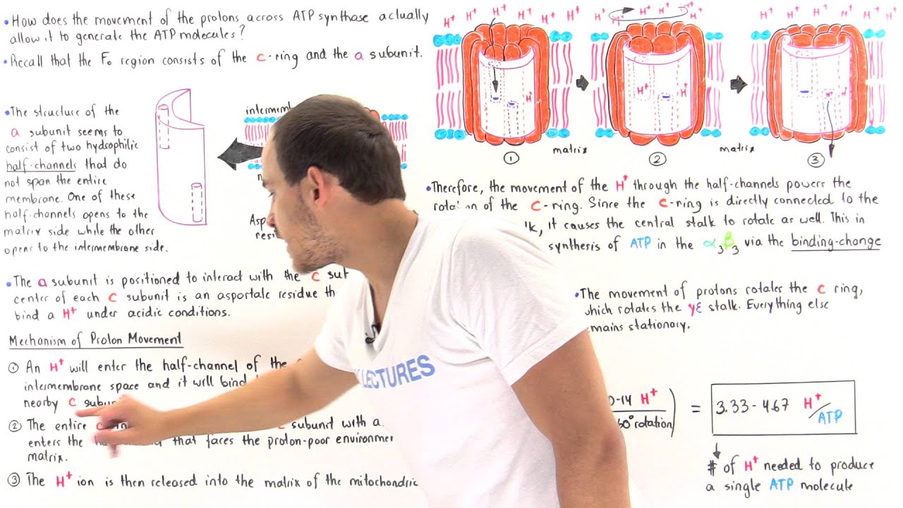
This video explains the molecular physics of proton translocation through the channel. It explains how protons enter half-channels in the -subunit to protonate specific aspartate residues on the -subunit ring, illustrating how electrostatic forces and protonation state changes drive the mechanical rotation of the -ring.
Knowledge Checkpoint
- Sketch the ATP Synthase structure, labeling the rotor, stator, -subunit, -ring, hexamer, and the central -spindle.
- Explain how protonation and deprotonation of the highly conserved aspartic acid (or glutamic acid) residues on the -ring drive rotational movement.
- Detail the physical and structural differences between the Open (O), Loose (L), and Tight (T) conformations of the -catalytic subunit, and explain how the rotating -subunit drives these conformational transitions.
- Design a step-by-step experiment based on Noji et al. (1997) to prove that the motor rotates in a clockwise direction when synthesizing ATP instead of running in reverse as an ATPase.
- Calculate the total ATP yield from the complete oxidation of one glucose molecule, using the precise mammalian P:O ratios (2.5 for NADH, 1.5 for ) rather than outdated integers.
Course Map
This map illustrates the physical and chemical sequence of oxidative phosphorylation. Note how the output of one module establishes the thermodynamic conditions required for the next.
Key People Index
| Scholar / Scientist | Context within Bioenergetics | Notable Contribution / Discovery |
|---|---|---|
| Hans Krebs | Module 2: Carrier Generation | Discovered the Citric Acid Cycle (Krebs Cycle), identifying how cells process acetyl-CoA to generate reducing equivalents. |
| Peter Mitchell | Module 4: Chemiosmotic Theory | Proposed the Chemiosmotic Hypothesis (1961), proving that ATP synthesis is driven by an electrochemical proton gradient across the inner membrane. |
| Paul Boyer | Module 5: ATP Synthase | Developed the "Binding Change Mechanism" model of ATP synthase, proving that conformational states (O, L, T) drive synthesis, earning him the Nobel Prize in 1997. |
| Masasuke Yoshida & Masahiro Noji | Module 5: ATP Synthase | Designed and executed the landmark 1997 single-molecule biophysics experiment using a fluorescent actin filament to visually confirm the physical rotation of the motor. |
Final Self-Assessment
Test your mastery of the material by completing the following checklist. If you cannot fully answer any item, review the corresponding module and recommended videos.
- I can write out the full, balanced biochemical equation for the oxidation of glucose through glycolysis and the Citric Acid Cycle, tracking the production of , ATP/GTP, and all reduced electron carriers.
- I can describe the structural features of ATP's phosphate groups that drive a high negative change in Gibbs free energy () upon hydrolysis.
- I can sketch the detailed paths of electrons through Complexes I, II, III, and IV, noting the role of cofactors like FMN, iron-sulfur clusters, ubiquinone, and Cytochrome .
- I can explain the thermodynamic mechanism of the Q-cycle in Complex III, detailing how it pumps protons across the inner mitochondrial membrane.
- I can state and derive the full thermodynamic Proton Motive Force (PMF) equation, detailing both the chemical concentration () and electrical potential () contributions.
- I can calculate the expected PMF of a mitochondrion given specific temperature, pH, and membrane potential values.
- I can describe the structural components of the and domains of ATP Synthase, identifying which parts rotate (rotor) and which remain stationary (stator).
- I can explain how protons move through the -subunit half-channels, how they protonate aspartate residues on the -ring, and how this driving force turns the rotor.
- I can explain Boyer's Binding Change Mechanism, detailing how Open, Loose, and Tight conformations cycle during a rotation to synthesize 3 ATP.
- I can calculate physiological P:O ratios for and using mammalian -ring stoichiometry (), accounting for the active transport costs of substrates.
- I can explain the single-molecule biophysics experiment by Noji et al. (1997), detailing how a fluorescent actin filament was used to observe the rotation of the domain.