CAR-T Manufacturing: Isolation, Vectors & QC
Learning Goal: Designing a clinical-grade CAR-T cell manufacturing workflow, including patient T-cell isolation, lentiviral vector transduction, ex vivo bioreactor expansion, and flow cytometry-based quality control testing.
Welcome to this comprehensive, video-based training curriculum on Chimeric Antigen Receptor (CAR) T-cell manufacturing. This curriculum bridges fundamental immunology with clinical bioprocessing engineering, taking you through the "vein-to-vein" process of developing clinical-grade cell therapies.
Prerequisites
- Basic Cell Biology & Immunology: Familiarity with cell culture concepts, adaptive immune responses, and basic molecular biology (DNA replication, transcription, translation).
- Bioprocess Basics: General understanding of cell culture vessels, media formulation, and sterile technique.
Estimated Total Study Time
- 16 Hours (incorporating video lectures, interactive analysis, and protocol design exercises).
Module 1: Immunology Foundations & CAR-T Fundamentals
This module establishes the biological foundation of T-cell immunology, cell surface marker classification, and the structural design of Chimeric Antigen Receptors (CARs). You will learn how endogenous T-cell receptor (TCR) signaling is mimicked and optimized using synthetic genetic constructs.
Recommended Videos

- Why this video: This deep-dive lecture establishes the definitive biological baseline. It covers adaptive immunity, V(D)J genetic rearrangement, MHC restriction, and endogenous T-cell receptor activation, providing the necessary immunological context before modifying these pathways synthetically.

- Why this video: Successful cell manufacturing depends on isolating the correct target populations. This video reviews cluster of differentiation (CD) markers, detailing how CD3, CD4, CD8, CD28, and tumor targets like CD19 are molecularly distinguished and leveraged.

- Why this video: This industry-level documentary connects T-cell immunology to therapeutic engineering. It explores the transition from first-generation CARs (possessing only CD3 signaling domains) to second/third-generation constructs (incorporating CD28 or 4-1BB co-stimulatory domains).
Module 1 Knowledge Checkpoint
- Diagram the structure of a second-generation CAR, clearly delineating the extracellular single-chain variable fragment (scFv), hinge region, transmembrane domain, and intracellular signaling domains ( and or ).
- Differentiate between helper and cytotoxic T-cell subpopulations and explain why their ratios matter in a final therapeutic formulation.
- Explain why a co-stimulatory signal (Signal 2, e.g., binding to ) is required alongside primary TCR signaling (Signal 1) to prevent T-cell anergy.
Module 2: Patient T-Cell Isolation and Activation
This module covers the primary steps of the clinical-grade CAR-T workflow: processing patient-derived leukapheresis material (Leukopaks), density gradient separation, washing protocols, and selective antibody-mediated magnetic bead cell enrichment and activation.
Recommended Videos
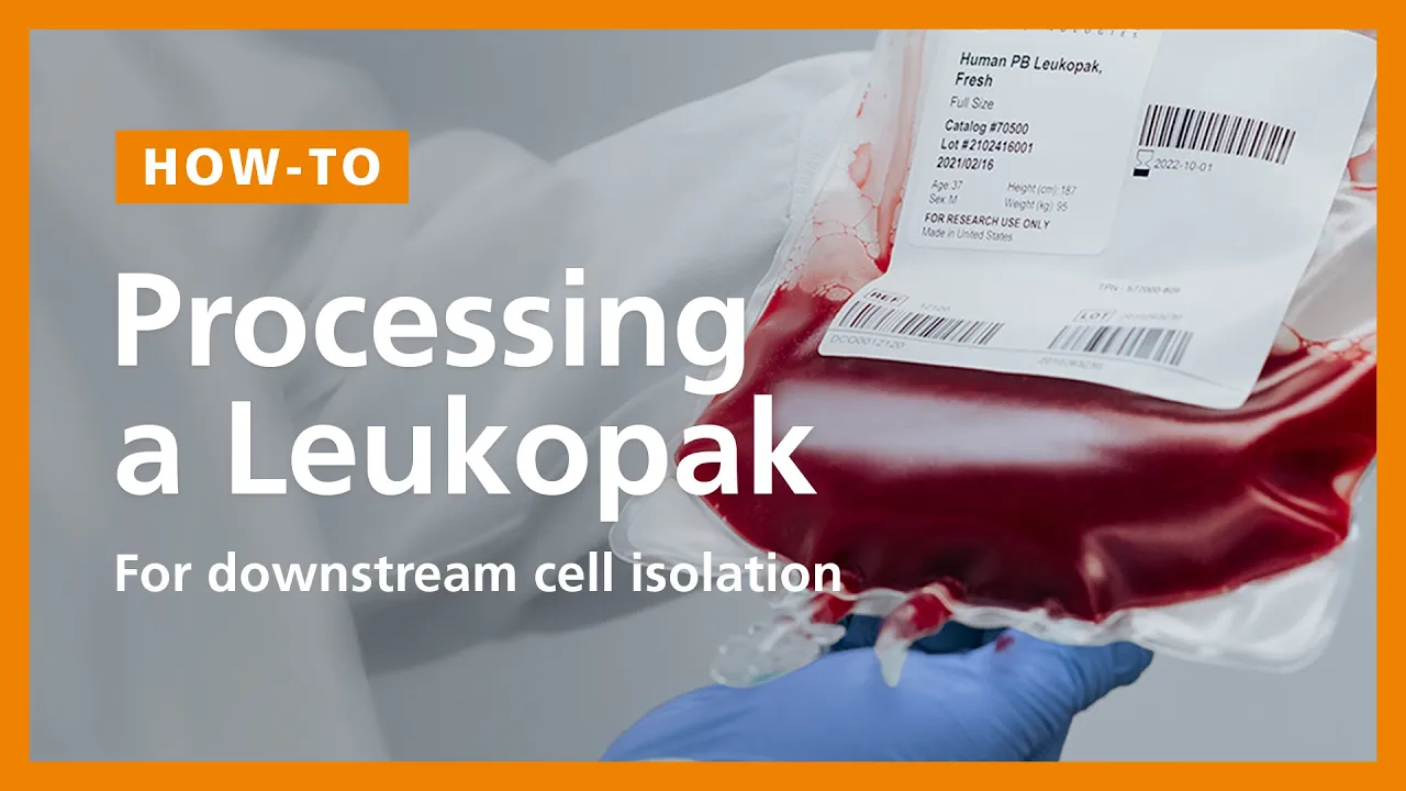
- Why this video: This protocol video provides hands-on instructions for processing clinical leukapheresis products. It details how to dilute raw materials, run density gradient separations (PBMC isolation), perform cell counts, and handle cell washing.
- Why this video: This video demonstrates Magnetic-Activated Cell Sorting (MACS) technology. It explains the mechanics of paramagnetic microbeads, ferromagnetic column matrix optimization, and the high-yield, high-purity selection of CD3+ T-cells.
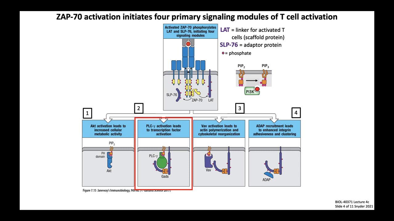
- Why this video: Explains the intracellular signaling pathways triggered during activation. Understanding the cascade from CD28 co-stimulation (including PI3K/PIP3 recruitment) explains why physical cross-linking with CD3/CD28 antibody-coated beads is used during manufacturing.
⚠️ Curriculum Note (Clinical Washing Gaps): Video demonstrations of automated clinical-scale washing systems (like Lovo or Sepax) are limited. In clinical GMP setups, density gradient separation is replaced with automated counter-flow centrifugation (elutriation) or closed-system washing bags. To supplement this, study standard operating procedures (SOPs) for the CliniMACS Cell Washer and phosphate-buffered saline (PBS) buffer formulations containing clinical-grade human serum albumin (HSA) to minimize cell clumping.
Module 2 Knowledge Checkpoint
- Calculate the target total nucleated cell (TNC) yield from a Leukopak sample based on hematology analyzer data.
- Explain the physical mechanism by which a ferromagnetic column amplifies external magnetic fields 10,000-fold during MACS processing.
- Contrast physical activation via magnetic beads with soluble cytokine-based activation methods in terms of signaling kinetics and downstream transduction susceptibility.
Module 3: Lentiviral Vector Transduction
In this module, you will master the genetic engineering phase where replication-incompetent lentiviral vectors deliver the synthetic CAR gene construct into the host T-cell genome. You will learn about transfection, transduction optimization parameters, and safety mechanisms.
Recommended Videos

- Why this video: Provides an overview of the lentiviral production timeline, including the co-transfection of packaging cells (HEK293T) and harvesting supernatants. This establishes the critical upstream parameters affecting vector quality.

- Why this video: A clear, schematized visual explanation of how a replication-deficient viral genome (carrying the gene of interest instead of viral structural genes) is packaged and used as a transduction engine.

- Why this video: Discusses experimental variables influencing transduction, such as cell density/confluency, virus-to-target-cell ratios (Multiplicity of Infection), and the use of chemical transduction enhancers.
⚠️ Curriculum Note (Transduction Protocol Gaps): Detailed clinical-grade physical parameters for spinoculation (centrifugal-assisted transduction) and the exact calculation of Vector Copy Number (VCN) are highly proprietary. In practice:
- Spinoculation: Typically run at to at for 90-120 minutes in the presence of retronecton-coated closed-system bags.
- VCN Calculations: Determined via droplet digital PCR (ddPCR) targeting specific viral elements (like WPRE or Psi packaging signals) relative to a reference housekeeping gene. Independent study should focus on FDA guidance recommending a VCN limit of copies per transduced cell genome to minimize insertional mutagenesis risks.
Module 3 Knowledge Checkpoint
- Formulate the mathematical equation used to calculate Multiplicity of Infection (), using variables for target cell count, viral titer (), and volume.
- Explain why modern clinical workflows utilize third or fourth-generation lentiviral systems (split-plasmid designs) instead of single-plasmid wild-type architectures.
- List two distinct strategies to optimize vector-cell contact during transduction in a clinical closed bag system.
Module 4: Ex Vivo Bioreactor Expansion
This module covers the scaling up of gene-modified CAR-T cells to therapeutic doses. You will explore automated clinical systems, rocking platform mechanics, gas exchange optimization, and critical process parameters in closed-system bioreactors.
Recommended Videos
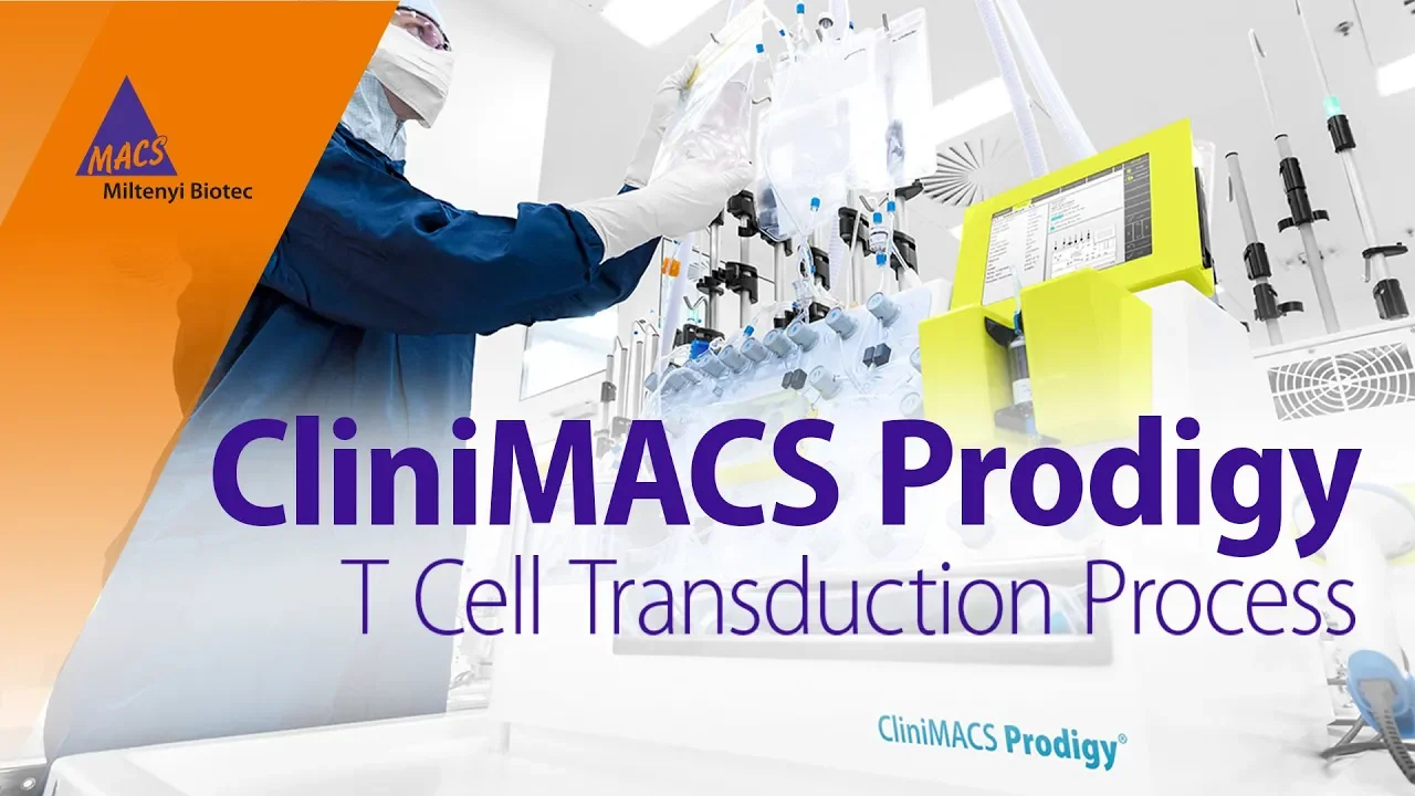
- Why this video: Shows the operation of the CliniMACS Prodigy system. This platform represents the industry standard for fully automated, closed "vein-to-vein" cell processing, integrating selection, activation, transduction, and expansion in a single-use tubing set.

- Why this video: This video details rocking-platform bioreactor kinetics. It shows how single-use bags on an automated rocking platform (using controlled angle and speed settings) optimize gas transfer and media perfusion without inducing shear-stress damage in T-cells.

- Why this video: Demonstrates the alternative stationary bioreactor design. G-Rex systems use a gas-permeable silicone membrane at the bottom, allowing cells to rest on a surface with high-density nutrients without rocking, followed by automated cell concentration harvesting.
- Why this video: Teaches bioprocess scaling principles. It explains critical engineering limitations such as oxygen transfer rates (OTR), carbon dioxide stripping, fluid shear stress, and mixing dynamics.
⚠️ Curriculum Note (Perfusion & Feeding Strategy Gaps): Bioreactor software-driven feeding profiles (continuous perfusion vs. automated bolus additions based on glucose/lactate ratios) are generally proprietary. When designing your process, plan for a baseline target glucose concentration and keep toxic lactate accumulations to prevent cellular exhaustion and preserve T-cell stemness.
Module 4 Knowledge Checkpoint
- Differentiate between the mechanical advantages of stationary, gas-permeable membrane expansion (G-Rex) and rocking-motion perfusion bag expansion (Xuri).
- Define the target critical process parameters (CPPs) for a T-cell expansion run: Temperature, setpoint, Dissolved Oxygen (), and Rocking Angle/Rate.
- Explain how perfusion-rate scaling (liters of media per day per cell density) changes the metabolic state and final viability of the expanded cell product.
Module 5: Quality Control & Flow Cytometry Testing
This final module focuses on clinical release criteria and analytical characterization. You will learn the mechanics of flow cytometry, fluorophore compensation, gating strategy design, and FDA-mandated Chemistry, Manufacturing, and Controls (CMC) release requirements.
Recommended Videos
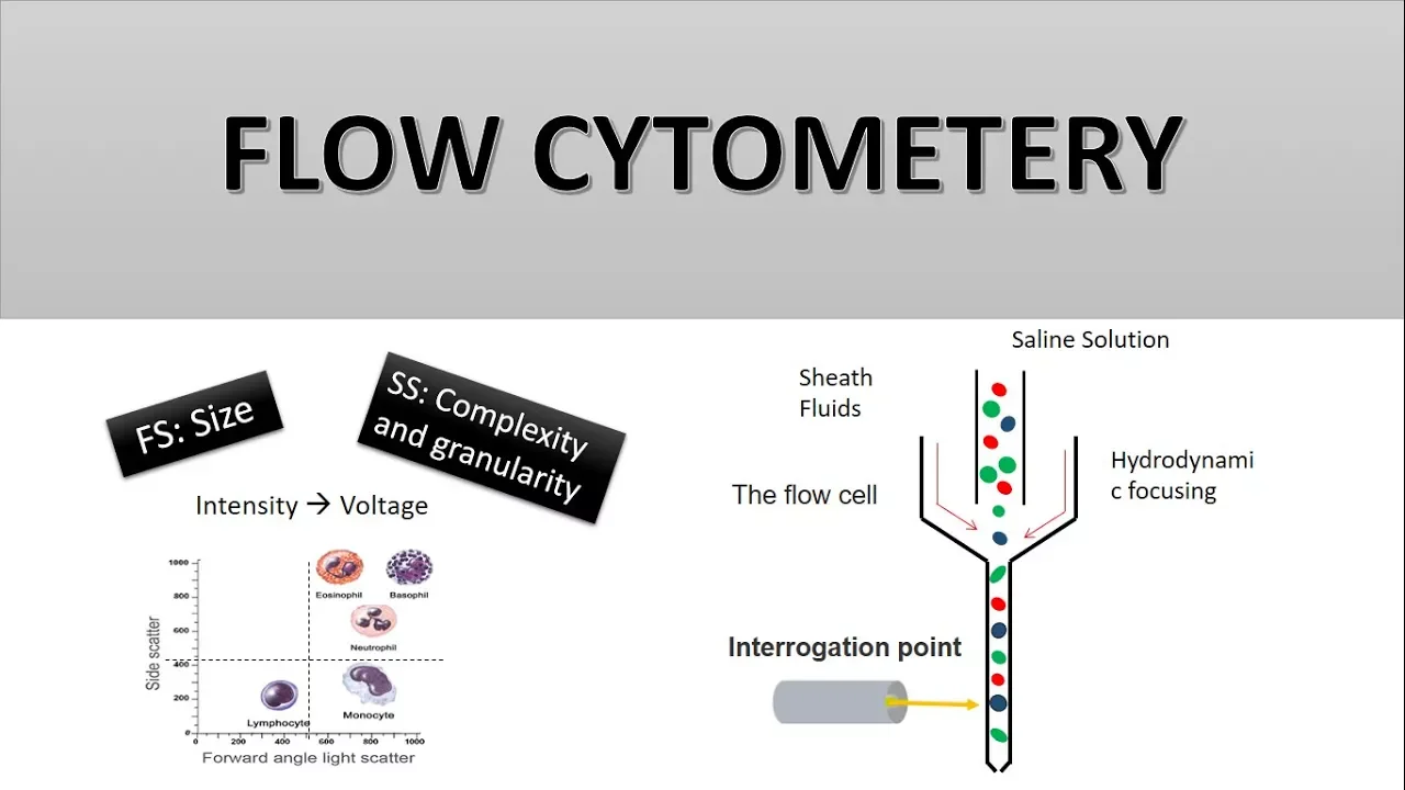
- Why this video: Explains the physical fundamentals of flow cytometry: hydrodynamic focusing, laser excitation, emission filtering, and the distinction between Forward Scatter (FSC - size) and Side Scatter (SSC - complexity/granularity).
- Why this video: Demonstrates analytical software gating. This tutorial shows how to construct scatter-gating, singlet filtering (using Area vs. Height plots), and marker thresholding, which are necessary steps for quantifying CAR expression and viability.
- Why this video: Highlights clinical release testing, covering FDA regulatory requirements for identity, purity, potency, viability, and microbiological safety (sterility, endotoxins, mycoplasma, and replication-competent retrovirus testing).
- Why this video: Discusses the logistics of rapid clinical release. It explores "just-in-time" testing strategies, including automated rapid sterility assays (such as BacT/Alert) that allow cell therapy products to be infused quickly before standard 14-day culture assays finish.
Module 5 Knowledge Checkpoint
- Design a standard flow cytometry gating hierarchy for CAR-T release, beginning with raw event acquisitions and ending with target therapeutic population quantifications.
- Explain how a cell singlet gate is resolved using Forward Scatter Height () vs. Forward Scatter Area () plotting, and why this is necessary to prevent false-positive CAR-positive readings.
- Contrast standard 14-day USP <71> compendial sterility testing with rapid alternative PCR-based methods in terms of operational turnaround times and validation requirements.
Course Map
Key People Index
- Dr. Carl June (University of Pennsylvania): Pioneer in the clinical application of CAR T-cell therapy. His team's trials using lentiviruses to reprogram patient T-cells to target CD19 led to the first FDA approvals for leukemia therapies.
- Dr. James Allison (MD Anderson Cancer Center): Awarded the Nobel Prize for identifying CD28 co-stimulation dynamics and CTLA-4 immune checkpoint inhibition, establishing the dual-signal paradigm for CAR design.
- Dr. Donald Kohn (UCLA): A leader in clinical gene therapy protocols, specializing in viral vector optimization parameters, transduction mechanics in hematopoietic lineages, and insertional mutagenesis profiles.
- Dr. Ronald Levy (Stanford University): Pioneer in the use of monoclonal antibodies and therapeutic strategies targeting malignant B-cell antigens, facilitating modern CAR targeting design.
Final Self-Assessment
To verify your readiness to design and oversee a clinical-grade CAR-T manufacturing campaign, confirm you can explain and perform each of the following:
- Leukopak Preparation: Detail the volume, cell concentration, and washing parameters used to prepare incoming material for selection.
- MACS Enrichment: Describe the mechanism of antibody-conjugated paramagnetic beads, column capture kinetics, and the eluting step used to isolate a pure population.
- Activation Signaling: Explain why stimulation alone causes cell anergy and list the intracellular targets downstream of signaling.
- Vector Kinetics: Differentiate between second, third, and fourth-generation replication-incompetent lentiviral helper plasmid configurations.
- Transduction Design: Calculate the volume of a viral vector stock () required to transduce cells at an of 5.
- Bioreactor Perfusion: Define the mechanical feedback loop that controls dissolved oxygen (), temperature (), and stability in a rocking closed-system bioreactor.
- Flow Cytometry Gating: Draw a visual gating layout showing how to transition raw sample scatter signals down to isolated live target cells.
- CMC Safety Release: List the five core FDA regulatory requirements for a cell therapy investigational new drug (IND) release, including the maximum acceptable vector copy number (VCN).