Auditory Neuroscience: Cochlea to Aphasia
Learning Goal: Analyze the neural mechanisms of auditory processing and language comprehension, tracing sound transduction from the cochlea to the auditory cortex, and detailing the functional roles of Broca's and Wernicke's areas in speech and aphasia.
- Prerequisites: Introductory biology, basic neuroanatomy (brain lobes, basic synaptic transmission).
- Estimated Total Study Time: 12 Hours
Module 1: Introduction to Sound and Ear Anatomy
This module establishes the physical foundation of auditory perception and the macroscopic anatomical structures designed to capture, focus, and amplify sound waves. You will study how pressure variations in air molecules are gathered by the pinna, channeled through the external auditory meatus, and transformed into mechanical vibrations by the tympanic membrane. We will pay special attention to the mechanical advantage of the middle ear ossicles (malleus, incus, and stapes) which solve the "impedance matching" problem when transferring sound from air into the fluid-filled cochlea.
Recommended Videos
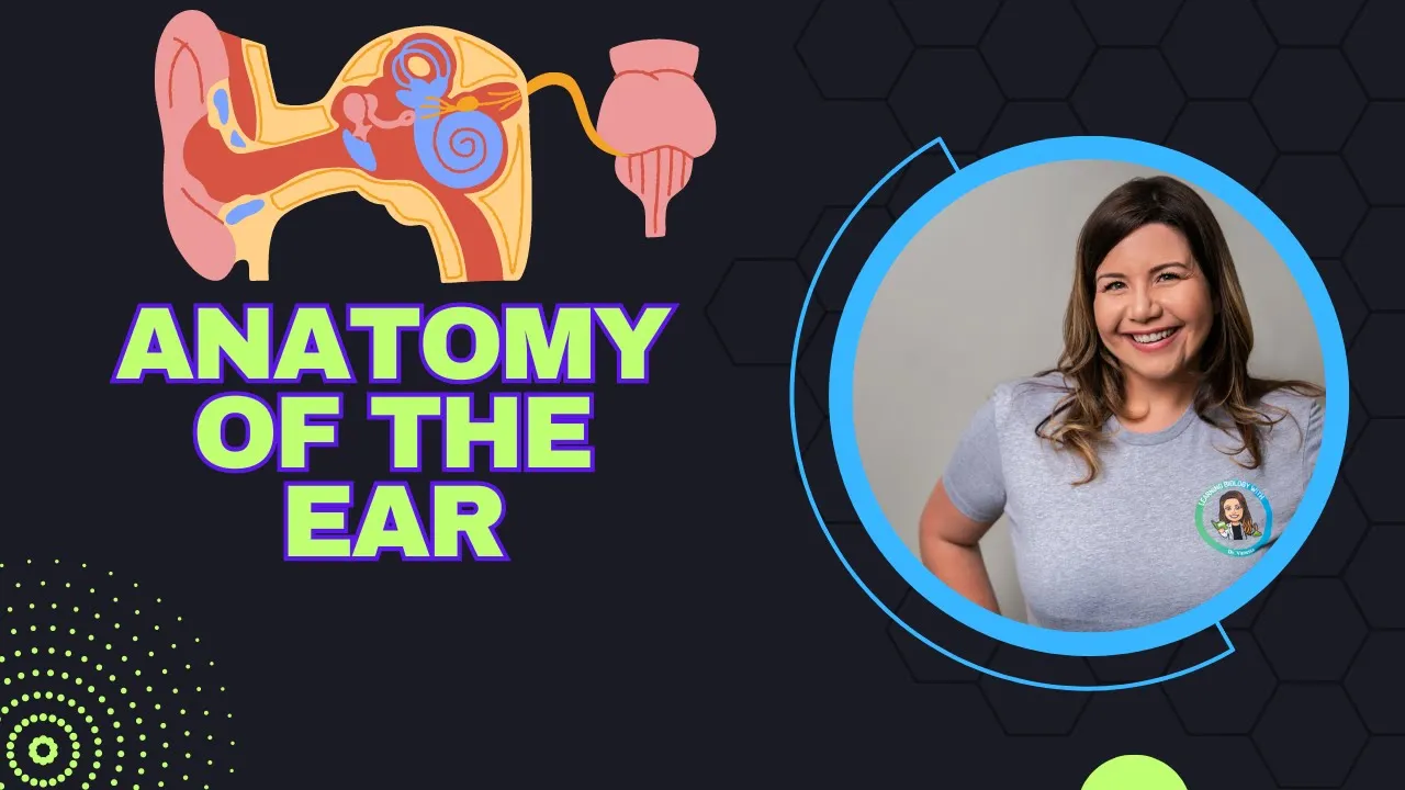
Why this video: This video provides an excellent 3D visual walkthrough of the macroscopic anatomy of the ear, dividing it clearly into external, middle, and inner sections. It is a perfect structural introduction to help you localize the exact boundaries of the tympanic membrane, the positioning of the ossicles, and their connection to the temporal bone.
- Knowledge Checkpoint:
- Identify the boundary dividing the external ear canal from the middle ear cavity.
- Name the three anatomical subdivisions of the human ear and their general functions.
- Trace the path of a sound wave as it enters the pinna and strikes the tympanum.
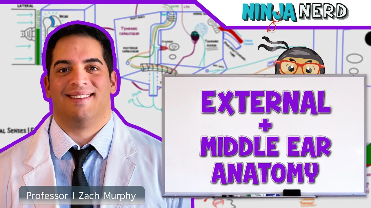
Why this video: For a deep, rigorous board-style lecture, Ninja Nerd details the physiological and structural characteristics of the outer and middle ear. This video is crucial for understanding the histological details (such as the stratified squamous epithelium of the auricle) and the operational physiology of structures like the Eustachian tube and tympanic musculature.
- Knowledge Checkpoint:
- Describe the protective role and glandular origin of cerumen (earwax) in the external acoustic meatus.
- Explain the clinical and mechanical significance of the Eustachian (auditory) tube in pressure equalization.
- Differentiate between the tissue layers making up the tympanic membrane.

Why this video: This video provides an exquisite, close-up anatomical demonstration of the three auditory ossicles—the malleus, incus, and stapes. Dr. Sam Webster explains how these microscopic bones form precise synovial joints with one another to efficiently bridge mechanical forces across the air-filled middle ear cavity.
- Knowledge Checkpoint:
- List the three auditory ossicles in order from lateral (anchored to the tympanum) to medial (resting in the oval window).
- Define the mechanical amplification role of the ossicular chain, and what happens when this chain is disrupted.
- Explain how the stapes footprint initiates fluid displacement within the inner ear.
Module 2: Cochlear Transduction: Converting Sound to Neural Signals
This module transitions from mechanical physics to sensory neurophysiology. You will explore the structure of the cochlea, focusing on its three fluid-filled chambers: scala vestibuli, scala tympani (both containing perilymph), and scala media (containing endolymph). You will study the Organ of Corti, detailing how mechanical shear forces displace the stereocilia of hair cells against the tectorial membrane. This displacement triggers mechanical gating of potassium channels, causing rapid depolarization and initiating sensory action potentials along the auditory branch of cranial nerve VIII.
Recommended Videos

Why this video: This state-of-the-art 3D animation visualizes the cellular architecture of the Organ of Corti. It showcases the exact spatial relationship between inner hair cells, outer hair cells, the basilar membrane, and the overlying tectorial membrane, modeling the fluid dynamics that trigger physical transduction.
- Knowledge Checkpoint:
- Identify the structural differences and relative populations of inner versus outer hair cells.
- Explain the mechanical function of the tectorial membrane during basilar membrane displacement.
- Map the flow of fluid pressure from the oval window, through the helicotrema, to the round window.

Why this video: This lesson outlines the physical mechanics underlying "tonotopic organization" across the basilar membrane. It clearly describes how the membrane's physical dimensions (narrow and stiff at the base, wide and floppy at the apex) dictate its resonant frequency responses.
- Knowledge Checkpoint:
- Locate where high-frequency versus low-frequency sounds are transduced along the basilar membrane.
- Contrast the physical properties of the basilar membrane at its base near the oval window versus its apex.
- Define the term "tonotopic mapping" and explain how it represents pitch.
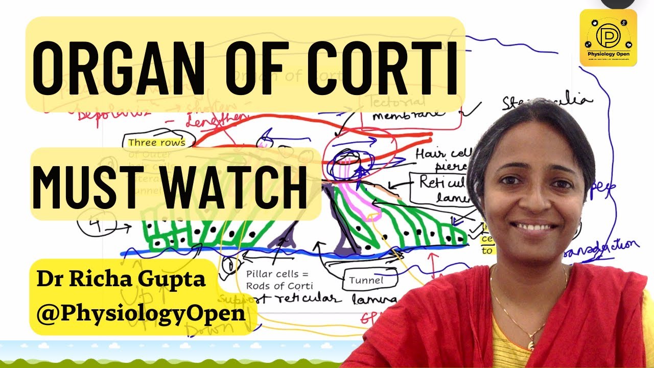
Why this video: This video delivers a clear physiological breakdown of sensory transduction at the hair cell level. It explains the mechanics of stereocilia bending, the opening of tip-links, potassium entry from endolymph, and the subsequent calcium-influx-mediated release of excitatory neurotransmitters.
- Knowledge Checkpoint:
- Explain why potassium (K+) influx depolarizes hair cells, referencing the unique high-potassium concentration of endolymph.
- Describe the role of tip links (cadherin-23 and protocadherin-15) in opening mechanosensitive ion channels.
- Identify the primary excitatory neurotransmitter released at the synapse of the inner hair cell and auditory nerve fiber.
Module 3: The Ascending Auditory Pathway to the Brain
This module maps the ascending pathway from the peripheral cochlear nerve to the primary auditory cortex (A1). You will trace the signal as it travels through the brainstem, pons, and midbrain, stopping at key synaptic relay stations: the Cochlear Nucleus, the Superior Olivary Complex (where bilateral sound localization is computed), the Lateral Lemniscus, the Inferior Colliculus, and the Medial Geniculate Body (MGB) of the thalamus.
Note on Video Coverage: Video resources tracing these specific brainstem nuclei can be brief. To supplement your learning, we recommend searching academic databases for diagrams of the "auditory brainstem response (ABR)" nuclei to solidify your visualization of these microscopic pathways.
Recommended Videos

Why this video: This video traces the primary stages of ascending auditory processing. It maps the pathway from the cochlea through the brainstem and thalamus up to the cortex, laying a strong foundation for understanding clinical audiological localization.
- Knowledge Checkpoint:
- Trace the path of auditory signals through the five primary relay centers of the ascending pathway (cochlea to A1).
- Explain the clinical significance of bilateral projections starting in the Superior Olivary Complex.
- Locate the Medial Geniculate Body (MGB) within the thalamus and describe its role as a gatekeeper to the cortex.
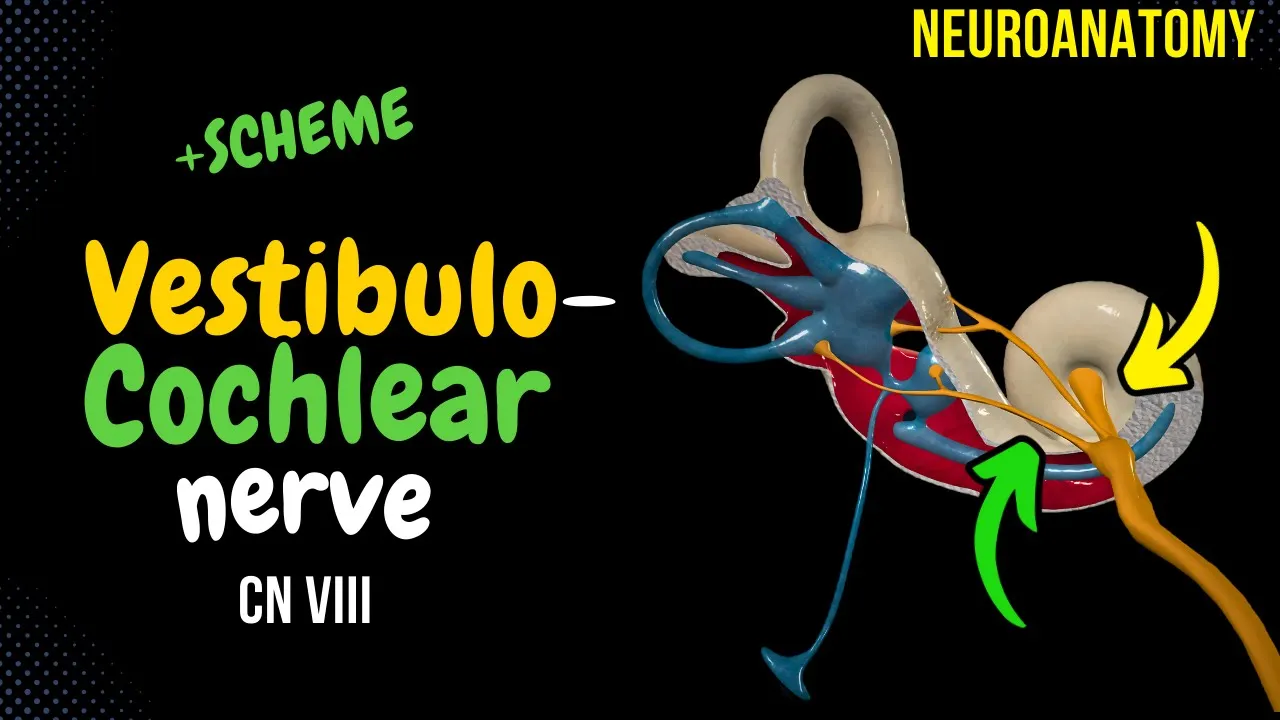
Why this video: This video offers a comprehensive neuroanatomical study of Cranial Nerve VIII (Vestibulocochlear Nerve). It illustrates how the cochlear branch exits the internal acoustic meatus of the temporal bone to synapse on the ipsilateral dorsal and ventral cochlear nuclei in the brainstem.
- Knowledge Checkpoint:
- Identify where the auditory branch of CN VIII enters the brainstem (the cerebellopontine angle).
- Differentiate between the vestibular and cochlear branches of CN VIII in both structure and function.
- Describe the path of first-order sensory neurons from the spiral ganglion to the cochlear nuclei.
Why this video: This presentation explains the mechanisms of directional hearing and spatial localization. It highlights how the Superior Olivary Complex calculates Interaural Time Differences (ITD) and Interaural Level Differences (ILD) to pinpoint sound sources in horizontal space.
- Knowledge Checkpoint:
- Explain how interaural time differences (ITD) help the brain locate low-frequency sounds.
- Describe how the "acoustic shadow" of the head creates interaural level differences (ILD) for high-frequency sounds.
- Predict how unilateral damage to the auditory cortex impacts sound localization versus sound detection.
Module 4: Cortical Language Processing: Broca's and Wernicke's Areas
This module shifts focus to higher-order cortical processing of language, primarily located in the dominant (usually left) hemisphere. You will study Wernicke's area (the posterior portion of the superior temporal gyrus), which is critical for language comprehension, and Broca's area (the pars opercularis and pars triangularis of the inferior frontal gyrus), which is essential for language planning and motor production. You will also examine the arcuate fasciculus, the primary white-matter tract that links these receptive and expressive language hubs.
Recommended Videos

Why this video: This high-resolution neurosurgical animation visualizes the arcuate fasciculus in situ. It clearly displays the white matter tracts connecting the temporal-parietal regions (Wernicke's) with the frontal region (Broca's), helping you visualize this essential pathway in 3D.
- Knowledge Checkpoint:
- Define the structural classification of the arcuate fasciculus (association fibers vs. projection fibers).
- Identify which cortical lobes are anatomically linked by the arcuate fasciculus.
- Explain the predicted functional deficit when this structural connection is severed.

Why this video: This historical and evolutionary perspective explores how the arcuate fasciculus developed into a "neural superhighway" in the human brain. It highlights how this fiber bundle became more robust in humans compared to other primates, facilitating our complex, dual-stream language system.
- Knowledge Checkpoint:
- Contrast the structural development of the arcuate fasciculus in humans versus non-human primates.
- Describe the evolutionary significance of connecting receptive and expressive language centers.
- Explain how a robust white-matter pathway supports real-time conversational processing.

Why this video: This video updates classic 19th-century models of language processing with modern neuroimaging insights. It shows that Broca's and Wernicke's areas are not isolated modules; instead, they serve as hubs within a distributed, bilateral network that manages complex syntax, semantics, and motor coordination.
- Knowledge Checkpoint:
- Explain why the classic "Broca-Wernicke-Lichtheim" model is considered an oversimplification.
- Describe the modern role attributed to Broca's area beyond basic motor speech production (e.g., syntactic processing).
- Discuss how right-hemisphere homologues contribute to overall language processing (e.g., prosody and metaphor).

Why this video: This advanced, expert-led lecture introduces the "disconnectome"—a modern framework that uses diffusion tensor imaging (DTI) and tractography to study brain networks. Stephanie Forkel explains how lesions in white matter pathways, rather than just cortical gray matter, can lead to persistent language deficits.
- Knowledge Checkpoint:
- Define what is meant by the "language disconnectome."
- Explain how diffusion tensor imaging (DTI) tractography maps white matter tracts in living patients.
- Contrast gray matter lesions with white matter tract lesions in terms of their long-term clinical prognosis.
Module 5: Clinical Neuropsychology: Aphasias and Language Deficits
This module connects neuroanatomy to clinical practice by examining aphasias—acquired language impairments resulting from brain injury. You will study Broca's (non-fluent) aphasia, Wernicke's (fluent) aphasia, and conduction aphasia. By reviewing real patient footage, you will learn to spot key diagnostic symptoms like speech non-fluency, impaired comprehension, paraphasias, neologisms, and selective deficits in repetition.
Recommended Videos

Why this video: This clinical video features a patient with Broca's aphasia. You will observe their halting, effortful speech production alongside their preserved comprehension, illustrating the classic presentation of a non-fluent aphasia.
- Knowledge Checkpoint:
- Describe the behavioral presentation of "non-fluent" speech (e.g., telegraphic speech, loss of function words).
- Evaluate the level of language comprehension in this patient when responding to conversational prompts.
- List two common emotional or behavioral reactions typical of patients with Broca's aphasia who are aware of their deficits.
Why this video: This clinical video showcases a patient with Wernicke's aphasia. You will hear fluent, effortless speech with normal prosody and grammar that nevertheless lacks meaning ("word salad"), paired with severely impaired language comprehension.
- Knowledge Checkpoint:
- Define "fluent aphasia" and describe how it sounds to an observer.
- Distinguish between literal paraphasias (swapping phonemes) and neologisms (inventing meaningless words).
- Explain why patients with Wernicke's aphasia often seem unaware of their communication difficulties (anosognosia).

Why this video: This video demonstrates a speech-language therapist working with a patient with fluent aphasia. It highlights the structured therapeutic techniques used to help patients manage comprehension deficits and reduce nonsensical verbal output.
- Knowledge Checkpoint:
- Identify two therapeutic techniques used to improve word retrieval and sentence structure in fluent aphasia.
- Explain how visual supports and gestures help bypass auditory comprehension deficits during rehabilitation.
- Describe how structured feedback from a therapist can help a patient monitor and correct their own speech.
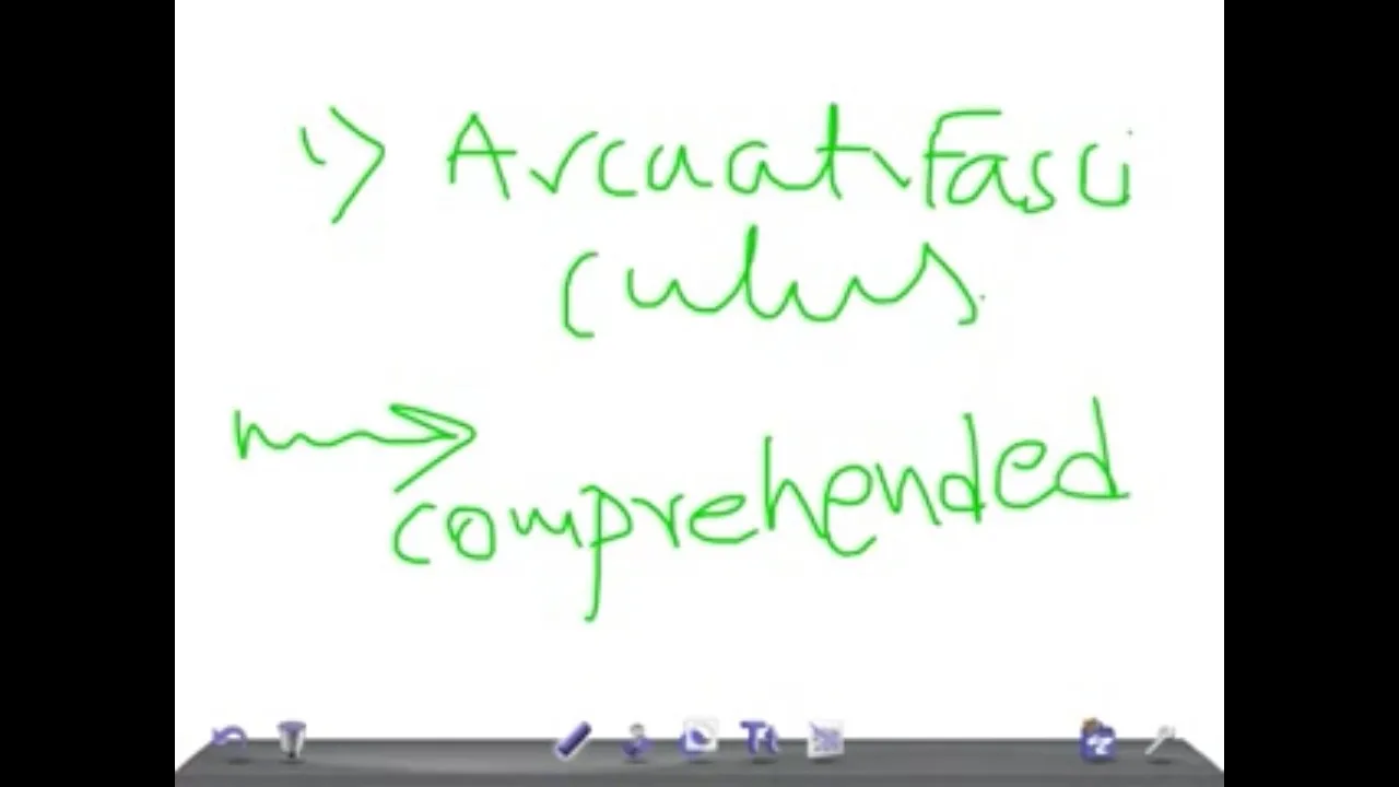
Why this video: This clinical review details conduction aphasia, which is caused by damage to the arcuate fasciculus. It explains why these patients maintain relatively normal comprehension and fluent speech but struggle to repeat spoken words.
- Knowledge Checkpoint:
- Identify the primary hallmark of conduction aphasia during clinical testing.
- Explain why language comprehension and spontaneous speech production remain mostly intact in conduction aphasia.
- Contrast the location of lesions in conduction aphasia with those in Broca's and Wernicke's aphasia.
Course Map
Key People Index
- Georg von Békésy (1899–1972): A Nobel laureate physicist who discovered the physical mechanics of the cochlear basilar membrane. He demonstrated how its traveling wave properties create a tonotopic map of pitch perception.
- Paul Broca (1824–1880): A French physician and anatomist who identified the left inferior frontal gyrus (specifically Brodmann areas 44 and 45) as critical for speech production. His findings were based on post-mortem examinations of patients with expressive language deficits, most notably his patient "Tan."
- Carl Wernicke (1848–1905): A German physician and neuropathologist who discovered that damage to the posterior superior temporal gyrus (Brodmann area 22) impairs language comprehension. He proposed the early "receptive-expressive" network model of language processing.
Final Self-Assessment
Complete this comprehensive self-assessment to verify your mastery of auditory neuroscience and clinical neuropsychology:
- Acoustic Physics: Explain how sound intensity (loudness) and sound frequency (pitch) are physically coded by the tympanic membrane.
- Impedance Matching: Describe how the middle ear ossicles overcome the acoustic transition from an air medium to a fluid-filled cochlear medium.
- Cochlear Chambers: Draw or describe the cross-sectional anatomy of the cochlea, labeling the scala vestibuli, scala tympani, scala media, Reissner's membrane, and the basilar membrane.
- Endocochlear Potential: State the electrical charge difference between endolymph and perilymph, and explain how this potential supports sensory transduction.
- Tonotopic Organization: Trace the tonotopic map from the base and apex of the basilar membrane up to the primary auditory cortex (A1).
- Ascending Auditory Pathway: List, in order, the five main synaptic relay stations from the vestibulocochlear nerve (CN VIII) to the temporal lobe.
- Binaural Integration: Identify the brainstem nuclei responsible for calculating interaural level differences (ILD) and interaural time differences (ITD).
- Broca's Area Anatomy: Pinpoint the exact anatomical boundaries of Broca's area, including its two main sub-regions (pars opercularis and pars triangularis).
- Wernicke's Area Anatomy: Define the location of Wernicke's area and trace the white-matter arcuate fasciculus that connects it to the frontal lobe.
- Aphasia Differentiation: Construct a diagnostic table comparing Broca's, Wernicke's, and conduction aphasia across three criteria: speech fluency, language comprehension, and repetition ability.
- Modern Connectomics: Discuss how modern diffusion tensor imaging (DTI) and tractography have refined classic models of language processing.