Neuronal Excitability: Biophysics & Synapses
Learning Goal: Analyze the biophysical principles of neuronal excitability, focusing on the ion channel dynamics of the action potential and the molecular machinery of calcium-dependent neurotransmitter release.
- Prerequisites: Basic college-level biology, general chemistry (thermodynamics and electrochemistry concepts), and introductory physics (electricity and magnetism, circuit basics).
- Estimated Total Study Time: 16 hours
Module 1: Cellular & Biophysical Foundations
This module establishes the structural and thermodynamic foundation of neurobiology. You will explore neuron morphology, membrane lipid bilayer physics, and the electrochemical forces (concentration gradients and electrostatic pressures) that drive passive ion transport.
Recommended Videos
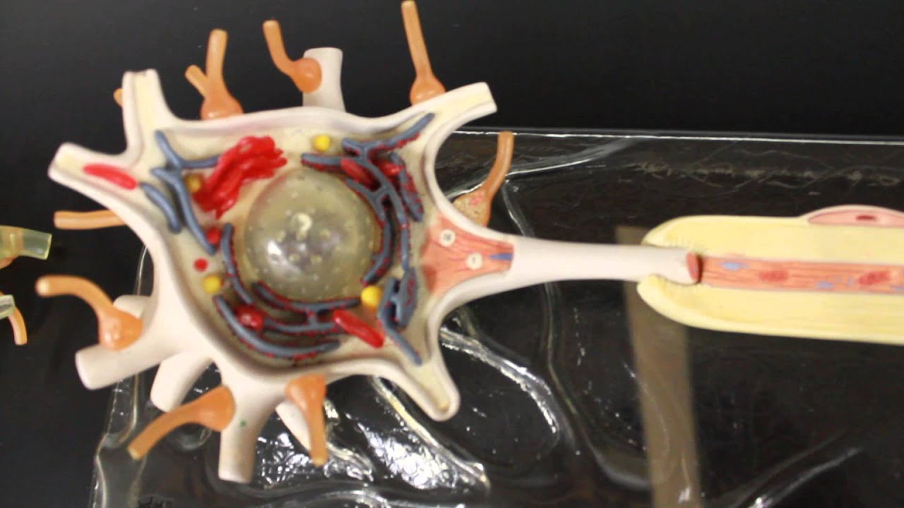
Why this video: This video provides an essential anatomical primer using a physical 3D model. It visualizes the spatial relationship between the soma, dendrites, myelin sheath, and axon hillock—the critical site of action potential initiation where high densities of voltage-gated channels reside.
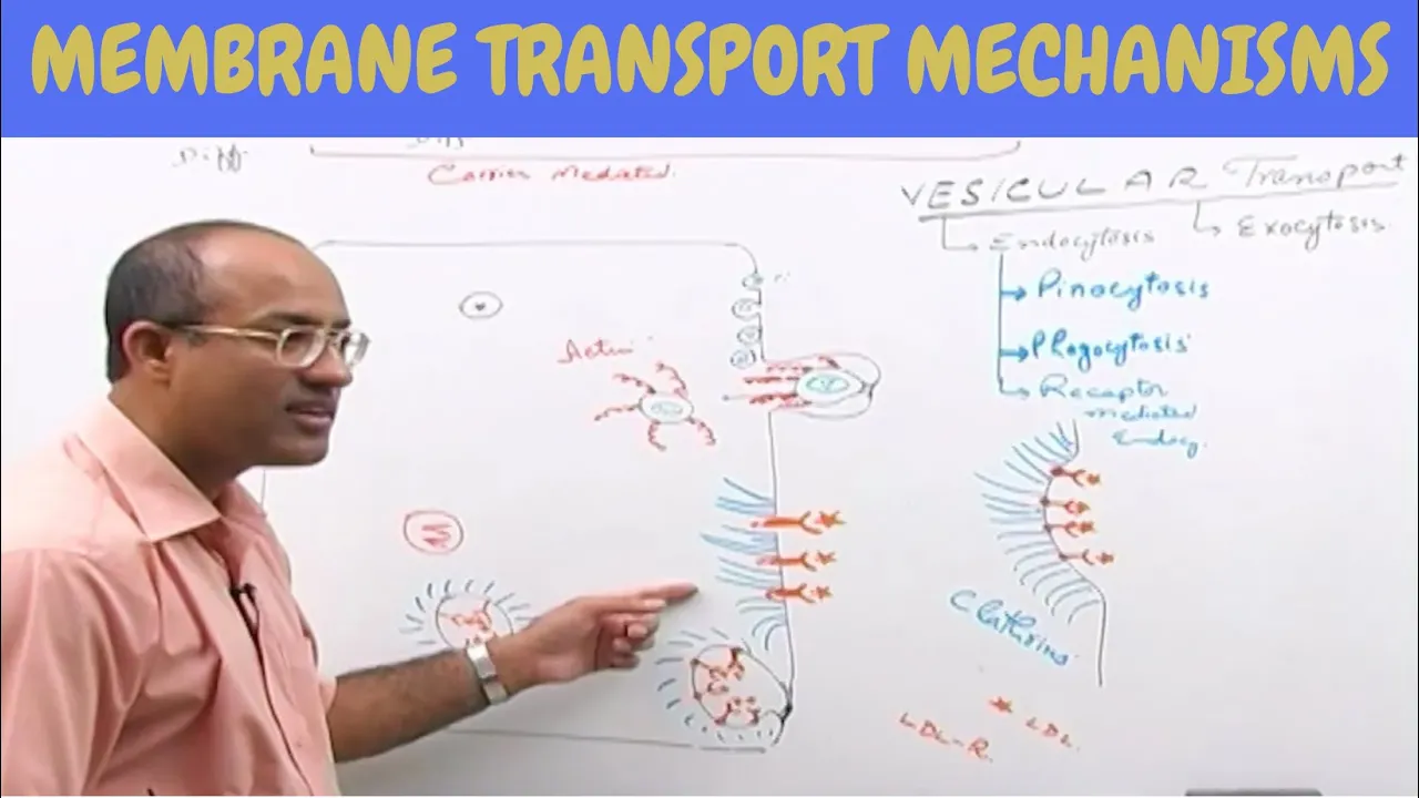
Why this video: Understanding passive transport is crucial before calculating membrane potentials. This video breaks down lipid bilayer permeability, simple diffusion, and facilitated diffusion via channel proteins, defining how ions cross the hydrophobic membrane core.

Why this video: This video provides the mathematical and physical foundations of electrochemical gradients. It details how the chemical gradient (entropy/concentration differences) and the electrical gradient (electrostatic forces) act in tandem, defining the net driving force () for ion movement.
Module 1 Knowledge Checkpoint
- Identify and describe the functional roles of the soma, dendrites, axon hillock, axon, and terminal bouton.
- Differentiate between simple and facilitated diffusion across the neuronal membrane.
- Mathematically state how the combination of chemical concentration differences and electrical potential differences creates a net electrochemical driving force.
- Explain how the lipid bilayer acts as a physical capacitor by separating charges across a thin, insulating boundary.
Module 2: The Resting Membrane Potential
This module focuses on how the resting membrane potential () is generated and maintained. You will study the quantitative contributions of individual ion species using the Nernst and Goldman-Hodgkin-Katz (GHK) equations, alongside the active electrogenic work of the ATPase pump.
Curriculum Note: General electrochemistry videos often focus on standard chemical cells. The selections below are specifically curated for neurobiology applications.
Recommended Videos
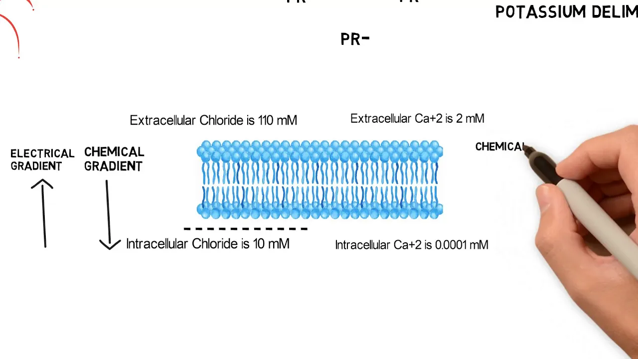
Why this video: This neurobiology-focused video explains the transition from the single-ion Nernst equilibrium potential () to the multi-ion GHK equation. It illustrates how resting membrane permeability is dominated by potassium leak channels, pulling close to .
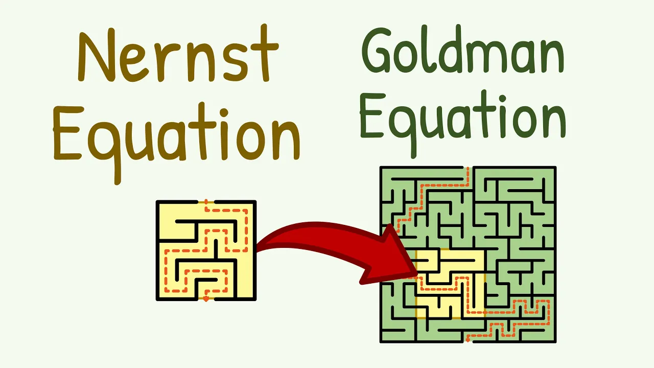
Why this video: This tutorial offers a highly detailed step-by-step breakdown of calculations using both the Nernst and GHK formulas. It explicitly handles the valence values () and explains why the chloride terms () in the GHK equation are inverted (out/in vs. in/out) due to chloride's negative charge.

Why this video: The ATPase is vital for preventing the dissipation of concentration gradients. This video details its active transport cycle, explaining how the pump uses ATP hydrolysis to export and import , generating an electrogenic hyperpolarizing current.
Module 2 Knowledge Checkpoint
- Write the Nernst equation and calculate the equilibrium potential for , , and given typical physiological concentration values.
- Explain why the Goldman-Hodgkin-Katz (GHK) equation is necessary to determine the overall resting membrane potential of a real, multi-permeable cell.
- Describe the stoichiometry and cycle of the ATPase pump and calculate its net charge impact per cycle.
- Predict how changing extracellular concentrations (e.g., hyperkalemia) shifts the resting membrane potential using GHK principles.
Module 3: Action Potential Dynamics & Ion Channels
This module covers the biophysics of the action potential. You will analyze the molecular gating of voltage-gated sodium () and potassium () channels, trace the phases of the action potential, and dissect the mathematical formulation of the classic Hodgkin-Huxley model.
Recommended Videos
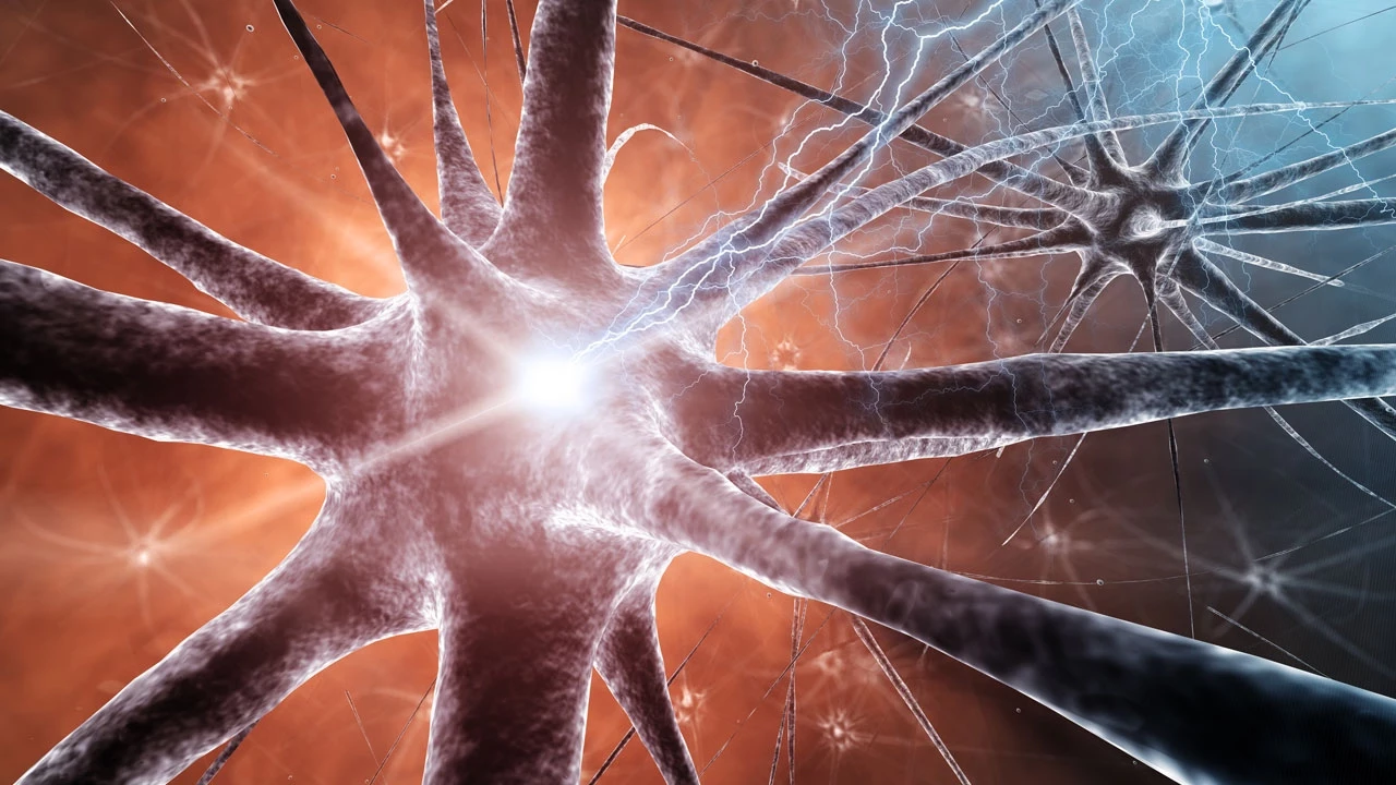
Why this video: This lecture highlights the distinct conformational states of voltage-gated channels. It describes the dual-gate mechanism of channels (activation and inactivation gates yielding closed, open, and inactivated states) versus the single-gate activation/deactivation mechanism of channels.
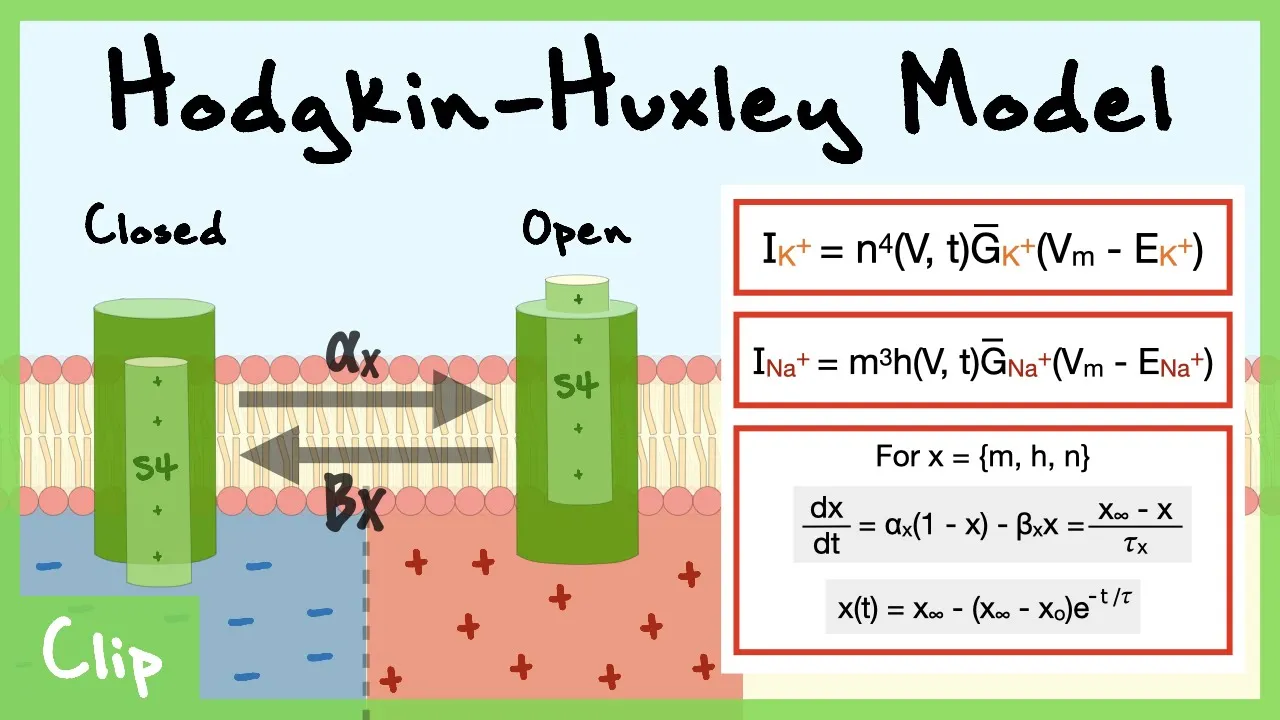
Why this video: This clip provides an in-depth explanation of the Hodgkin-Huxley gating variables (, , and ). It walks through the differential equations representing the probability of channel opening and closing as a function of membrane voltage and time.
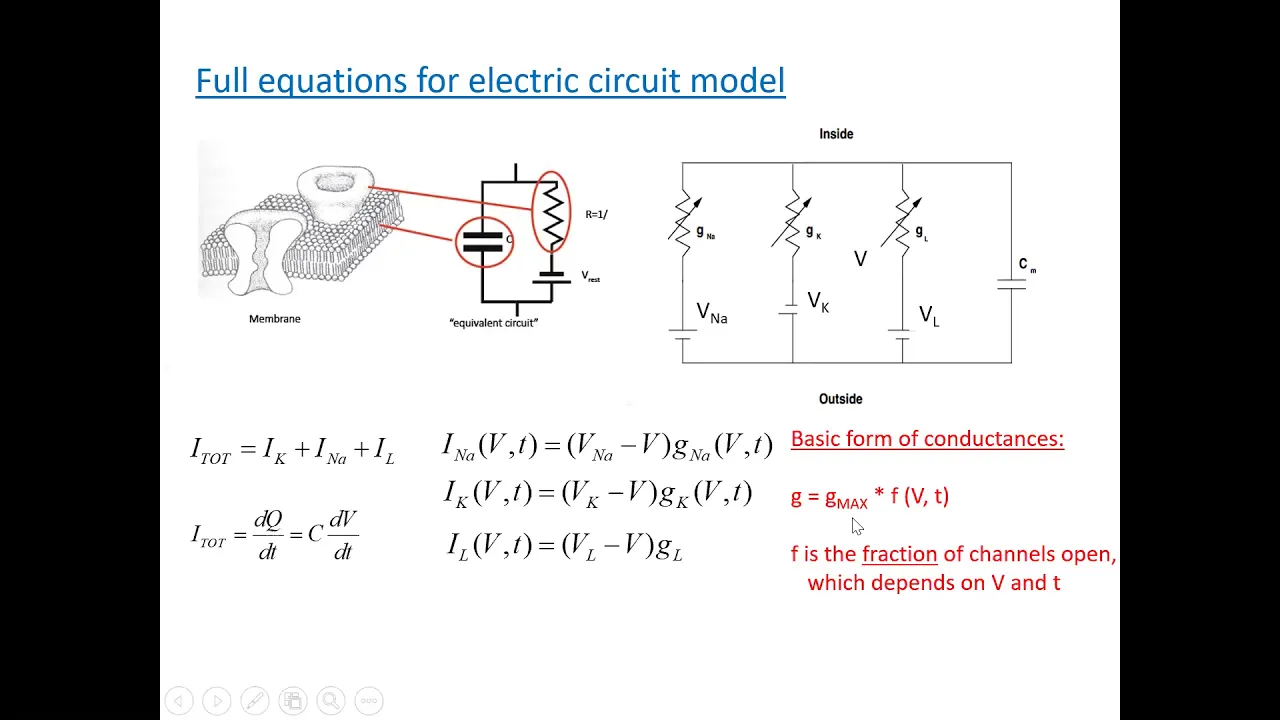
Why this video: This advanced lecture models the neuronal membrane as an equivalent electrical circuit. It maps the lipid bilayer to a capacitor, ion channels to variable resistors (conductances), and equilibrium potentials to batteries, providing the full mathematical framework of the Hodgkin-Huxley equations.
Module 3 Knowledge Checkpoint
- Detail the physical and functional differences between the activation (-gate) and inactivation (-gate) gates of voltage-gated sodium channels.
- Trace the ionic currents ( and ) during the rising, peak, falling, and undershoot (hyperpolarization) phases of an action potential.
- Define the Hodgkin-Huxley gating variables , , and , including their exponents in the conductance equations ( and ).
- Differentiate between the absolute and relative refractory periods in terms of channel state inactivation.
Module 4: Passive Membrane Properties & Cable Theory
Curriculum Correction: This module has been renamed to emphasize the quantitative biophysical properties that govern electrical signaling. We focus here on resistance, capacitance, and spatial-temporal decay constants.
This module details how subthreshold electrical signals propagate passively along the axon and dendrites. Using passive cable theory, you will learn to calculate the length () and time () constants, and study how myelin and saltatory conduction overcome passive decay.
Recommended Videos
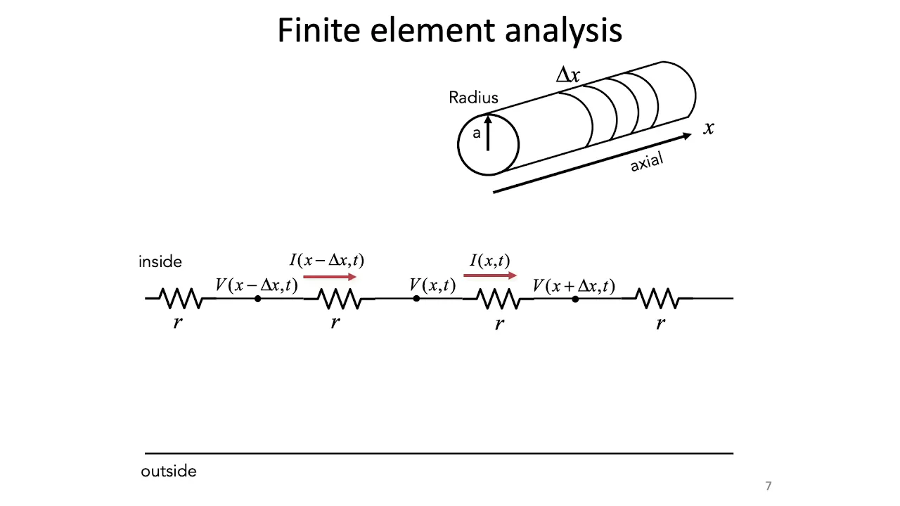
Why this video: An excellent, highly technical introduction from MIT to the mathematics of passive cable properties. It derives the length constant and time constant , explaining how they dictate signal decay over distance and time.
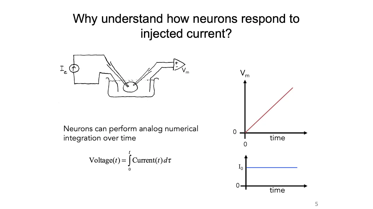
Why this video: This brief video demonstrates that the membrane time constant () is a fundamental property of the membrane material, showing mathematically that the membrane area cancels out when calculating .

Why this video: This comprehensive lecture explores the physical parameters of membrane resistance (), longitudinal/axial resistance (), and membrane capacitance (). It explains how cell diameter and myelin systematically alter these variables to optimize conduction velocity.

Why this video: This video bridges passive cable theory with active propagation. It explains how myelin decreases membrane capacitance and increases membrane resistance, enabling action potentials to travel passively through internodes and regenerate at the high-conductance nodes of Ranvier.
Module 4 Knowledge Checkpoint
- Define the membrane time constant () and describe its effect on temporal summation of synaptic inputs.
- Define the membrane length constant () and describe how changes in membrane resistance () and axial resistance () affect spatial signal propagation.
- Explain physically why myelination decreases total membrane capacitance () and increases total membrane resistance ().
- Describe the process of saltatory conduction and explain why it is energetically and kinetically superior to continuous propagation.
Module 5: Calcium-Dependent Synaptic Transmission
This final module focuses on the presynaptic active zone. You will analyze the molecular mechanics of neurotransmitter exocytosis, tracing how action potential arrival triggers voltage-gated calcium () influx, which initiates the assembly of the SNARE-synaptotagmin complex.
Recommended Videos

Why this video: This video provides an excellent visual overview of the entire synaptic release sequence. It explicitly highlights how incoming binds to Synaptotagmin, triggering the conformational change that drives the final fusion step.

Why this video: This video details the assembly of the core SNARE complex. It categorizes the primary molecular players, describing the roles of the vesicle-associated SNARE (v-SNARE: Synaptobrevin) and target-membrane SNAREs (t-SNAREs: Syntaxin and SNAP-25).

Why this video: This advanced seminar clip focuses on the physical assembly and structural organization of the SNARE complex. It details the molecular interactions of the 4-helix bundle during vesicle docking, priming, and fusion.
Module 5 Knowledge Checkpoint
- Outline the sequence of events from action potential arrival at the presynaptic terminal to vesicle membrane fusion.
- Identify the protein subunits of the core SNARE complex, categorizing them into v-SNAREs (Synaptobrevin) and t-SNAREs (Syntaxin-1 and SNAP-25).
- Explain the role of Synaptotagmin-1 as the primary calcium sensor, detailing how calcium binding triggers vesicle exocytosis.
- Contrast the concepts of vesicle "priming" and vesicle "fusion" at the active zone.
Course Map
This map outlines the ideal path through the biophysical and molecular concepts of this curriculum.
Key People Index
- Alan Hodgkin & Andrew Huxley: Pioneered the voltage-clamp technique on the squid giant axon, formulating the mathematical model of the action potential that earned them the 1963 Nobel Prize.
- Walther Nernst: Developed the Nernst equation (1889) relating chemical concentrations to electrical potentials at thermodynamic equilibrium.
- David E. Goldman, Alan Hodgkin, & Bernard Katz: Developed the GHK equation, extending the Nernst equation to account for multi-ion systems with varying membrane permeabilities.
- Thomas C. Südhof: Awarded the 2013 Nobel Prize for identifying the molecular machinery of vesicle release, including the calcium-sensing function of Synaptotagmin-1 and the assembly of SNARE proteins.
Final Self-Assessment
Use this assessment to test your synthesis of the biophysical principles of neuronal excitability and synaptic transmission.
- Electrochemical Driving Force: Can you calculate the net direction of ion flow given a cell's membrane potential () and the ion's equilibrium potential ()?
- GHK Sensitivity: Can you predict how a sudden 10-fold increase in sodium permeability () changes ?
- Pump Contribution: Can you explain why disabling the pump causes gradual depolarization?
- Hodgkin-Huxley Gates: Can you describe the mathematical meaning of the gating parameters , , and , and explain why the sodium conductance utilizes ?
- Refractory Periods: Can you explain how the inactivation gate of the channel establishes the absolute refractory period?
- Cable Theory Derivation: Can you derive how a cell's physical diameter affects internal axial resistance () vs. membrane resistance (), and how this changes ?
- Myelin Biophysics: Can you describe how myelin increases the speed of electrical transmission without violating the physical constraints of membrane capacitance?
- Calcium Influx: Can you explain why extracellular calcium concentration is the primary rate-limiting factor for neurotransmitter release?
- SNARE Assembly: Can you draw a diagram of the core 4-helix bundle of the SNARE complex, labeling Synaptobrevin, Syntaxin-1, and SNAP-25?
- Synaptotagmin-1: Can you describe the conformational change that occurs when binds to the C2 domains of Synaptotagmin-1?