Neuroplasticity: Synaptic & Motor Learning
Learning Goal: Understand the neurobiological mechanisms of neuroplasticity and motor learning, focusing on how synaptic plasticity (LTP/LTD), the basal ganglia, and the cerebellum coordinate to acquire, refine, and habituate new motor skills.
- Prerequisites: Basic biology/physiology background recommended. No prior neuroscience experience required.
- Estimated Total Study Time: 14 hours
Module 1: Foundations of Neuroanatomy and Neural Communication
This module establishes the foundational principles of neuroscience necessary to understand higher-level plastic changes. You will explore the structure of the neuron, the mechanical and electrochemical generation of the action potential, and the precise steps of chemical synaptic transmission that allow neurons to communicate.
Recommended Videos
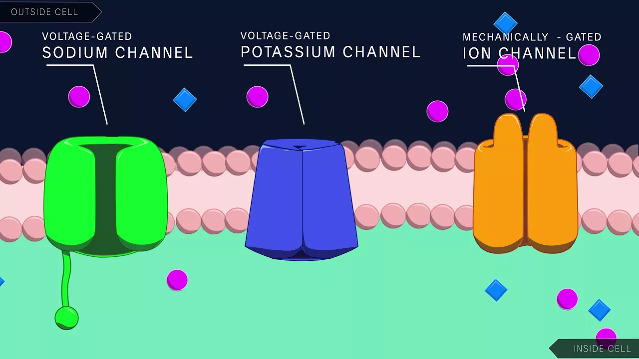
Why this video: This video offers an elegant, highly visual explanation of the electrochemical properties of the neuronal membrane. It walks through the resting potential, the gating mechanism of voltage-gated sodium and potassium channels, and how the threshold of excitation (-55mV) triggers an all-or-none action potential down the axon.
- Knowledge Checkpoint:
- Explain how the sodium-potassium pump ( ATPase) maintains the resting membrane potential at approximately -70mV.
- Describe the structural state of activation and inactivation gates of voltage-gated channels during depolarization, repolarization, and hyperpolarization.
- Define the difference between the absolute and relative refractory periods in terms of ion channel gating.
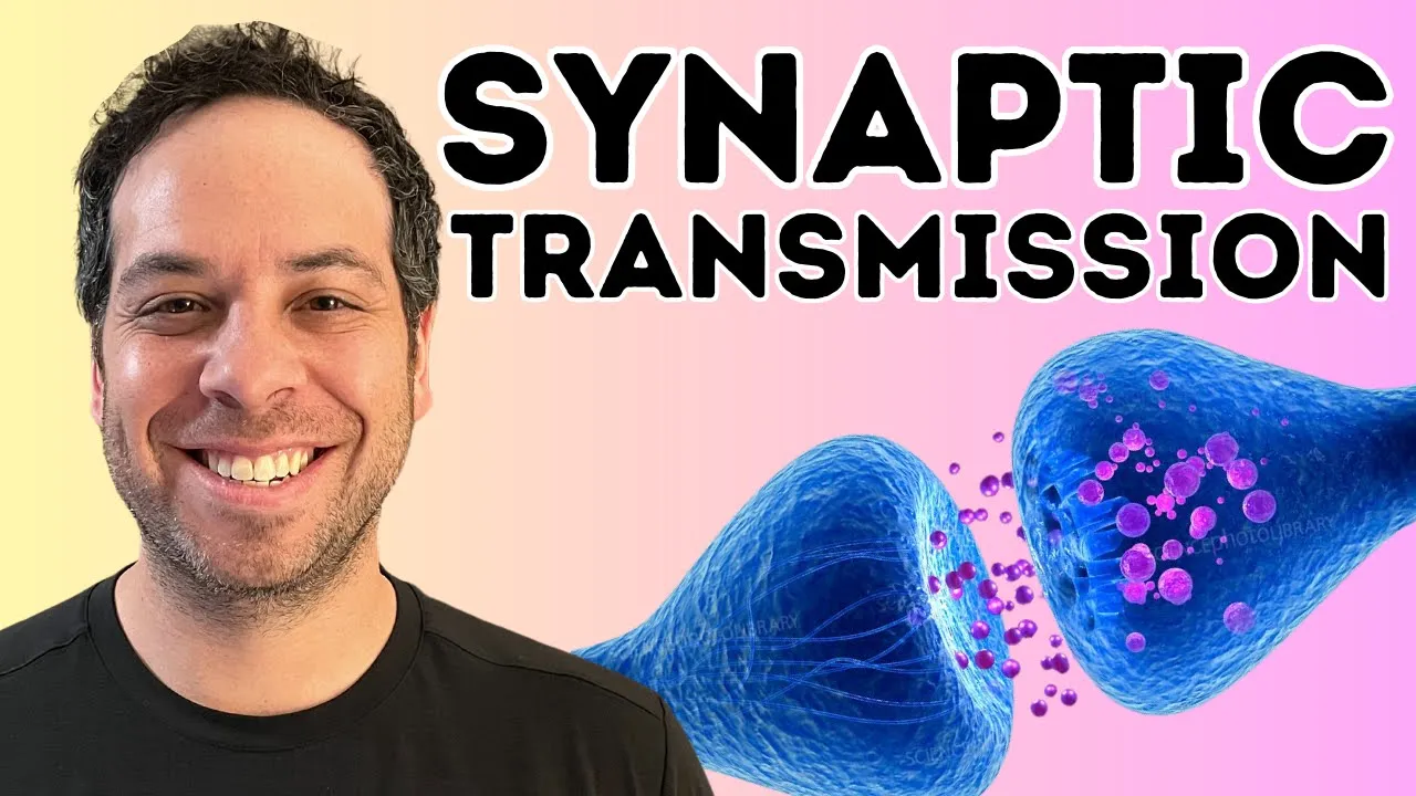
Why this video: Understanding neuroplasticity requires a flawless grasp of synaptic transmission. This video breaks down the micro-events at the synaptic cleft, focusing on calcium influx, neurotransmitter vesicle docking, exocytosis, and receptor binding on the postsynaptic membrane.
- Knowledge Checkpoint:
- Outline the sequential steps from the arrival of an action potential at the presynaptic terminal to transmitter release.
- Explain the role of voltage-gated calcium () channels in triggering vesicle fusion.
- Contrast ionotropic and metabotropic receptors in terms of response latency and downstream intracellular signaling.
Module 2: Synaptic Plasticity: Mechanism of LTP and LTD
This module explores the molecular and cellular mechanisms of Long-Term Potentiation (LTP) and Long-Term Depression (LTD). We will analyze how the pattern of presynaptic stimulation determines whether a synapse is strengthened or weakened, focusing specifically on the biophysical properties of AMPA and NMDA glutamate receptors.
Recommended Videos
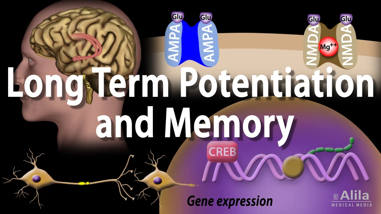
Why this video: An exceptionally clear, step-by-step animation detailing the transition from low-frequency baseline transmission to high-frequency tetanic stimulation that induces LTP. It clearly shows how depolarization ejects the block from the NMDA receptor pore.
- Knowledge Checkpoint:
- Describe why the NMDA receptor acts as a molecular "coincidence detector."
- Explain the roles of Calcium/Calmodulin-dependent protein kinase II (CaMKII) and retrograde messengers (like nitric oxide) in early-phase LTP.
- Distinguish between early LTP (receptor trafficking) and late LTP (gene transcription via CREB and structural remodeling).
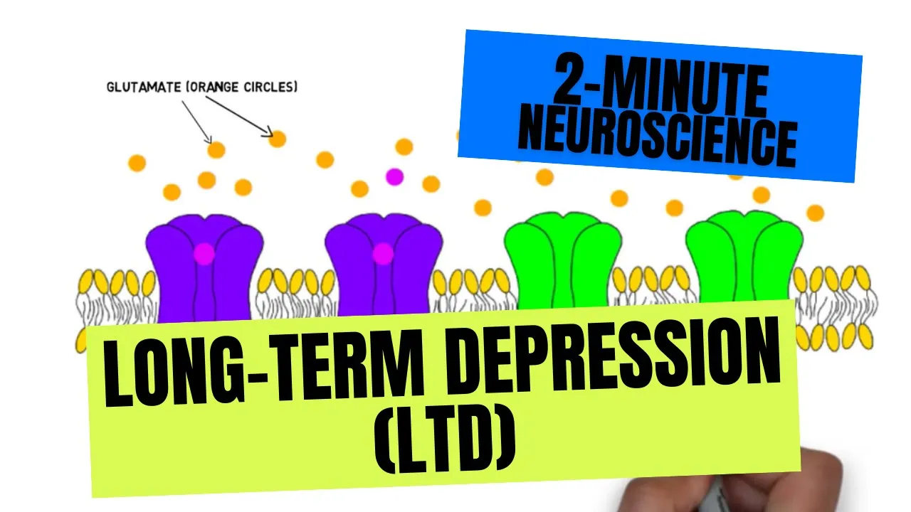
Why this video: While LTP strengthens synapses, LTD is essential for weakening underutilized pathways, preventing synaptic saturation. This short, high-yield video contrasts the low-frequency stimulation patterns that trigger LTD with the high-frequency patterns of LTP.
- Knowledge Checkpoint:
- Explain how a slow, prolonged rise in intracellular concentration activates phosphatases rather than kinases.
- Describe the process of AMPA receptor internalization (endocytosis) during LTD.

Why this video: This video bridges the gap identified in the review feedback by presenting a comprehensive comparison of LTP and LTD cellular pathways side-by-side. It highlights how the magnitude and kinetics of calcium entry through NMDA receptors act as the switch between synaptic potentiation and depression.
- Knowledge Checkpoint:
- Contrast the specific enzymatic pathways activated by high calcium spikes (kinases like CaMKII/PKC) versus low calcium trickles (phosphatases like calcineurin/PP1).
- Understand how these cellular changes alter the post-synaptic density (PSD-95) structurally.
Module 3: The Motor System: Planning and Executing Movement
This module examines how the brain constructs and sends motor commands. We analyze the functional divisions of the motor cortex—namely, the primary motor cortex (M1), premotor cortex (PMC), and supplementary motor area (SMA)—and trace the descending pyramidal tract responsible for voluntary muscle activation.
Recommended Videos
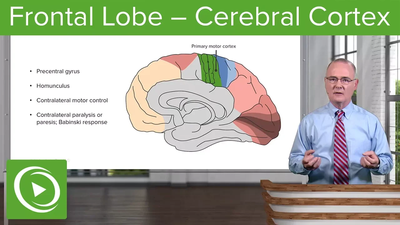
Why this video: This lecture clarifies the functional anatomy of the frontal lobe, drawing clear boundaries between the motor, premotor, and prefrontal areas. It directly addresses the review feedback by clarifying how motor plans are formulated before being sent to the primary motor cortex for execution.
- Knowledge Checkpoint:
- Distinguish the role of the supplementary motor area (SMA) in executing internally generated, well-learned sequences from the premotor cortex's role in sensory-cued planning.
- Explain the layout and proportional distortion of the motor homunculus in the precentral gyrus.

Why this video: This clinical review video goes deep into the Brodmann areas (Area 4 vs Area 6) and discusses the functional deficits seen when these specific cortices are damaged. It is highly useful for mapping anatomical boundaries to direct physiological movement roles.
- Knowledge Checkpoint:
- Identify Brodmann Area 4 and Brodmann Area 6 on a cortical map.
- Explain how damage to the premotor cortex affects complex movement sequences while leaving simple voluntary motor execution partially intact.
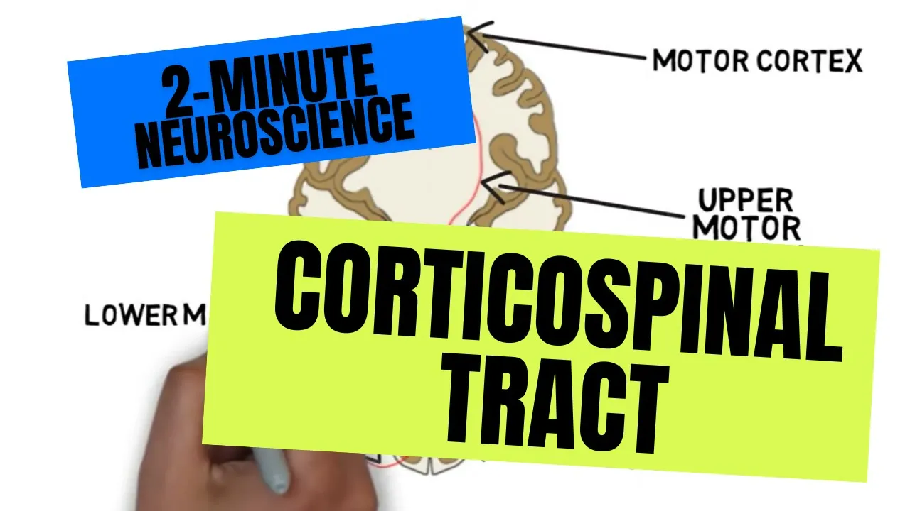
Why this video: A concise, clean spatial map of the corticospinal tract. It traces the axons of upper motor neurons as they descend from the cortex through the internal capsule, brainstem, and spinal cord.
- Knowledge Checkpoint:
- Trace the corticospinal pathway from the precentral gyrus down to the ventral horn of the spinal cord.
- Identify where the lateral corticospinal tract decussates (crosses over) in the caudal medulla.
- Explain the functional difference between the lateral corticospinal tract and the anterior corticospinal tract.
Module 4: The Basal Ganglia: Action Selection and Habit Formation
This module details how the basal ganglia gate voluntary movement, choose appropriate motor programs, and transition behaviors from goal-directed actions to automated habits. We will break down the direct and indirect pathways and study the role of dopamine in modulating striatal plasticity.
Recommended Videos
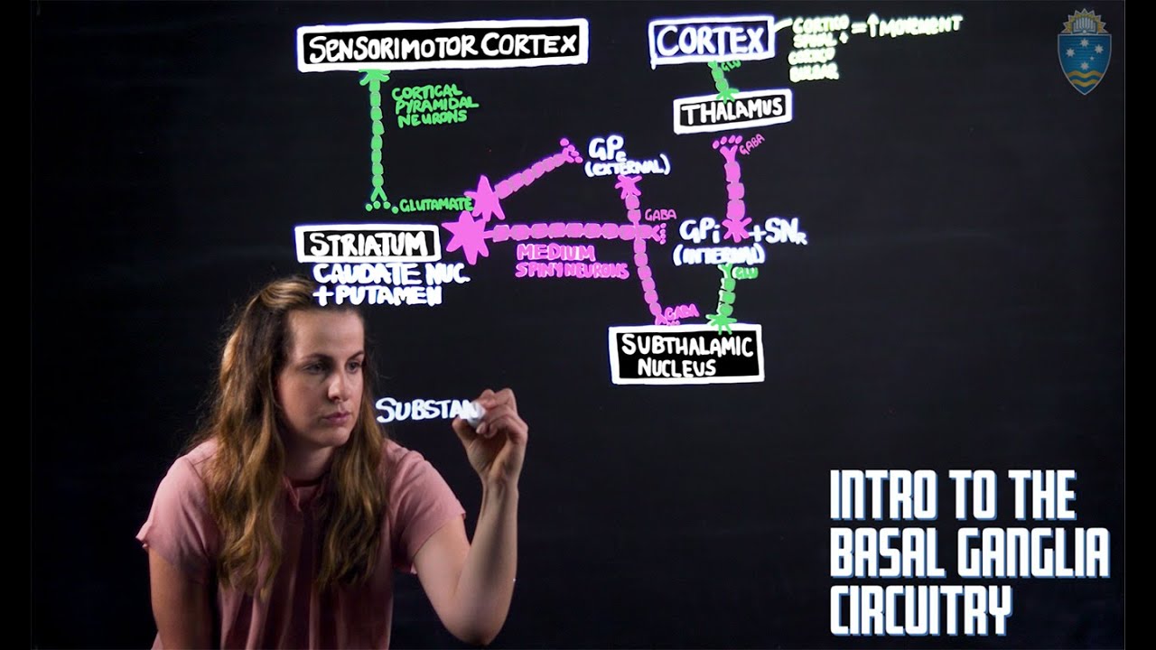
Why this video: A comprehensive, hand-drawn dissection of the basal ganglia loop. It covers the specific neurotransmitters used at each synapse (GABA, Glutamate) and explains how the direct pathway disinhibits the thalamus to allow movement, while the indirect pathway inhibits it.
- Knowledge Checkpoint:
- Map out the step-by-step neural connections of the direct pathway (Cortex Striatum GPi/SNr Thalamus Cortex).
- Map out the steps of the indirect pathway, including the role of the subthalamic nucleus (STN) and Globus Pallidus externus (GPe).
- Explain how dopamine released from the substantia nigra pars compacta () differentially affects the direct (D1 receptors) and indirect (D2 receptors) pathways.
Why this video: This researcher-led video addresses the critical review gap on habituation. Dr. Lerner explains the neural mechanism of habits, how behaviors shift over time, and how different dopamine signals operate on discrete circuits.
- Knowledge Checkpoint:
- Define a "habit" neurobiologically using the outcome devaluation test.
- Explain how motor control transitions from the associative striatum (caudate nucleus in primates) to the sensorimotor striatum (putamen) as a skill is overlearned.

Why this video: Explores the functional integration of specific basal ganglia nuclei (specifically the caudate) in memory consolidation and spatial mapping during movement loops.
- Knowledge Checkpoint:
- Describe the functional outcomes of caudate lesions on short-term motor recall.
- Discuss how the striatum serves as a central hub where cortical sensory inputs meet dopaminergic reward-prediction signals.
Module 5: The Cerebellum: Motor Calibration and Error Correction
This module focuses on the cerebellum, the brain's real-time calibration computer. We will explore its micro-circuit architecture—comprising mossy fibers, climbing fibers, granule cells, and Purkinje cells—and analyze how the cerebellum uses sensory feedback to adjust motor output and perform error correction.
Recommended Videos
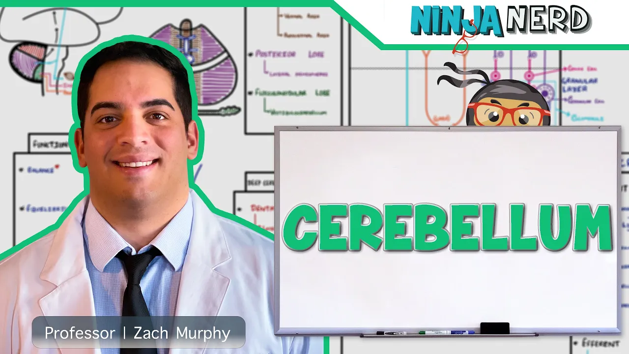
Why this video: An incredibly detailed, world-class whiteboard lecture that traces the afferent and efferent projections of the cerebellum. It is essential for understanding how the cerebellum receives motor copies from the cortex and sensory feedback from the spinocerebellar tract to compute adjustments.
- Knowledge Checkpoint:
- Identify the three functional divisions of the cerebellum (vestibulocerebellum, spinocerebellum, cerebrocerebellum) and their primary roles.
- Name the deep cerebellar nuclei and explain which cerebellar zones project to them.
- Trace the path of the "efference copy" (correlative motor plan) from the cortex to the cerebellum.
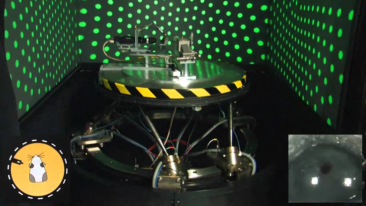
Why this video: Dr. de Zeeuw, a leading neuroscientist, challenges classical models of cerebellar learning (which prioritized LTD at Purkinje synapses) and introduces modern findings showing that LTP at parallel fiber-to-Purkinje synapses acts as a major driver for motor adjustments.
- Knowledge Checkpoint:
- Explain how classical Albus-Marr-Ito models of cerebellar learning relied on long-term depression (LTD) to explain motor adaptation.
- Summarize the modern research showing why potentiation (LTP) at Purkinje cell synapses is highly active during real-time calibration.

Why this video: An advanced, research-level seminar tracking the temporal consolidation of motor memories inside Purkinje cells. It explains how optogenetics is used to study the post-training window during which motor adaptations are solidified.
- Knowledge Checkpoint:
- Explain the role of Purkinje cells as the sole inhibitory output of the cerebellar cortex.
- Explain how the timing of climbing fiber inputs (carrying sensory "error signals") and parallel fiber inputs (carrying context) changes Purkinje cell firing rates.
- Define the consolidation window (0-90 minutes post-training) for structural cerebellar changes.
Module 6: Integrated Motor Learning: From Acquisition to Mastery
This final module integrates the preceding modules. You will study how the cortex, basal ganglia, and cerebellum coordinate as a dynamic, unified system. Over time, motor control shifts from a highly conscious, cerebellum-heavy corrective loop to an automated, basal ganglia-driven habit.
Recommended Videos

Why this video: Directly addresses the "system-level integration loop" gap. It details how the cerebellum, basal ganglia, and motor cortex are structurally interconnected via direct subcortical loops, and how they synchronize their oscillatory activity during movement.
- Knowledge Checkpoint:
- Describe the reciprocal, subcortical anatomical pathways linking the cerebellum and the basal ganglia (thalamic-mediated and disynaptic pathways).
- Explain how abnormal synchronization across this loop can lead to motor dysfunction.

Why this video: Outlines the classical Fitts and Posner model of motor learning. Understanding these cognitive stages provides the psychological scaffolding that maps onto the neurobiological shifts occurring in the brain.
- Knowledge Checkpoint:
- Name and define the three stages of motor learning: Cognitive, Associative, and Autonomous.
- Identify which brain structures are most active during the cognitive stage versus the autonomous stage.

Why this video: Summarizes how repetition, error-generation, and cognitive focus trigger rapid systemic plasticity. It provides a practical framework for consolidating motor memory based on systems-level neuroscience.
- Knowledge Checkpoint:
- Explain how generating physical movement "errors" acts as a cellular cue to increase neuroplasticity.
- Explain the role of post-training resting states in facilitating immediate neural replay of the learned skill.
💡 Supplementary Synthesis Note: The Dynamic Motor Loop Shift
Because system-level synthesis is challenging to capture in individual videos, study the following paradigm carefully:
[Early Acquisition Stage]
- High conscious control (Prefrontal Cortex active).
- High sensory-error correction (Cerebellum active; compares intent to execution).
- Associative Basal Ganglia active (Caudate / Medial Striatum). │ ▼ (Repeated practice & LTP/LTD structural consolidation) │ [Late Autonomous Stage]
- Shift from conscious correction to automatic execution.
- Sensorimotor Basal Ganglia dominant (Putamen / Lateral Striatum).
- Motor program is consolidated as a habit; prefrontal and cerebellar cortex activation decreases.
Course Map
Key People Index
- Dr. Andrew Huberman (Stanford University)
Featured in Module 6. Well-known for translating systems-level neuroscience into actionable protocols for learning and neuroplasticity. - Prof. Dr. Chris de Zeeuw (Erasmus MC / Netherlands Institute for Neuroscience)
Featured in Module 5. A pioneering cerebellar neuroscientist who redefined modern views of cerebellar motor learning and Purkinje cell plasticity. - Dr. Talia Lerner (Northwestern University)
Featured in Module 4. Researcher focusing on the neurobiology of dopamine, striatal circuits, and habit formation pathways. - Dr. Daniela Popa (Institut de Biologie de l'ENS)
Featured in Module 6. Investigator of subcortical networks, specializing in the functional connections between the cerebellum and the basal ganglia.
Final Self-Assessment
Test your synthesis of the material by verifying you can explain and check off each item:
- I can trace a motor signal from its initial planning phases in the SMA/PMC to its descent through the pyramids, detailing where it crosses over.
- I can explain the cellular biophysics of how the NMDA receptor blocks calcium entry under resting conditions and how it is expelled during high-frequency depolarization.
- I can describe the structural and enzymatic differences between long-term potentiation (LTP) and long-term depression (LTD).
- I can map the entire direct pathway of the basal ganglia and explain how it facilitates motor program selection by disinhibiting the thalamus.
- I can map the indirect pathway and explain its role in suppressing competing motor movements.
- I can explain how dopamine interacts with vs metabotropic receptors to bias basal ganglia output toward action selection.
- I can explain the cellular mechanism of the "climbing fiber" error signal in the cerebellum and how it induces synaptic plasticity in Purkinje cells.
- I can describe the structural transition of a motor skill as it shifts from the caudate (associative striatum) to the putamen (sensorimotor striatum) during habituation.
- I can explain how the cerebellum and basal ganglia are connected subcortically without relying entirely on relays through the cerebral cortex.
- I can identify the 3 psychological stages of motor learning (Fitts & Posner) and correlate them with their primary neurobiological networks.