Sleep & Circadian Biology: Brain Waves & Memory
Learning Goal: Understand the neurobiological mechanisms of sleep and circadian rhythms, focusing on how the suprachiasmatic nucleus, key neurotransmitters, and neural oscillations during REM and NREM sleep regulate memory consolidation and brain waste clearance.
- Prerequisites: Basic high school-level biology.
- Estimated Total Study Time: 22 Hours (includes video viewing, active note-taking, and supplementary reading/hands-on review of the concepts).
Module 1: Foundational Neuroscience: Neurons, Synapses, and Brain Structure
This module establishes the foundational cellular and structural neuroscience required to understand sleep neurobiology. You will learn the anatomy of a neuron, how action potentials trigger chemical synapses, and the primary lobes of the human brain.
Recommended Videos
1. Synaptic Transmission: Chemical Synapses & Neurotransmitters Explained

Why this video: This 3D animation clearly shows how an action potential triggers the release of neurotransmitters across the synaptic cleft. It provides a visual framework for understanding the chemical signals that regulate sleep switches and neural oscillations.
- Knowledge Checkpoint:
- Explain how an electrical action potential translates into chemical signaling at the synaptic cleft.
- Describe the role of calcium influx in neurotransmitter exocytosis.
- Differentiate between excitatory and inhibitory postsynaptic potentials (EPSPs and IPSPs).
2. Chemical Synapse Animation
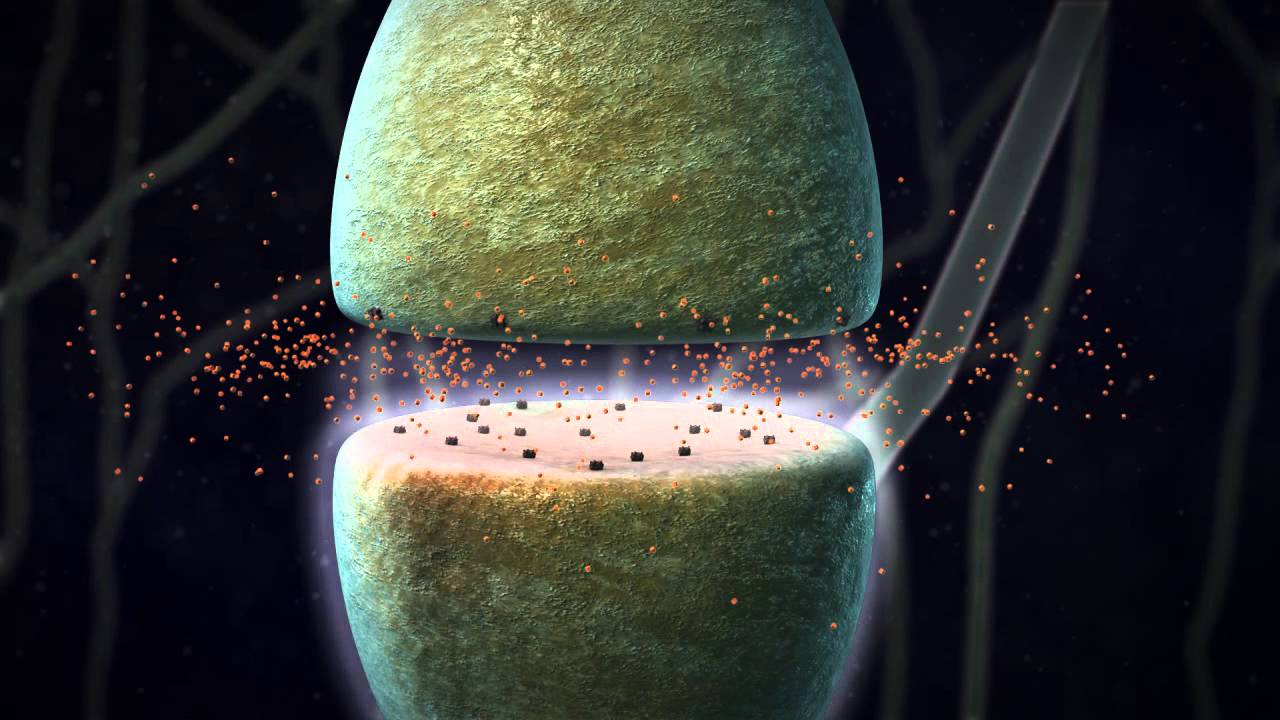
Why this video: This brief, highly focused animation isolates the micro-mechanics of the chemical synapse, displaying vesicle docking and receptor binding in clear, uncluttered detail.
- Knowledge Checkpoint:
- Identify where neurotransmitter vesicles are stored prior to release.
- Define the synaptic cleft and describe the pathway of a neurotransmitter across it.
3. Anatomy - Brain (Circle of Willis and Stroke)
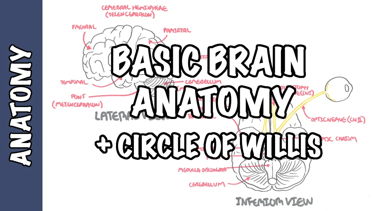
Why this video: To comprehend sleep centers, you must first master the macroscopic geography of the brain. This video breaks down the major lobes (frontal, parietal, temporal, occipital) and key brainstem structures (pons, medulla) involved in sleep control.
- Knowledge Checkpoint:
- Locate the four main lobes of the cerebrum.
- Identify the positions of the brainstem and cerebellum relative to the cerebrum.
Module 2: The Circadian Clock: The Suprachiasmatic Nucleus (SCN) and Light
This module covers the core circadian clock mechanism. You will study how external photic cues (zeitgebers) hit the retina, travel via the retinohypothalamic tract (RHT) to the suprachiasmatic nucleus (SCN), and trigger the intracellular CLOCK/BMAL1 and PER/CRY molecular transcription-translation feedback loop (TTFL).
Recommended Videos
1. 2024.2.13 - CWRU SMGR: Dr. Mark Wu - Circadian Rhythm Disorders
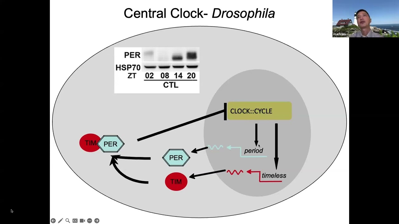
Why this video: This medical lecture specifically addresses the genetic and molecular feedback loops regulating circadian biology. It provides the deep academic mechanism of the mammalian CLOCK and BMAL1 heterodimer driving the cyclic transcription of Period (PER) and Cryptochrome (CRY) proteins.
- Knowledge Checkpoint:
- Define the Transcription-Translation Feedback Loop (TTFL) of the circadian clock.
- Detail how CLOCK and BMAL1 proteins interact with PER and CRY inside SCN cells.
- Explain the consequences when genes governing these molecular feedback loops are mutated.
2. Retina | Few Important Points | Part 3
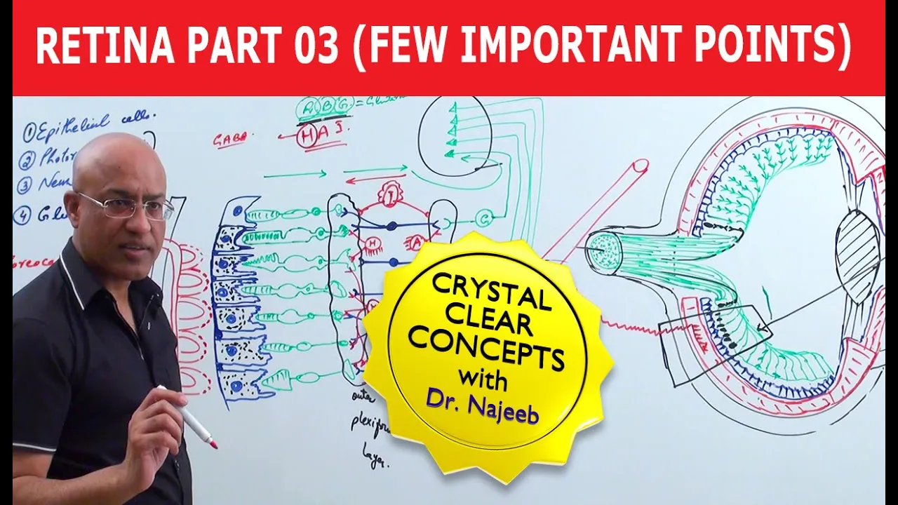
Why this video: This anatomical overview highlights the subpopulation of intrinsically photosensitive retinal ganglion cells (ipRGCs) that contain melanopsin. These cells project directly to the hypothalamus to communicate ambient light levels.
- Knowledge Checkpoint:
- Distinguish ipRGCs from traditional photoreceptors (rods and cones).
- Describe the anatomical target of the projections leaving these ganglion cells.
3. Pay attention to your body's master clock
Why this video: Emily Manoogian explains how the central pacemaker in the SCN coordinates peripheral molecular clocks residing in almost every cell type in the body, ensuring temporal alignment of physiological systems.
- Knowledge Checkpoint:
- Describe the difference between the master clock (SCN) and peripheral tissue clocks.
- Explain how mismatched peripheral rhythms (e.g., in liver or gut tissue) affect metabolic health.
Gap Note on Circadian Pathways: To fully understand this module, you should also independently research the melatonin pathway. When light decreases, SCN neural outputs to the paraventricular nucleus, superior cervical ganglion, and pineal gland change, stimulating the conversion of serotonin into melatonin (as alluded to in nutritional pathways in Video 59).
Module 3: Sleep Architecture: Deciphering NREM, REM, and EEG Waves
This module dives into sleep stages. You will learn to identify different physiological states—from awake beta waves, relaxed alpha waves, light NREM (N1, N2 with sleep spindles and K-complexes), deep slow-wave sleep (N3 delta waves), to the highly active, paradoxically desynchronized REM state.
Recommended Videos
1. 7 EEG in Normal Sleep

Why this video: This academic presentation offers real-world EEG trace analysis. It is critical for learning the exact visual biomarkers of drowsiness, loss of alpha rhythm, theta onset, sleep spindles, K-complexes, and slow wave progression.
- Knowledge Checkpoint:
- Identify the change in dominant wave patterns as a patient shifts from "eyes-closed awake" to "drowsiness."
- Visually identify a sleep spindle and a K-complex on an EEG trace.
- Quantify the frequency range (Hz) characteristic of theta and delta waves.
2. Sleep Cycle, EEG Waves Mnemonics , NON REM Versus REM Sleep

Why this video: This lecture provides structural mnemonics for memorizing sleep stages (N1 through N3, and REM) along with their corresponding electroencephalographical signals, muscle tone shifts, and eye movement signatures.
- Knowledge Checkpoint:
- List the order of stages in a standard 90-minute sleep cycle.
- Contrast REM sleep with N3 deep sleep in terms of muscle tone (atonia) and brain activity.
3. Sleep stages and circadian rhythms | Processing the Environment | MCAT | Khan Academy
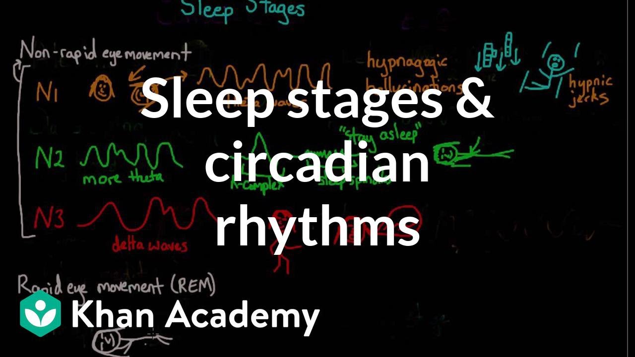
Why this video: This standard medical preparation video quickly defines the specific roles of sleep spindles and K-complexes during Stage 2 NREM sleep, helping contextualize how these bursts of activity defend the brain against external sensory arousal.
- Knowledge Checkpoint:
- Explain the hypothetical function of sleep spindles in sensory gating.
- Describe the shape and characteristics of a K-complex.
Module 4: Chemical Switches: Neurotransmitters Regulating Sleep and Wakefulness
This module covers the neural circuitry driving arousal and sleep. You will learn about the Ascending Reticular Activating System (ARAS) in the brainstem, homeostatic sleep pressure governed by adenosine, and the mutually inhibitory "flip-flop" switch between sleep-promoting centers (like the VLPO) and wake-promoting monoaminergic/orexinergic systems.
Recommended Videos
1. Sleep 4.2 | Sleep Neuroscience
Why this video: This video walks through historical and modern neuroscience experiments (including Moruzzi and Magoun's 1949 work) that mapped the reticular activating system (RAS) in the upper pons and midbrain, demonstrating its necessity for maintaining consciousness.
- Knowledge Checkpoint:
- Identify the anatomical structures comprising the ascending reticular activating system (ARAS).
- Contrast the two pathways of the ascending arousal system (thalamic vs. extrathalamic/cortical).
2. Introduction to Neurophysiology II, Areas of the brain and Sleep wake cycle

Why this video: This clinical neurophysiology resource outlines the functional anatomy of the brainstem and hypothalamus as they coordinate sleep-wake transitions.
- Knowledge Checkpoint:
- Detail how brainstem arousal centers interact with hypothalamic nuclei.
- Explain what happens to consciousness when the reticular formation is damaged.
3. Caffeine and Adenosine Receptors
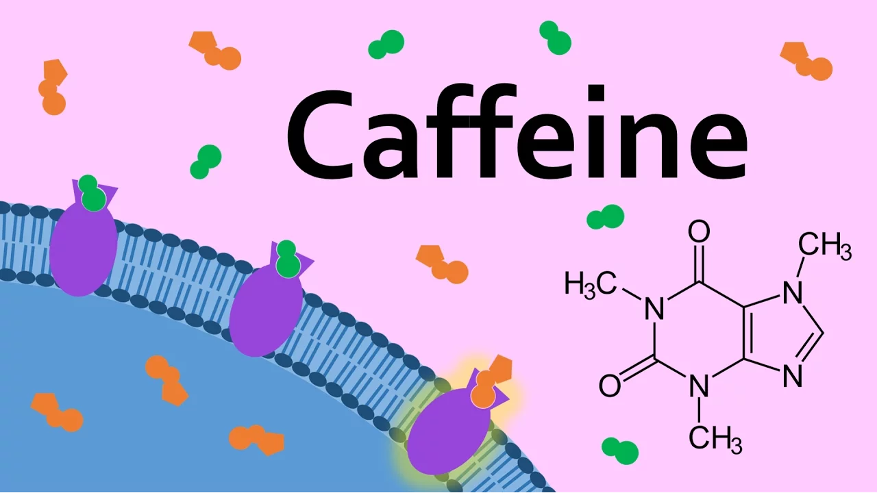
Why this video: This visual overview focuses on adenosine, the cellular byproduct of ATP breakdown that builds up in the basal forebrain and cortex during waking hours. It explains how caffeine binds competitively to adenosine receptors to temporarily block homeostatic sleep pressure.
- Knowledge Checkpoint:
- Explain how ATP breakdown generates adenosine during wakefulness.
- Describe the molecular mechanism of competitive antagonism by caffeine on adenosine receptors.
4. Why you're always tired

Why this video: This science communication piece highlights the orexinergic (hypocretin) system located in the lateral hypothalamus. It details how orexin acts as a vital stabilizer of the wakefulness-promoting side of the flip-flop circuit, preventing sudden transitions into sleep.
- Knowledge Checkpoint:
- Locate where orexinergic neurons originate in the hypothalamus.
- Describe the stabilizing role of orexin in the sleep-wake flip-flop model.
- Explain how a loss of orexin-producing neurons leads to narcolepsy.
Module 5: Cognitive Consolidation: How Sleep Processes and Stores Memory
This module explores the active, offline processing of memory. We analyze how sleep spindles and slow oscillations synchronize to facilitate hippocampal-neocortical dialogue, transferring memories from temporary storage (hippocampal RAM) to permanent neocortical structures (hard drive), alongside Giulio Tononi's Synaptic Homeostasis Hypothesis (SHY).
Recommended Videos
1. Lecture 1.7: Hippocampus, Memory, & Sleep, Part 2
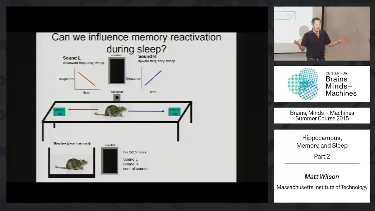
Why this video: An MIT course video detailing the exact neurophysiology of place cells, sharp-wave ripples (SWRs), and hippocampal spatial replay during sleep. It shows the computational evidence of cellular replay driving the transfer of episodic representations to the cortex.
- Knowledge Checkpoint:
- Define hippocampal "replay" and identify which sleep phase it typically occurs in.
- Describe the physical occurrence and physiological role of sharp-wave ripples (SWRs).
- Explain how replay leads to synaptic restructuring within neocortical circuits.
2. Giulio Tononi: 2011 Allen Institute for Brain Science Symposium

Why this video: Giulio Tononi presents his groundbreaking Synaptic Homeostasis Hypothesis (SHY). He outlines how wakefulness builds net synaptic potentiation across the brain, and how the slow waves of NREM sleep serve to systematically downscale synaptic strength to prevent metabolic exhaustion and signal saturation.
- Knowledge Checkpoint:
- State the primary premise of the Synaptic Homeostasis Hypothesis (SHY).
- Contrast synaptic changes during active wakefulness (learning) with those during slow-wave sleep.
- Explain how global synaptic downscaling preserves signal-to-noise ratio and energy.
3. Dr. Matt Walker: Using Sleep to Improve Learning, Creativity & Memory

Why this video: This clip highlights the relationship between NREM stage 2 sleep spindles and the physical shifting of files from short-term memory (hippocampus) to the cortical "vault" of long-term storage.
- Knowledge Checkpoint:
- Detail the correlation between sleep spindle frequency/density and memory retention.
- Contrast hippocampal capacity restrictions with neocortical long-term storage capacity.
Module 6: The Glymphatic System: Waste Clearance and Brain Health
This final module focuses on brain maintenance. You will explore the glymphatic system, a glial-mediated fluid transport pathway. You will study how astrocytic Aquaporin-4 (AQP4) water channels facilitate convective cerebrospinal fluid (CSF) flow to flush metabolic byproducts, such as beta-amyloid and tau proteins, during deep slow-wave sleep.
Recommended Videos
1. Improve Your Lymphatic System for Overall Health & Appearance

Why this video: Dr. Andrew Huberman explains the mechanics of the glymphatic system, focusing on astrocytes. During sleep, these glial cells shrink, expanding the perivascular space by roughly 60% and allowing CSF to flow through and clear accumulated toxins.
- Knowledge Checkpoint:
- Explain how astrocytes regulate the size of the interstitial/perivascular space between wakefulness and sleep.
- Define the convective fluid movement of CSF through brain tissue.
- Describe the 60% expansion of interstitial space during slow-wave sleep and its physical impact.
2. How Does Your Brain Clean Itself?
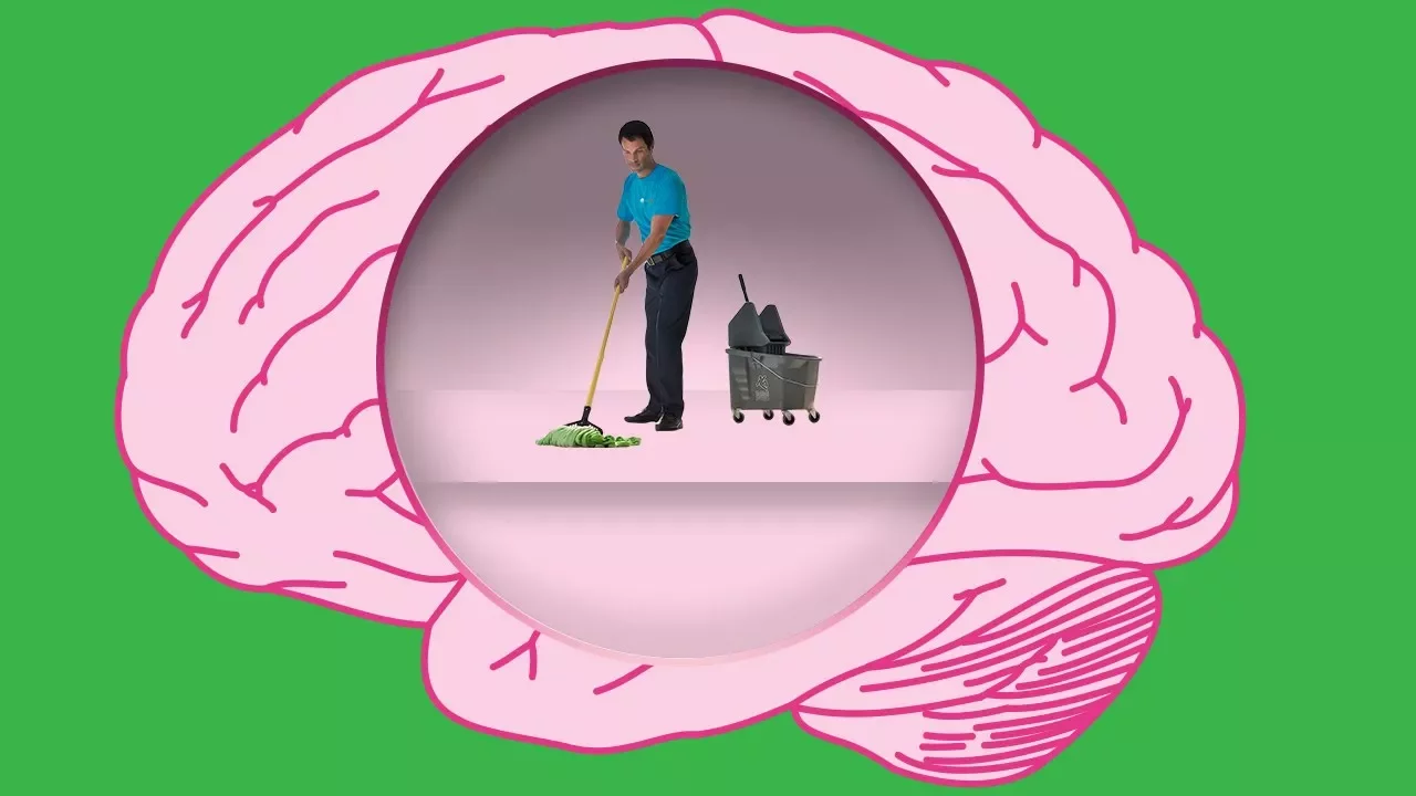
Why this video: This short overview summarizes Dr. Maiken Nedergaard's 2012 discovery of the glymphatic system using two-photon imaging in mice, demonstrating how the brain compensates for its lack of a traditional lymphatic lymphatic system.
- Knowledge Checkpoint:
- Identify who discovered and named the glymphatic system.
- Distinguish the glymphatic system's operations from the lymphatic system of the rest of the body.
3. We finally know why it's so good for you
Why this video: This highly visual explanation reinforces how the contraction of neurons/glial cells during deep sleep allows CSF to flow freely like a dishwashing cycle, picking up beta-amyloid plaques along the way.
- Knowledge Checkpoint:
- Explain why waste clearance is less efficient during waking states.
- Name the principal neurodegenerative biomarkers (e.g., amyloid-beta) cleared by this process.
Gap Note on Aquaporin-4 (AQP4): While the videos visually demonstrate glial contraction and CSF flow, they omit the molecular "pump" itself. To fill this gap, research how Aquaporin-4 (AQP4) water channels, localized heavily on astrocytic endfeet lining the vasculature, facilitate the high-rate convective movement of CSF into the brain parenchyma to mix with interstitial fluid (ISF).
Course Map
This flowchart outlines your path through this curriculum, from cellular foundations to complex neurological outcomes.
Key People Index
| Researcher / Expert | Affiliation / Context | Contribution to Sleep Science |
|---|---|---|
| Dr. Maiken Nedergaard | University of Rochester Medical Center | Discovered and characterized the glymphatic system in 2012, mapping glial waste clearance of toxic proteins. |
| Dr. Giulio Tononi | University of Wisconsin–Madison | Formulated the Synaptic Homeostasis Hypothesis (SHY), showing that sleep is the price we pay for neural plasticity. |
| Dr. Matthew Walker | University of California, Berkeley | Prolific sleep author and researcher; specializes in the mapping of NREM sleep spindles to cognitive performance and memory. |
| Dr. Mark Wu | Johns Hopkins University | Physician-scientist researching genetic molecular clocks and circadian rhythm disorders. |
Final Self-Assessment
Review this list to test your comprehension of the material.
- Action Potentials & Synapses: Can you explain the sequential ionic and chemical events required for a presynaptic terminal to pass a message to a postsynaptic dendrite?
- Circadian TTFL: Can you draw the feedback loop showing how CLOCK/BMAL1 transcription factors drive PER/CRY production, and how these products subsequently inhibit CLOCK/BMAL1?
- Retinal Pathways: Can you map the exact pathway of photic signals from the melanopsin-containing ipRGCs, along the retinohypothalamic tract, to the SCN?
- Sleep Oscillations: Can you visually differentiate theta waves, alpha waves, delta waves, K-complexes, and sleep spindles on an EEG reading?
- The Wake Arousal System: Can you describe the neurotransmitter systems (including orexin, histamine, acetylcholine) and anatomical routes that make up the ARAS?
- The Sleep-Wake Switch: Can you explain the mutual inhibition of the VLPO (GABAergic) and the monoaminergic arousal centers that creates the "flip-flop" switch?
- Hippocampal Dialogue: Can you trace how spatial representations stored in the hippocampus are reactivated via sharp-wave ripples and moved to the neocortex during sleep?
- Synaptic Homeostasis: Can you explain the metabolic and cognitive reasons why global synaptic downscaling is necessary during sleep?
- Glymphatic Mechanics: Can you describe the cellular changes that allow the perivascular space to expand by 60% during deep NREM sleep, and how astrocytic Aquaporin-4 (AQP4) water channels facilitate this?