Epigenetics: Methylation, Histone & Chromatin
Learning Goal: Investigate the biochemical mechanisms of epigenetic regulation, focusing on how DNA methylation, histone modification, and chromatin remodeling control eukaryotic gene expression.
- Prerequisites: Basic college-level general chemistry and introductory cell biology.
- Estimated Total Study Time: 18 Hours
Module 1: Foundations of Genetics & Gene Expression
This module establishes the core molecular biology framework required to understand epigenetics. Before examining how gene expression is modified by epigenetic marks, you must master the classic Central Dogma: how double-stranded DNA stores genetic information, how RNA Polymerase transcribes it into messenger RNA (mRNA), and how transcription factors assemble at promoter regions to recruit transcription machinery.
Recommended Videos
-
Why this video: This video provides an energetic, visually engaging refresher on the biochemistry of DNA. It details the antiparallel double-helix structure, the composition of nucleotides (phosphate backbones, deoxyribose sugars, and nitrogenous bases), and the hydrogen-bonding rules of complementary base-pairing (A-T and C-G).
-
Knowledge Checkpoint:
- Explain the structural orientation of the antiparallel 5' to 3' DNA strands.
- Identify the structural differences between purines and pyrimidines.
- Describe how hydrogen bonding maintains the stability of the double helix.
-
Why this video: Ninja Nerd delivers an incredibly rigorous, whiteboard-style lecture detailing the step-by-step enzymatic mechanics of eukaryotic and prokaryotic transcription. It explains how RNA Polymerase II finds promoter sites, synthesizes single-stranded mRNA, and relies on transcription factors to coordinate transcription initiation.
-
Knowledge Checkpoint:
- Differentiate between the initiation, elongation, and termination phases of eukaryotic transcription.
- Outline the subunit composition and role of RNA Polymerase II.
- Explain the biochemical processing of pre-mRNA (capping, tailing, splicing) before nuclear export.

-
Why this video: Dr. Najeeb breaks down the complex anatomy of promoter proximal elements, enhancers, silencers, and general transcription factors. This biochemical understanding is essential because epigenetic modifications function primarily by blocking or facilitating the assembly of these transcription factors at promoter regions.
-
Knowledge Checkpoint:
- Define the roles of the TATA box and CAAT box in eukaryotic promoter regions.
- Contrast general transcription factors with cell-type-specific transcriptional activators.
- Explain how transcription factors physically bind DNA sequences through specific motifs (e.g., zinc fingers, leucine zippers).
Module 2: Chromatin Structure and DNA Packaging
Eukaryotic DNA does not exist as naked double helices; it is wound, folded, and packaged into chromatin. This module investigates the structural hierarchy of chromatin, beginning with the octameric histone core that forms the nucleosome "beads-on-a-string" structure, up to the highly condensed mitotic chromosomes. You will learn to contrast transcriptionally active, loosely packed euchromatin with transcriptionally silent, dense heterochromatin.
Recommended Videos
-
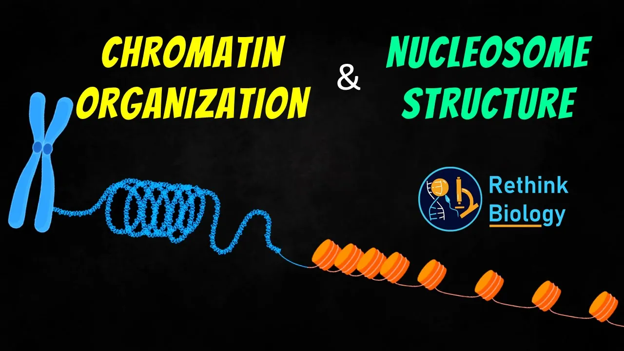
Why this video: This animation offers a clean, 3D visualization of nucleosome assembly and chromatin looping. It demonstrates how a ~147-base-pair segment of negatively charged DNA wraps approximately 1.65 times around a positively charged histone octamer core, acting as the fundamental packaging unit.
-
Knowledge Checkpoint:
- Draw the basic structural unit of a nucleosome, indicating the position of the core histone octamer and linker DNA.
- State the exact length of DNA (in base pairs) wrapped around a single nucleosome core.
- Explain how linker histones (such as H1) help stabilize higher-order chromatin structures.

-
Why this video: This video focuses on the biochemical properties of histones. It explains the core composition of the histone octamer (two copies each of H2A, H2B, H3, and H4) and reveals how basic, positively charged amino acid residues (lysine and arginine) mediate the strong electrostatic attraction to the acidic phosphate backbone of DNA.
-
Knowledge Checkpoint:
- Name the four core histone proteins and identify their assembly into a dimer/tetramer configuration.
- Describe the chemical nature of the electrostatic attraction between histone tails and DNA.
- Identify where the highly conserved, unstructured "histone tails" project relative to the nucleosome core.
-
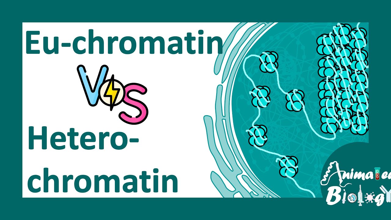
Why this video: Understanding the functional states of chromatin is critical to epigenetic regulation. This presentation uses clear molecular comparisons to show why euchromatin permits transcription (loose packing, high acetylation) while heterochromatin prevents it (dense compaction, high methylation).
-
Knowledge Checkpoint:
- Differentiate between constitutive and facultative heterochromatin.
- Contrast euchromatin and heterochromatin based on DNA density, transcriptional activity, and typical localization within the nucleus.
- Explain how chromatin compaction prevents RNA Polymerase and general transcription factors from accessing target promoters.
Module 3: DNA Methylation and Gene Silencing
DNA methylation is one of the most stable and well-characterized epigenetic modifications. This module explores the biochemical addition of a methyl group to the 5th carbon of cytosine rings, specifically within CpG dinucleotide clusters known as CpG islands. You will dive deep into the enzymatic actions of de novo DNA methyltransferases (DNMT3A/DNMT3B) that establish methylation patterns, and maintenance DNA methyltransferases (DNMT1) that preserve these patterns during DNA replication.
   Cytosine base                 5-Methylcytosine
        NH2                            NH2
       /                             /
  C---CH                        C---C---CH3  <-- Epigenetic Mark Added
 //   \\                       //   \\
HN     CH                     HN     CH
 \     /                       \     /
  C===N                         C===N
Recommended Videos

-
Why this video: This video bridges the molecular and clinical realms. It clearly contrasts the enzymes responsible for maintaining existing methylation patterns on hemimethylated DNA strands (DNMT1) during cell division with those that establish new, de novo methylation patterns (DNMT3A and DNMT3B).
-
Knowledge Checkpoint:
- Explain the enzymatic division of labor between DNMT1 (maintenance) and DNMT3A/DNMT3B (de novo).
- Detail how hemimethylated DNA acts as a substrate for DNMT1 during S-phase replication.
- Discuss the pathological consequences of aberrant hypermethylation at tumor-suppressor gene promoters.
-
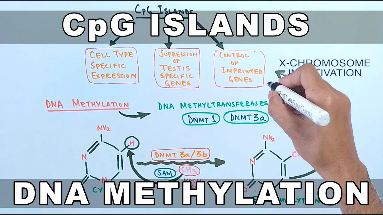
Why this video: This video focuses on CpG islands—regions of the genome rich in cytosine-guanine dinucleotide pairs, often located within promoter regions. It explains why these regions are typically unmethylated in transcriptionally active genes but become methylated when a gene is destined for stable, long-term silencing.
-
Knowledge Checkpoint:
- Define a "CpG Island" in terms of length, G-C content, and expected-to-observed CpG ratio.
- Explain why the human genome is generally depleted of CpG dinucleotides due to evolutionary deamination.
- Describe how CpG methylation directly blocks transcription factor binding and recruits repressive protein complexes.
-
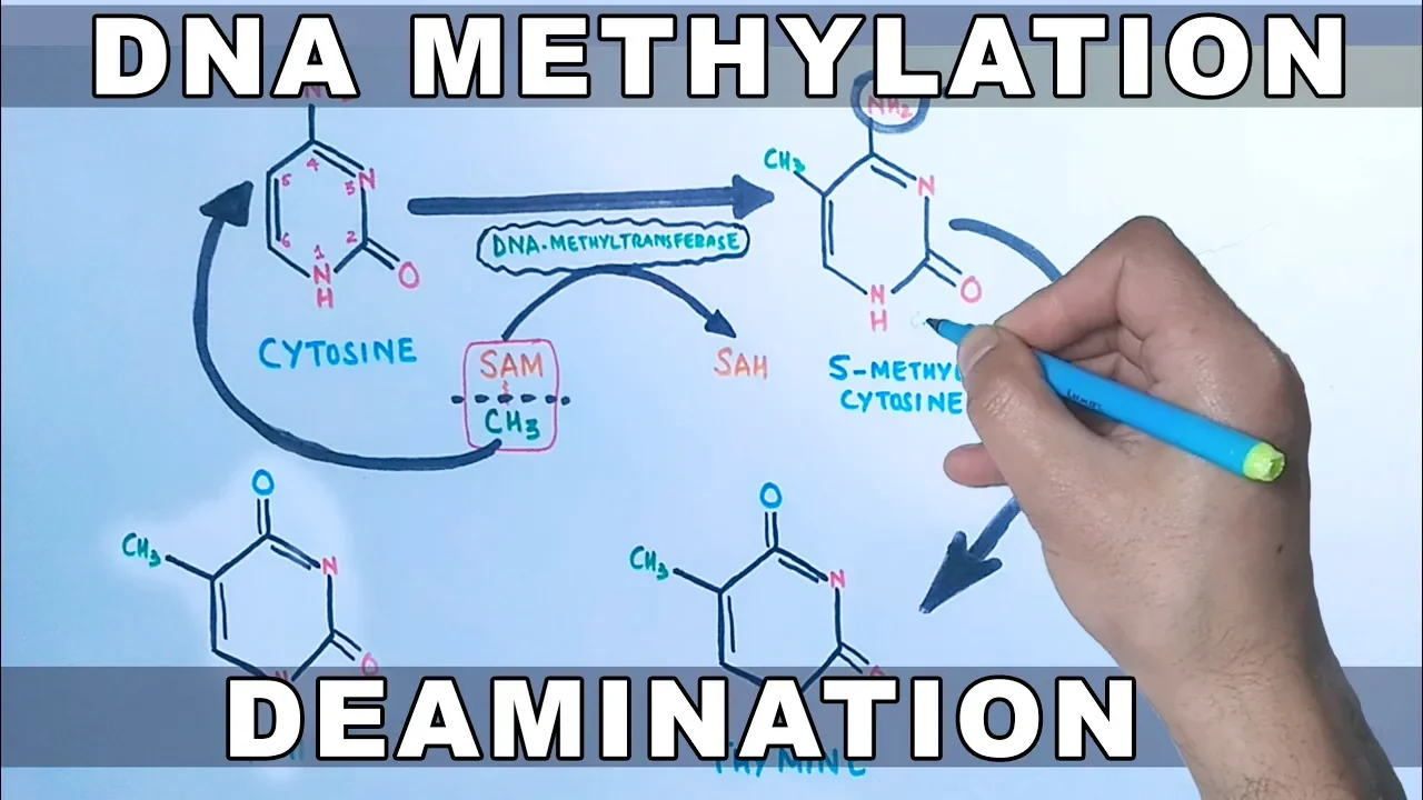
Why this video: This presentation covers the biochemical mechanism of DNA methyltransferase enzymes. It details the molecular donation of a methyl group from S-adenosyl methionine (SAM) to the 5th carbon of the cytosine pyrimidine ring, resulting in 5-methylcytosine (5mC).
-
Knowledge Checkpoint:
- Identify S-adenosyl methionine (SAM) as the universal methyl donor in epigenetic methylation reactions.
- Outline the chemical modification that converts a cytosine base to 5-methylcytosine.
- Explain how Methyl-CpG-binding domain proteins (MBDs) recognize 5mC and recruit histone deacetylases to reinforce silencing.
Module 4: The Histone Code: Acetylation and Methylation
While DNA methylation modifies the genome itself, the "Histone Code" hypothesis suggests that post-translational modifications (PTMs) on the unstructured, N-terminal tails of histone proteins write a complex regulatory overlay. This module examines the biochemical properties of these modifications, focusing on the dynamic, enzymatic balance between Histone Acetyltransferases (HATs), Histone Deacetylases (HDACs), and Histone Methyltransferases (HMTs).
HISTONE TAIL MODIFICATION DYNAMICS
[Lysine Tail] === HAT (Writers) ===> [Acetylated Lysine] (Active Gene) (+ charge) Adds Acetyl Groups (Neutralized charge, loose DNA)
[Lysine Tail] <== HDAC (Erasers) === [Acetylated Lysine] (Silent Gene) (+ charge) Removes Acetyl Groups (Re-established charge, tight DNA)
Recommended Videos

-
Why this video: This video focuses on the chemical mechanisms of histone acetylation. It explains how Histone Acetyltransferases (HATs) transfer an acetyl group from acetyl-CoA to positively charged lysine residues, neutralizing their positive charge. This reduces the histone's electrostatic affinity for DNA, leading to chromatin relaxation.
-
Knowledge Checkpoint:
- Explain how HATs neutralize the positive charge of lysine residues on histone tails.
- Describe the source of the acetyl donor (acetyl-CoA) and how cellular metabolism can impact epigenetic states.
- Detail how open, acetylated chromatin (euchromatin) facilitates transcription factor binding.

-
Why this video: This short, focused 3D animation visualizes the action of Histone Deacetylase (HDAC) enzymes. It demonstrates the removal of acetyl groups from histone lysine residues, restoring their positive charge and causing the chromatin to re-condense into a transcriptionally repressed heterochromatin state.
-
Knowledge Checkpoint:
- Describe the enzymatic reaction catalyzed by Histone Deacetylases (HDACs).
- Explain how HDACs contribute to gene silencing and chromatin compaction.
- Name the class of therapeutic drugs designed to inhibit HDACs (HDAC inhibitors) and their general clinical objectives in cancer therapy.

-
Why this video: This academic lecture introduces the concept of molecular "writers," "readers," and "erasers" in the context of the Histone Code. It details how chemical marks are written (by HATs and HMTs), interpreted by specialized reader proteins (possessing bromodomains or chromodomains), and erased (by HDACs and HDMs) to control gene expression dynamically.
-
Knowledge Checkpoint:
- Define and provide examples of epigenetic "Writers," "Readers," and "Erasers."
- Explain how bromodomains recognize acetylated lysine residues, while chromodomains recognize methylated lysine residues.
- Contrast the downstream gene-regulatory effects of histone H3 lysine 4 methylation (H3K4me3) with H3 lysine 9 methylation (H3K9me3).
Module 5: ATP-Dependent Chromatin Remodeling Complexes
Chemical modifications to DNA and histones are not always sufficient to clear physical obstructions from a promoter site. This module introduces the active, ATP-dependent mechanical motors that slide, evict, or restructure nucleosomes to expose critical DNA regulatory sequences. You will study chromatin remodeling complexes, specifically focusing on the SWI/SNF family and the RSC complex, to understand how these molecular machines use ATP hydrolysis to remodel chromatin.
Recommended Videos

-
Why this video: This video offers a clear explanation of nucleosome remodeling mechanisms. It shows how specialized remodeling complexes use energy from ATP hydrolysis to dynamically manipulate chromatin. The video details three primary mechanisms: nucleosome sliding (sliding the octamer along the DNA), histone eviction (completely removing histones to expose a promoter), and histone replacement (swapping core histones for variant histones).
-
Knowledge Checkpoint:
- Describe how ATP hydrolysis is coupled to mechanical work in nucleosome repositioning.
- Define nucleosome "sliding" and describe how it exposes previously hidden promoter regions.
- Compare the processes of histone eviction and variant histone substitution (e.g., swapping H3 for H3.3).
-
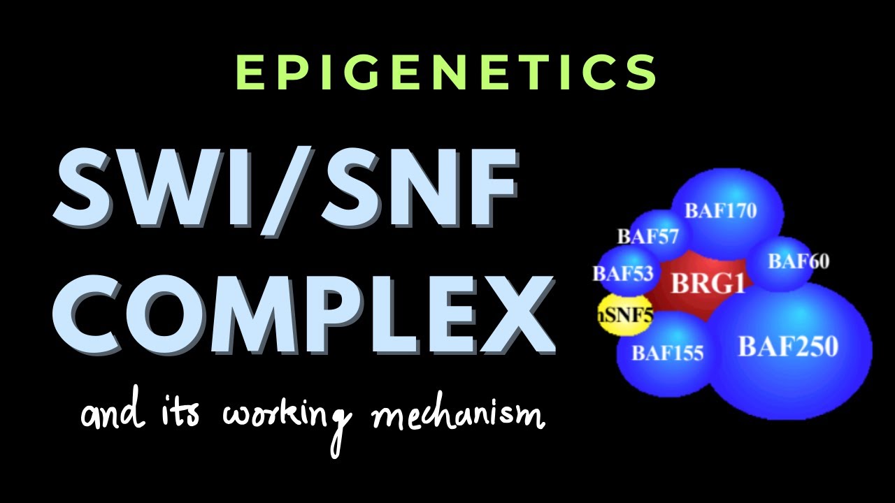
Why this video: This video provides an in-depth look at the SWI/SNF multi-subunit chromatin remodeling complex. It explains its structural composition, binding properties, and how its ATP-dependent activities regulate transcription by physically altering nucleosome positioning near active gene promoters.
-
Knowledge Checkpoint:
- Identify the core catalytic ATPase subunit of the SWI/SNF remodeling complex.
- Explain how SWI/SNF is recruited to targeted gene promoters via transcription factor interactions.
- Describe the clinical relevance of mutations in SWI/SNF subunit genes (such as SMARCA4) in human cancers.
-
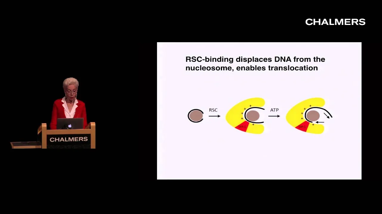
Why this video: Delivered by prominent researcher Dr. Yahli Lorch, this academic seminar offers a deep dive into the biochemistry of the RSC (Remodels the Structure of Chromatin) complex. Dr. Lorch details the experimental evidence showing how chromatin remodeling complexes physically reposition histone octamers, slide nucleosomes, and relieve gene repression.
-
Knowledge Checkpoint:
- Summarize the experimental biochemical assays used to observe nucleosome sliding in vitro.
- Explain the structural changes the nucleosome core undergoes during interaction with the RSC complex.
- Discuss how AT-rich DNA sequences can stimulate chromatin remodeling activity.
Gap Note & Independent Study Advice: Detailed structural models of SWI/SNF-nucleosome interactions are a highly specialized area of structural biology, and video coverage can be highly academic. To supplement this module, we recommend independently searching Google Scholar for recent cryo-EM structures of the SWI/SNF-nucleosome complex to help visualize the physical DNA translocation process.
Module 6: Epigenetics in Development, Disease, and Inheritance
This module integrates your biochemical understanding of DNA methylation, histone modifications, and chromatin remodeling to explain physiological processes. You will examine the molecular basis of X-chromosome inactivation (mediated by the non-coding RNA Xist), genomic imprinting (parent-of-origin-specific gene expression), and how epigenetic dysregulation can contribute to chronic diseases and cancer.
Recommended Videos
-
Why this video: Veritasium explains the biological concept of random X-chromosome inactivation in female mammals. This highly engaging video serves as a great introduction to how massive heterochromatic silencing is initiated, maintained, and physically represented as mosaic patterns in adult tissue.
-
Knowledge Checkpoint:
- Define the biological purpose of dosage compensation in mammalian organisms.
- Describe how a Barr body represents a physical, highly condensed heterochromatic X chromosome.
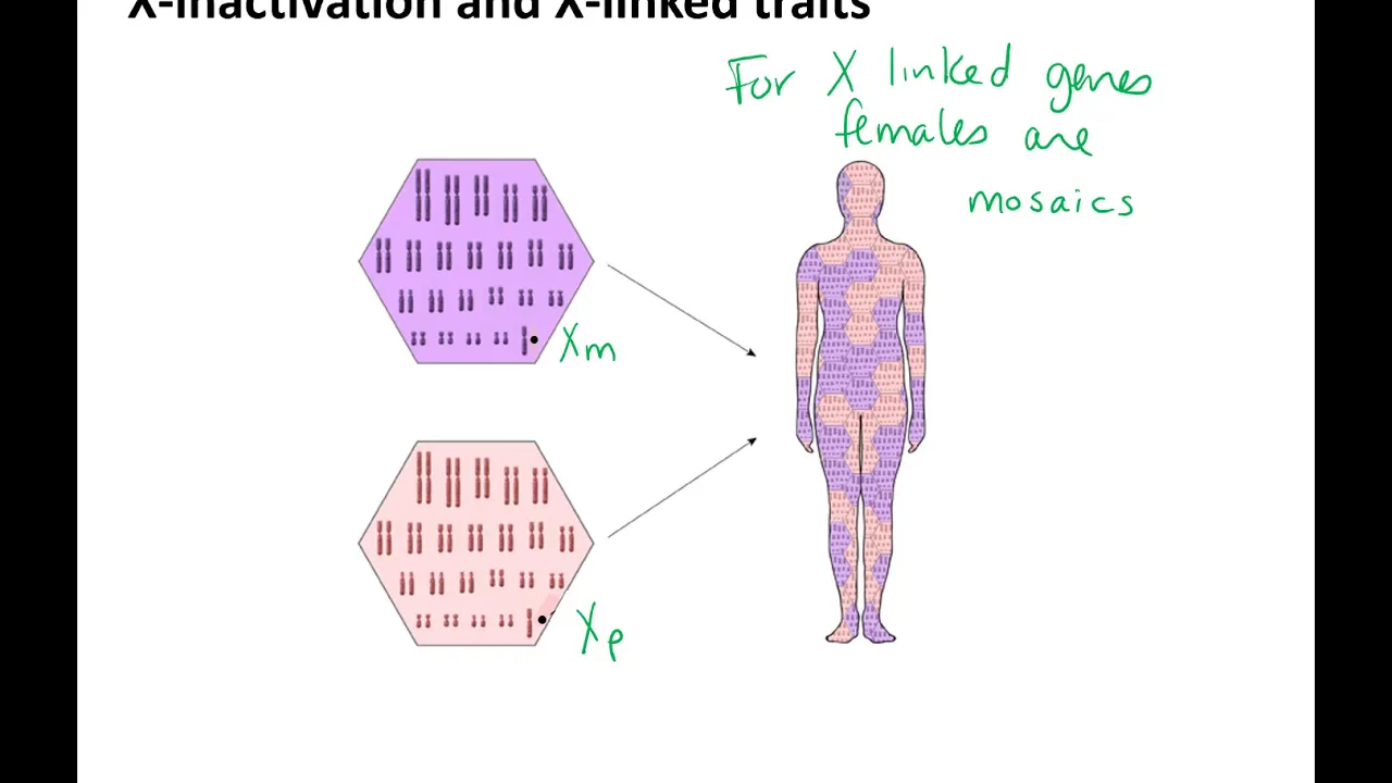
- Explain why females are genetic mosaics for any heterozygous gene located on the X chromosome.
-
Why this video: This TED-Ed lesson dives into the epigenetic mechanics of X-inactivation. It explains how the long non-coding RNA (lncRNA) Xist acts as an epigenetic architect by physically coating one of the two X chromosomes, recruiting polycomb repressive complexes, driving DNA methylation, and inducing histone deacetylation to permanently silence the chromosome.
-
Knowledge Checkpoint:
- Describe the transcription site and mechanism of action of the Xist long non-coding RNA.
- Explain the cascade of chromatin modifications that occur following Xist coating of the inactive X chromosome.
- Clarify how random inactivation of an X chromosome in early development is clonal across all daughter cells.
-
Why this video: This college-level biology lecture explores genomic imprinting—where genes are expressed in a parent-of-origin-specific manner. The lecture details how epigenetic marks are selectively erased and rewritten during gametogenesis, and explains the clinical outcomes of imprinting errors, such as Prader-Willi and Angelman syndromes.
-
Knowledge Checkpoint:
- Define genomic imprinting and state why it violates Mendelian genetic inheritance rules.
- Explain the lifecycle of imprinting marks: how they are erased in primordial germ cells, established during gametogenesis, and maintained throughout somatic life.
- Describe the genetic basis of Prader-Willi and Angelman syndromes on chromosome 15, focusing on how uniparental disomy or deletion of active alleles leads to disease.
Course Map
Below is the recommended pathway through the modules. While the initial modules follow a linear progression, Modules 3 and 4 run in parallel to build the foundational biochemical components of epigenetic modifications, which then feed directly into chromatin remodeling and physiological phenotypes.
Key People Index
Throughout this curriculum, you will encounter the work of notable researchers who have helped define the field of epigenetics:
- C. David Allis (1951–2023): Groundbreaking biochemist who pioneered the "Histone Code" hypothesis. He identified Tetrahymena protozoa with transcriptionally active macronuclei and transcriptionally silent micronuclei, paving the way for the discovery of histone acetyltransferases and demonstrating that histone modifications directly regulate transcription.
- Yahli Lorch: Renowned structural and biochemist at Stanford University. Her work with yeast chromatin remodeling complexes helped prove that ATP-dependent remodelers like SWI/SNF and RSC mechanically slide and evict nucleosomes to regulate transcription.
- Moshe Szyf: A pioneer of environmental epigenetics, Szyf's research shows how maternal behavior, stress, and environmental exposures can establish lasting DNA methylation changes that alter behavior and physiology over a lifetime.
Final Self-Assessment
This comprehensive self-assessment covers the key concepts presented in this curriculum. Use it to test your understanding of the biochemical mechanisms of epigenetic regulation.
- Draw a DNA double helix, label the 5' and 3' ends of both strands, and identify where methyl groups are attached during DNA methylation.
- Detail the composition of a histone octamer and explain how its overall charge interacts with the phosphate backbone of DNA.
- List the dynamic differences between euchromatin and heterochromatin, including typical levels of histone acetylation, histone methylation, and DNA methylation.
- Contrast the biochemical functions of DNMT1, DNMT3A, and DNMT3B, and explain why hemimethylated DNA is a crucial intermediate in epigenetic inheritance.
- Detail how HATs reduce the affinity of histones for DNA, and how HDACs restore that affinity.
- Define the terms "Writer," "Reader," and "Eraser" within the Histone Code framework, and provide at least two protein or enzyme examples of each.
- Explain how chromatin remodeling complexes couple ATP hydrolysis to physical changes in chromatin structure (e.g., nucleosome sliding and histone eviction).
- Describe the biochemical mechanism by which the lncRNA Xist mediates X-chromosome inactivation.
- Outline the cycle of genomic imprinting, explaining how parental imprints are erased in the germline and re-established based on the sex of the individual.
- Explain the molecular basis of Prader-Willi and Angelman syndromes as examples of imprinting defects in humans.
- Explain how environmental factors (like maternal nutrition or stress) can leave chemical marks on the epigenome that persist into adulthood.