Gastrulation & Neural Crest in Embryology
Learning Goal: Trace the embryonic development of vertebrates, focusing on the cellular movements of gastrulation, germ layer differentiation, and neural crest cell migration.
- Prerequisites: Basic understanding of cell biology (mitosis, cell-to-cell adhesion, and cell signaling pathways) and general genetics.
- Estimated Total Study Time: 9 hours
Module 1: Foundations of Early Embryology: Fertilization to Blastula
This module introduces the initial stage of vertebrate development. You will follow the path from the fusion of sperm and egg (fertilization) to the rapid mitotic cell divisions of cleavage (which bypass cell growth), culminating in the formation of the hollow, fluid-filled sphere of cells known as the blastula (or blastocyst in mammals). This stage establishes the cellular pool and spatial layout required for subsequent morphogenetic movements.
Recommended Videos

Why this video
This video provides an excellent high-level overview of early embryogenesis. It clearly maps out the sequential transitions from a single-celled zygote to a multi-celled morula (16–32 cell stage), explaining how the process of compaction drives the formation of the blastula. It sets a solid foundation for terminology before diving deeper into molecular biology.

Why this video
AK Lectures offers a detailed academic explanation of zygotic cleavage and blastulation. The video explains the unique mitotic mechanics where cells divide rapidly without an intervening G1 or G2 phase, resulting in smaller blastomeres within the constant volume of the zona pellucida. It details the structural differences between the trophoblast and the inner cell mass (embryoblast).
Why this video
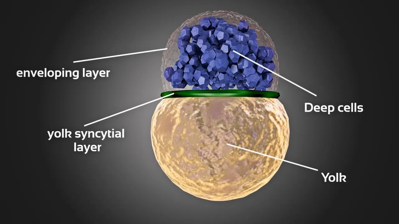
Using zebrafish (Danio rerio) as a comparative non-human vertebrate model, this video visualizes meroblastic (incomplete) cleavage. Unlike mammalian holoblastic cleavage, zebrafish cleavage is confined to the blastodisc at the animal pole due to the large yolk mass at the vegetal pole. Studying this variation is critical for a complete comparative embryology perspective.
Knowledge Checkpoint
- Explain the difference between holoblastic (complete) and meroblastic (incomplete) cleavage patterns.
- Describe the physical role of the zona pellucida during early cleavage divisions and why blastomeres decrease in size.
- Identify the structural components of a mammalian blastocyst and their respective fates (trophoblast vs. inner cell mass).
Module 2: Gastrulation: The Orchestrated Cellular Movements
Gastrulation is the defining event of early development, where a simple blastula transforms into a highly structured, three-layered gastrula. This module explores the physical and biophysical mechanics driving these changes—specifically focusing on five classic morphogenetic movements: invagination, involution, epiboly, convergent extension, and ingression.
Note: In alignment with review feedback, this module places special emphasis on comparative non-human vertebrate models (specifically Xenopus and chick embryos) where these mechanics have been best characterized.
Recommended Videos
Why this video
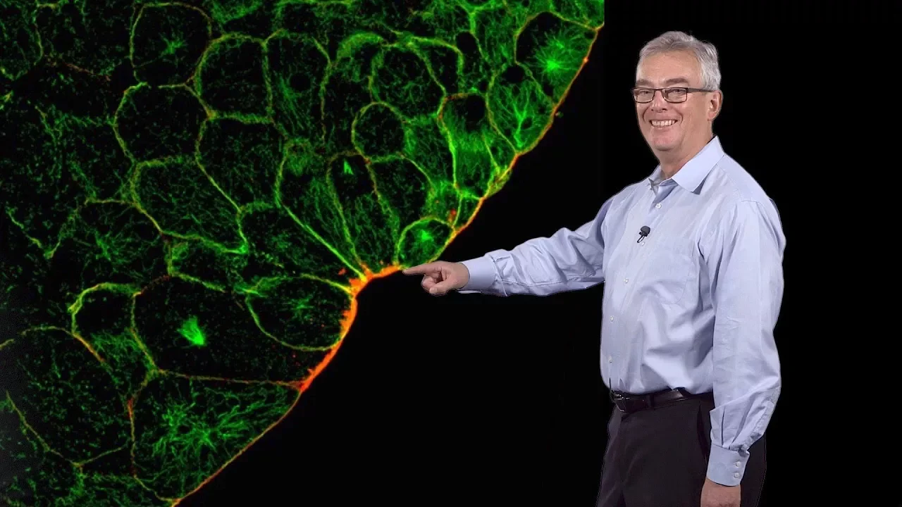
Delivered by developmental biologist Dr. Richard Harland, this video targets a major gap highlighted in the review feedback: the specific cellular and biophysical mechanics of gastrulation in Xenopus laevis. Dr. Harland breaks down the precise coordination of seven distinct cell movements—including vegetal rotation, bottle cell invagination, epiboly of the ectoderm, and convergent extension of the mesoderm.

Why this video
This focused animation provides a clear visual breakdown of frog gastrulation. It explicitly details how bottle cells at the dorsal lip of the blastopore initiate invagination, how the marginal zone cells undergo involution over the blastopore lip, and how epiboly spreads ectodermal cells to envelop the entire yolk plug.

Why this video
This seminar presents high-end biophysical and computational modeling of avian (chick) gastrulation. It explores how large-scale tissue flows, epiboly, convergent extension (via mesodermal cell intercalation), and primitive streak ingression cooperate to sculpt the flat blastodisc of a bird into three distinct layers.

Why this video
This lecture provides an in-depth mechanical breakdown of "bottle cells." It explains how their dramatic apical constriction drives the localized bending of the epithelial sheet, creating the initial groove of the blastopore lip that guides the migrating mesoderm and endoderm inward.
Knowledge Checkpoint
- Define and draw schematics for: invagination, involution, epiboly, ingression, and convergent extension.
- Detail how bottle cells physically alter their cytoskeleton (actin filaments and myosin) to achieve apical constriction.
- Contrast the gastrulation mechanics of a spherical amphibian embryo (Xenopus) with those of a flat avian/mammalian blastodisc (primitive streak formation).
Module 3: Germ Layer Differentiation and Axis Specification
Once cellular migration is complete, the embryonic tissues settle into three primary germ layers: ectoderm, mesoderm, and endoderm. This module focuses on the molecular pathways that dictate these cells' fates. You will study embryonic induction, the seminal Spemann-Mangold organizer experiments, and the signaling networks (including BMP, Wnt, Chordin, and Noggin) that establish the primary body axes.
Recommended Videos
Why this video
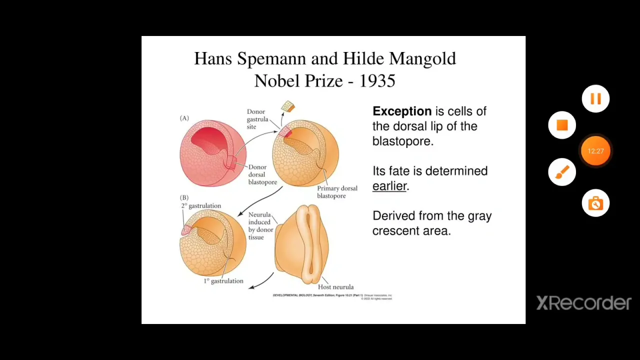
This video breaks down Hans Spemann and Hilde Mangold's classic 1924 transplantation experiment. By transplanting the dorsal blastopore lip of a pigmented newt gastrula to the ventral region of an unpigmented host, they induced a secondary, complete body axis. This lecture explains the core concepts of "induction" and "competence."

Why this video
Addressing a critical feedback gap, this video honors molecular embryologist Edward De Robertis, who isolated the molecular elements of the Spemann organizer. The presentation highlights how molecular signaling pathways—specifically the secretion of BMP antagonists like Chordin, Noggin, and Goosecoid—underlie the organizer's ability to neuralize the ectoderm and pattern the dorsal-ventral axis.
Why this video
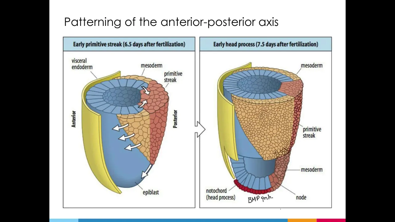
This concise lecture shifts focus to mammalian axis specification. It explains the dual-organizer system in mammals: the node (which acts as the equivalent of the Spemann-Mangold organizer to pattern the trunk and nervous system) and the anterior visceral endoderm (AVE), which is crucial for establishing head development and the anterior-posterior axis.
Knowledge Checkpoint
- What is the "default" state of the ectoderm, and how do BMP signaling and its inhibitors (Chordin, Noggin) regulate its fate?
- Describe the Spemann-Mangold experiment and outline why the host embryo developed a secondary central nervous system composed of host cells.
- Map the roles of the Node and the Anterior Visceral Endoderm (AVE) in mammalian anterior-posterior and left-right symmetry axes.
Module 4: Neurulation: Folding the Central Nervous System
Neurulation is the stage where the newly specified ectodermal "neural plate" is transformed into the neural tube—the precursor to the brain and spinal cord. This module details the mechanical folding process (primary neurulation) and the condensing of mesenchymal cells to form a tube (secondary neurulation), while identifying the critical roles played by the underlying notochord.
Recommended Videos
Why this video
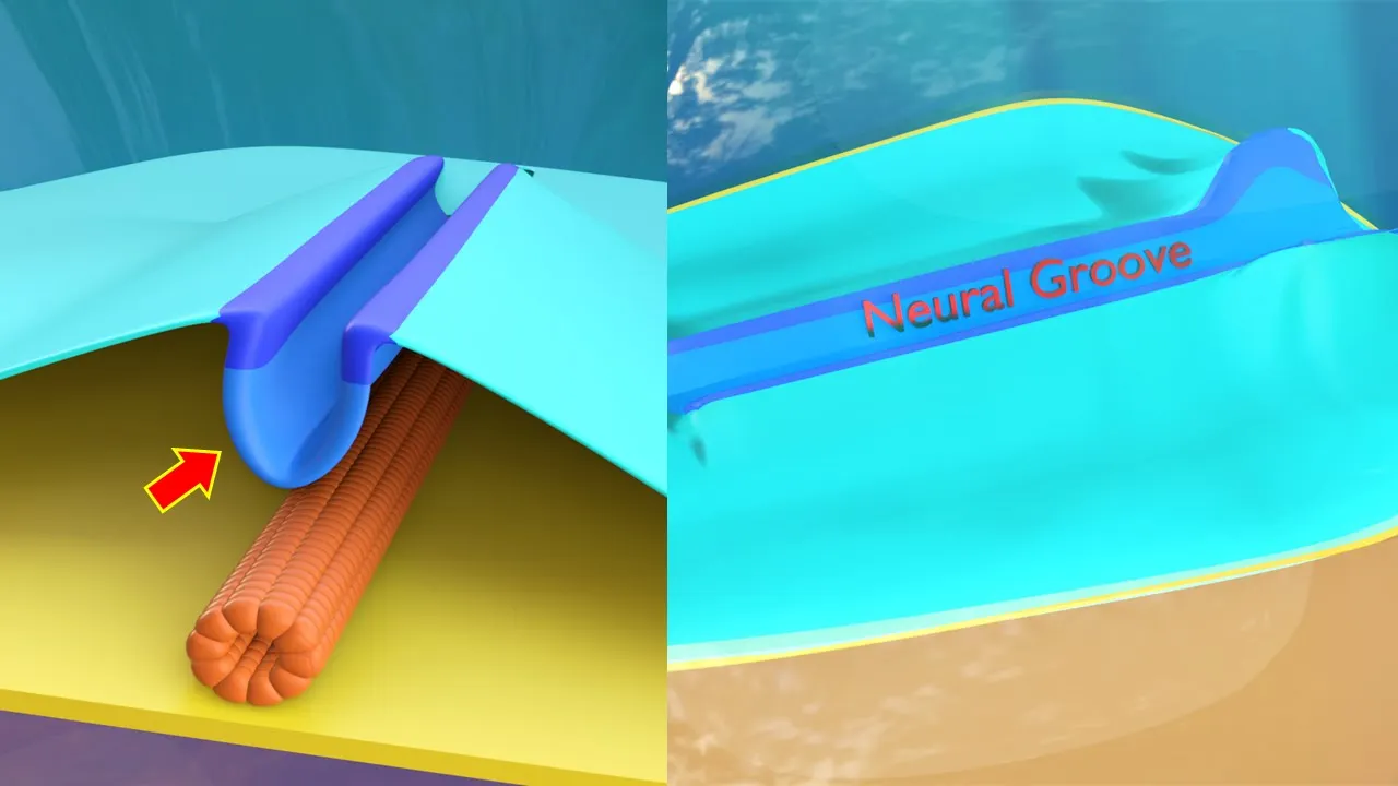
This video features superb 3D medical animations that help viewers visualize primary and secondary neurulation. It traces the thickening of the ectoderm to form the neural plate, the elevation of neural folds at the lateral boundaries, and their subsequent midline fusion. It also highlights the caudal-most formation of the neural tube via secondary neurulation.

Why this video
AK Lectures provides a highly structured breakdown of the inductive signals flowing from the mesodermally derived notochord to the overlying ectoderm. It introduces the mechanical hinges (median hinge point and dorsolateral hinge points) that facilitate the bending of the neural plate.
Why this video
This classic university-level embryology lecture delves deeply into clinical correlates of neurulation. Beyond the mechanical steps, it explains the timing of cranial and caudal neuropore closures and the developmental pathologies—such as Anencephaly and Spina Bifida—that arise when these folding processes fail.
Knowledge Checkpoint
- Differentiate between primary neurulation (folding) and secondary neurulation (medullary cord canalization) regarding the region of the body they form.
- Explain how the notochord chemically induces neurulation in the overlying ectoderm.
- Describe the physical steps of neural fold fusion and the role of differential cell-adhesion molecules (E-cadherin vs. N-cadherin) in separating the neural tube from the future epidermis.
Module 5: Neural Crest Cells: Migration and Fate
Often called the "fourth germ layer," neural crest cells (NCCs) arise at the border of the neural plate and non-neural ectoderm. Upon neural tube closure, these cells undergo an extraordinary Epithelial-to-Mesenchymal Transition (EMT), detach from the neuroepithelium, and migrate throughout the embryo to differentiate into a diverse array of lineages.
Recommended Videos

Why this video
Presented by world-renowned developmental neurobiologist Chaya Kalcheim, this seminar addresses a major curriculum gap by focusing on the mechanics of Epithelial-to-Mesenchymal Transition (EMT) in neural crest cells. The lecture explores the cell-cycle transitions, loss of cell polarity, cytoskeletal remodeling, and matrix metalloproteinase secretion that allow these cells to delaminate from the dorsal neural tube.

Why this video
This seminar provides a deep, molecular investigation of neural crest migration. It focuses on how tight temporal control of EMT is maintained, the transcriptional regulators involved (such as Snail, Slug, and Twist), and the chemotactic cues (such as SDF-1/CXCR4) that direct migration pathways.
Why this video
Dr. Robert Weinberg, a pioneer in cancer biology, explains how the molecular machinery of developmental EMT (normally used by migrating neural crest cells) is co-opted by adult epithelial cancer cells to undergo metastasis and colonize distant organs. This comparison illustrates why understanding embryological EMT is vital for modern pathology.
Why this video
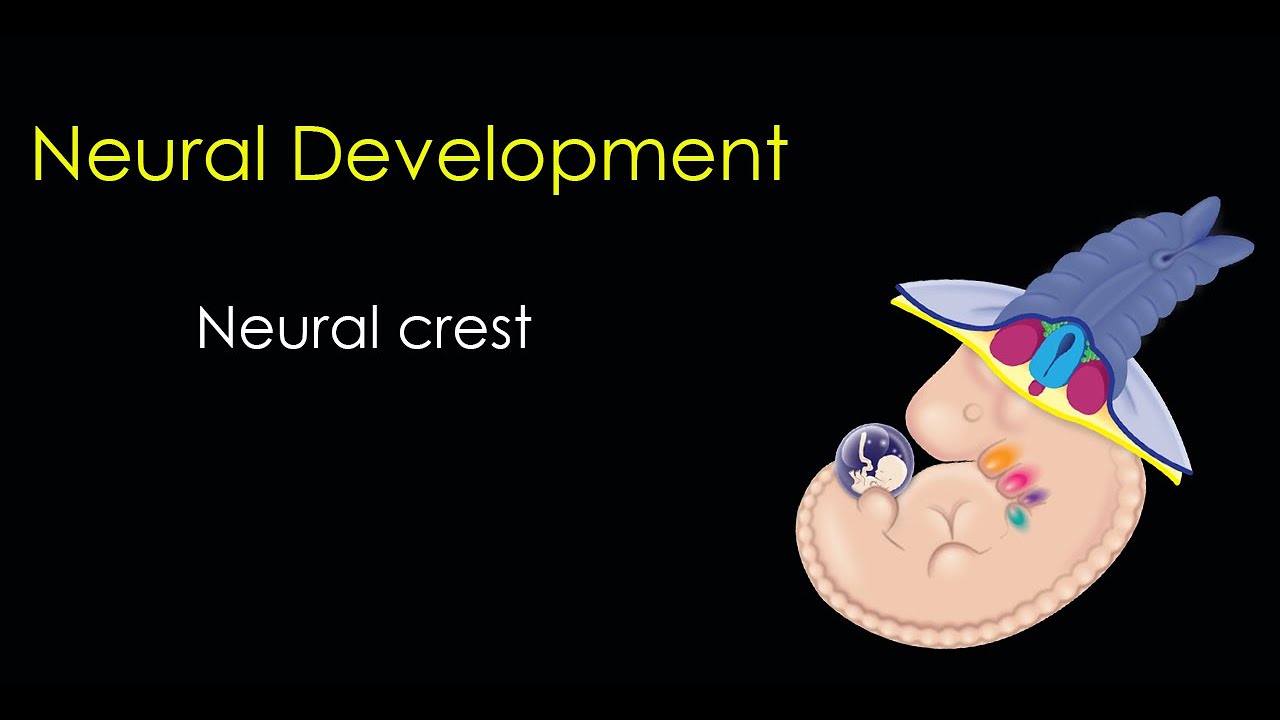
This tutorial lists and classifies the diverse derivatives of neural crest cells. It categorizes them into cranial, cardiac, vagal, and trunk crest populations, detailing how they give rise to structures as varied as melanocytes, craniofacial cartilage, Schwann cells, and the enteric nervous system.
Knowledge Checkpoint
- Detail the molecular events of Epithelial-to-Mesenchymal Transition (EMT), including the downregulation of E-cadherin and upregulation of N-cadherin/vimentin.
- List the transcription factors (e.g., Snail, Slug, Twist, Sox10) that serve as master regulators of the neural crest lineage.
- Identify four main anatomical sub-regions of the neural crest (cranial, cardiac, vagal, trunk) and name at least two major cellular derivatives of each.
Course Map
Below is a flowchart representing the sequential progression of vertebrate development covered in this curriculum, illustrating how early physical events directly gate subsequent tissue movements and cell fate specifications.
Key People Index
- Hans Spemann & Hilde Mangold
Pioneered the concept of embryonic induction. Hilde Mangold’s PhD thesis (under Spemann’s supervision) demonstrated that a specific region of the early embryo (the dorsal lip of the blastopore) was capable of dictating the axis of the entire organism, establishing the primary organizer. Hans Spemann was awarded the Nobel Prize in Physiology or Medicine in 1935. - Edward De Robertis
A leading molecular embryologist who systematically cloned the genes expressed in the Spemann-Mangold organizer region. His work identified key molecular factors like Chordin and Noggin, proving that the organizer works by secreting antagonists that neutralize ventralizing BMP signals. - Richard Harland
UC Berkeley professor who discovered Noggin, a key protein secreted by the organizer that induces the ectoderm to form neural tissue. His research has been instrumental in characterizing the structural and cellular basis of amphibian gastrulation. - Chaya Kalcheim
Professor at the Hebrew University of Jerusalem and a pioneer in spinal cord and neural crest development. Her lab discovered the molecular triggers regulating the timing of neural crest EMT and delamination. - Robert Weinberg
An internationally recognized cancer biologist at MIT/Salk Institute. He linked basic developmental biology to clinical oncology by showing that cancer metastasis reactivates the embryonic EMT program, allowing carcinoma cells to leave primary tumors.
Final Self-Assessment
Test your understanding of the entire vertebrate embryology curriculum by verifying your ability to answer the following key conceptual questions:
- Explain how synchronous, rapid cleavage divisions occur in the absence of G1 and G2 cell-cycle phases, and why this stage is transcriptionally silent until the Mid-Blastula Transition (MBT).
- Contrast how epiboly spreads cell sheets over an egg in Xenopus versus how tissue flow/convergent extension operates within the avian epiblast.
- Detail the physical role of bottle cells in initiating invagination: What structural change occurs at their apical membranes, and how does this affect the surrounding tissue sheet?
- Explain how the Spemann-Mangold organizer organizes the dorsal-ventral axis: What is the relationship between BMP-4, Wnt signaling, and organizer factors like Chordin and Noggin?
- Draw a cross-section of a neurulating embryo, labeling the neural ectoderm, non-neural ectoderm, neural fold, neural crest, notochord, and paraxial mesoderm.
- Explain the molecular mechanism of homophilic cell adhesion (such as Cadherins) and how a switch from E-cadherin to N-cadherin/E-cadherin segregation allows the neural tube to separate from the overlying ectoderm.
- Describe the complete transcriptional cascade (Snail, Slug, Twist) that shuts down epithelial traits (apical-basal polarity, tight junctions, desmosomes) and activates mesenchymal traits during neural crest EMT.
- Contrast the migration pathways of trunk neural crest cells (the dorsolateral pathway vs. the ventromedial pathway) and explain how these pathways determine their eventual differentiation into melanocytes vs. sensory/sympathetic ganglia.