3D Bioprinting: Vascular Scaffolds & iPSCs
Learning Goal: Formulating and executing a 3D bioprinting protocol for vascularized tissue scaffolds using bioinks and induced pluripotent stem cell (iPSC)-derived vascular cells, including CAD modeling and perfusion-based maturation.
- Prerequisites: Basic undergraduate-level molecular biology, organic chemistry (understanding polymers), and introductory physics.
- Estimated Study Time: 50 hours
Module 1: Foundations of Cell Biology and Stem Cells
This module covers the core biological foundations necessary to understand cell architecture, pluripotency, and the cellular reprogramming breakthrough of induced pluripotent stem cells (iPSCs). You will learn about cellular structure and the mechanics of turning somatic cells back into embryonic-like master stem cells using defined transcription factors.
Recommended Videos
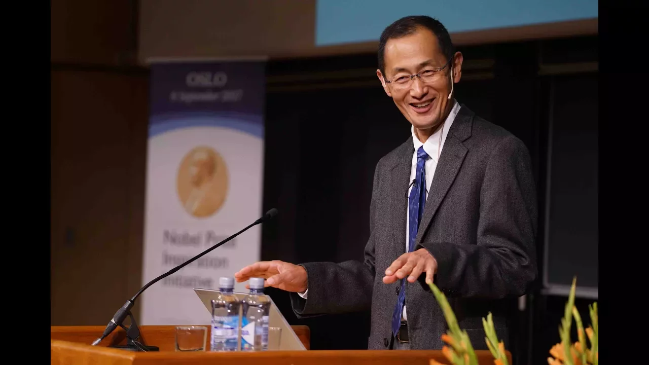
Why this video is valuable: This is a definitive, high-level keynote lecture delivered by Shinya Yamanaka himself, explaining the Nobel Prize-winning discovery of iPSCs. It provides peerless historical and functional context regarding how Yamanaka factors (Oct3/4, Sox2, Klf4, and c-Myc) reset adult cell epigenetic states.
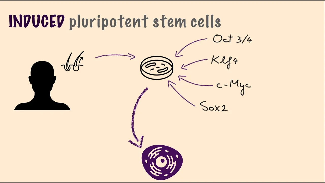
Why this video is valuable: This video serves as an accessible conceptual overview of the reprogramming process. It breaks down the difference between somatic adult cells, embryonic stem cells, and iPSCs, serving as an excellent visual summary to cement Yamanaka's longer lecture.

Why this video is valuable: A quick refresher on foundational cell biology principles from MIT. It establishes the cellular paradigm that "structure dictates function," which is a core tenet of tissue engineering.
Knowledge Checkpoint
- Explain the epigenetic mechanism by which the Yamanaka factors (OSKM) revert a differentiated fibroblast to pluripotency.
- List the primary differences in genomic stability and tumor risk (teratoma formation) between using three vs. four Yamanaka factors.
- Define cell pluripotency and contrast it with totipotency and multipotency.
Module 2: Bio-CAD and Slicing for Bioprinting
Special Curricular Warning
The video pool contains numerous mechanical engineering CAD tutorials (e.g., AutoCAD Civil 3D pipe systems for infrastructure). Do not use civil engineering pipe design strategies for biological systems; biological vascular networks are non-linear, multi-scale, and require specialized hydrogel flow kinetics. This module transitions generic CAD and slicer knowledge into biological hydrogel extrusion settings.
Recommended Videos
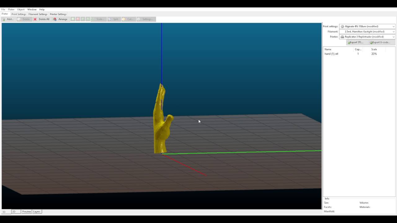
Why this video is valuable: This is a vital tutorial on slicing parameters tailored specifically for biological hydrogels printed via Freeform Reversible Embedding of Suspended Hydrogels (FRESH). It discusses layer height relative to needle diameter, perimeter settings to prevent over-extrusion, and rectifying slicing errors for soft matter.

Why this video is valuable: This video introduces the basics of parametric 3D modeling using FreeCAD. Understanding coordinate systems, sketches, extrusions, and boolean operations is critical before attempting to design biological channels.
Addressing the Slicing/CAD Gap
To model functional vascular networks:
- Software selection: Standard CAD packages (FreeCAD, Fusion360) are useful for modeling basic perfusion molds and chambers. However, chaotic and biomimetic microvasculature models require procedural generation or specialized medical imaging (DICOM) conversions (via software like 3D Slicer).
- Hydrogel parameters: Unlike plastics, hydrogels have a yield stress. In Slic3r, you must adjust flow rates, set the infill speed low (10–20 mm/s), and ensure your layer height is exactly 40%–50% of the internal needle diameter to prevent shearing the gel matrix.
Knowledge Checkpoint
- Calculate the target layer height for a 150μm bioprinting needle using the 40% rule.
- Identify why standard infill pattern speeds for PLA/PETG will cause structural failure in a gelatin-alginate bioink.
- Explain how parametric constraints in a CAD environment can be used to alter the lumen diameter of a vascular tube model.
Module 3: Bioinks and Bioprinting Physics
This module addresses the materials science of biopolymers, the fluid mechanics of extrusion, and the physical/chemical crosslinking reactions that allow soft, cell-laden gels to hold their shape post-printing.
Recommended Videos
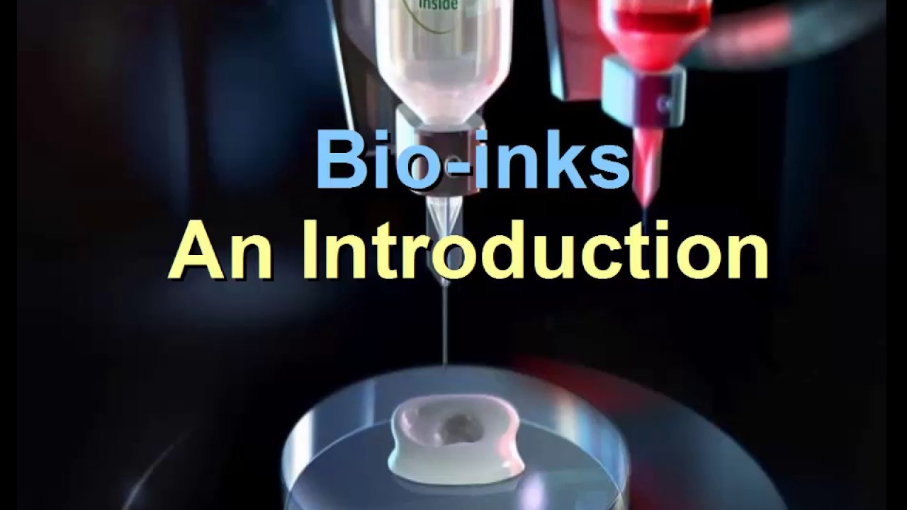
Why this video is valuable: This presentation provides a strong academic survey of the primary natural polymers used in bioinks—collagen, gelatin, hyaluronic acid, alginate, and nanocellulose—and analyzes their biocompatibility and mechanical trade-offs.
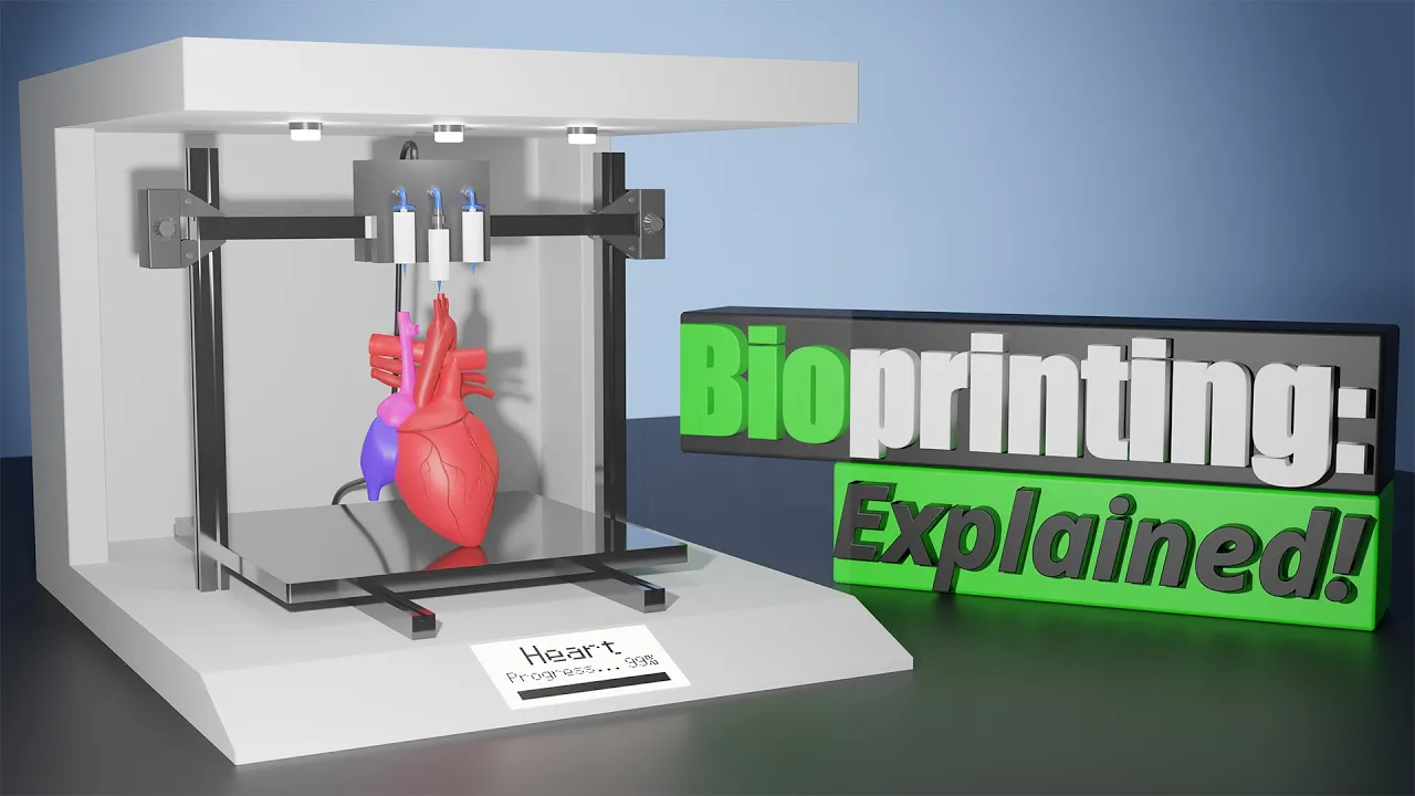
Why this video is valuable: A deep dive into the primary printing physics: extrusion-based, inkjet, and stereolithography (SLA). It explains how fluid shear during extrusion impacts cell viability and how viscosity dictates print resolution.
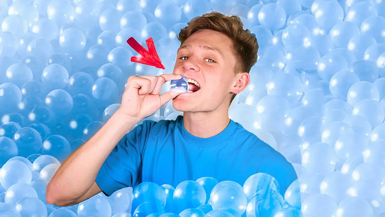
Why this video is valuable: Though framed as an entertainment video, this displays the exact chemistry of ionic crosslinking between sodium alginate and calcium ions. It serves as an intuitive visual demonstration of polymer coordination and gelation.

Why this video is valuable: A rapid laboratory overview of photo-crosslinking, demonstrating the assembly of a polymer (PEGDA) with a photo-initiator under UV exposure to build stable structures.
Knowledge Checkpoint
- Describe the chemical bond formation difference between ionic crosslinking (e.g., alginate + ) and covalent photo-crosslinking (e.g., GelMA + photoinitiator + UV light).
- Explain shear-thinning behavior in hydrogels and why it is crucial for preserving cell viability during extrusion.
- Define the mechanical trade-off between bioink stiffness (elastic modulus) and the rate of cell proliferation/migration within the printed matrix.
Module 4: Differentiating iPSCs into Vascular Lineages
This module explains the conversion of pluripotency into highly specialized somatic vascular lineages: endothelial cells (ECs) to line the vessel lumen and smooth muscle cells (SMCs) to provide structural support.
Recommended Videos
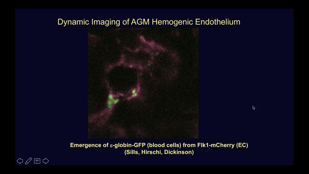
Why this video is valuable: Discusses the in vitro differentiation of embryonic and induced pluripotent stem cells into hemogenic endothelial cells without using mouse feeder layers, illustrating the biochemical signaling pathways involved.

Why this video is valuable: A concise overview showing how chemical cocktails applied over a 12-day protocol steer pluripotent stem cells into specified vascular lineages.

Why this video is valuable: Detail-oriented analysis of isolating and characterizing functional vascular cells via flow cytometry, using markers such as CD31 and PECAM-1.
Protocol Gaps & Manual Study
Because detailed laboratory differentiation protocols are highly proprietary and sparse in general video repositories, you must supplement your study:
- The Directed Differentiation Protocol: iPSCs are typically grown to 60-80% confluence. On Day 0, activate the Wnt pathway using CHIR99021 in LaSR medium for 2 days to induce mesoderm lineage. On Day 2, transition cells to medium supplemented with VEGF-A and bFGF to drive endothelial differentiation.
- Purification: On Day 5-7, use Magnetic-Activated Cell Sorting (MACS) or Flow Cytometry (FACS) to isolate cells expressing CD31 (PECAM-1) and CD144 (VE-Cadherin).
Knowledge Checkpoint
- What is the function of CHIR99021 in early mesodermal commitment during iPSC differentiation?
- Identify the specific cell-surface markers used to verify endothelial differentiation versus smooth muscle differentiation via flow cytometry.
- Explain why separating the differentiated endothelial cells from undifferentiated iPSCs is critical prior to printing (hint: teratoma prevention).
Module 5: Formulating Bioinks and Executing the Print
This module brings together your vascular cells, bioink chemistry, and G-code designs to perform cellularized bioprinting. We focus on multi-material extrusion and co-axial needle systems designed to print hollow tubular vessels directly.
Recommended Videos

Why this video is valuable: This video shows the precise mechanics of co-axial (concentric) microfluidic printing. A dual-layer printhead dispenses a core crosslinking solution surrounded by a sheath of bioink to print continuous hollow channels in a single pass.

Why this video is valuable: A demonstration of how to blend cells with hydrogel matrices while maintaining sterile conditions and high cell viability, highlighting extracellular matrix (ECM) digests.
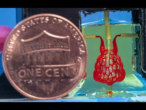
Why this video is valuable: Visually demonstrates open-source, projection-based stereolithographic bioprinting of highly complex, entangled vascular networks using biocompatible photo-crosslinkable hydrogels.
Knowledge Checkpoint
- Explain the flow velocity profile inside a co-axial nozzle and how core flow rate vs. sheath flow rate affects the wall thickness of a printed vessel.
- What temperature ranges must be maintained when mixing cell suspensions with gelatin-based bioinks to prevent cell lysis or premature gelation?
- Contrast projection-based light stereolithography with traditional pneumatic extrusion bioprinting in terms of print resolution and localized shear stress on the cells.
Module 6: Bioreactor Setup and Perfusion-Based Maturation
Simply printing a cellularized tube is not enough; without perfusion, the inner cells will die from a lack of oxygen and nutrient diffusion. This module explains how to design and assemble fluidic loops that apply shear stress to align and mature your newly printed vascular channels.
Recommended Videos

Why this video is valuable: Explains the core physics of fluid shear stress, demonstrating how viscosity and velocity gradients generate shear forces. This physics is directly applicable to the wall shear stress experienced by endothelial cells lining a printed vessel.
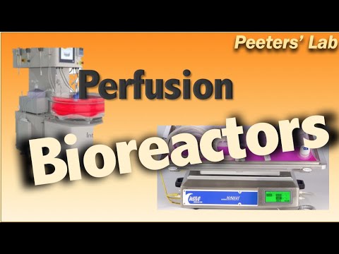
Why this video is valuable: Highlights the paradigm shift toward continuous culture and perfusion bioreactors, showing how dynamic media exchange preserves the viability of 3D scaffolds.

Why this video is valuable: Explains the physiological perspective of vascular shear stress as a frictional drag on endothelial cell linings, explaining its role in cellular signaling and maturation.

Why this video is valuable: Despite being focused on algae, this video provides an excellent structural teardown of dynamic bioreactor assembly, displaying the use of 3D-printed modular couplers, silicone tubings, O-ring seals, and aseptic fluidic connections.
Knowledge Checkpoint
- Write the mathematical formula for fluid shear stress and identify what variables a tissue engineer can adjust to optimize perfusion maturation.
- Why do endothelial cells require shear stress to form tight junctions (e.g., VE-cadherin expression) and achieve proper barrier function?
- Detail the aseptic steps required to connect a newly printed hydrogel scaffold to a peristaltic pump loop without introducing bacterial contamination.
Course Map
The following flowchart details the suggested progression of modules and dependencies. Notice how the biological track (M1 M4) and engineering/physics track (M2 & M3) converge directly at Module 5 before entering the final bioreactor maturation stage (M6).
Key People Index
- Dr. Shinya Yamanaka (Kyoto University / Gladstone Institutes): Winner of the 2012 Nobel Prize in Physiology or Medicine. Discovered that the expression of four specific transcription factors (Oct4, Sox2, Klf4, c-Myc) can reprogram any adult somatic cell into an embryonic-like pluripotent stem cell (iPSC).
- Dr. Anthony Atala (Wake Forest Institute for Regenerative Medicine): Pioneer in the translation of 3D bioprinting technologies for clinical applications, famous for constructing and transplanting bio-engineered tissues and organs.
- Dr. Jordan Miller (Rice University): Highly prominent open-source bioprinting researcher who cleared major structural hurdles in SLA printing of highly complex, entangled vascular networks inside protective hydrogels.
Final Self-Assessment
Complete this comprehensive self-assessment to verify your mastery of the curriculum material.
- I can describe the exact molecular mechanism of how the Yamanaka transcription factors induce epigenetic remodeling in adult human fibroblasts.
- I can calculate appropriate layer heights and volumetric flow rates in a slicer for extrusion printing of biological hydrogels based on nozzle gauges.
- I can identify the physical and chemical differences between physical gelation (such as thermal gelation of gelatin) and chemical/ionic crosslinking (such as alginate-calcium coordination).
- I can map the biochemical differentiation path of iPSCs into CD31+ and CD144+ hemogenic endothelial cells.
- I understand the use of magnetic-activated cell sorting (MACS) to purify differentiated vascular populations prior to bioink preparation.
- I can explain the physical operation of a microfluidic co-axial nozzle for the direct, continuous printing of hollow vascular conduits.
- I can compute fluid shear stress within a printed channel given the media's viscosity, channel dimensions, and pump volumetric flow rates.
- I can assemble a sterile perfusion loop containing a reservoir, peristaltic pump, and 3D printed chamber for long-term scaffold culture.
- I understand how physiological shear stress drives morphological changes (such as cellular alignment and elongation) in endothelial cell linings.