Neurodevelopment: Tubes, Migration & Guidance
Learning Goal: Trace the stages of embryonic neurodevelopment, detailing the molecular cues and cellular mechanisms governing neural tube formation, neuronal migration, axon guidance, and synaptogenesis.
- Prerequisites: Basic knowledge of cellular biology, genetics, and general eukaryotic cell structure.
- Estimated Total Study Time: 12 Hours
Module 1: Foundations of Embryology and Neural Induction
This module introduces the crucial structural transitions of the early embryo. You will explore gastrulation—the morphogenetic process that transforms a simple, single-layered blastula into a trilaminar embryonic disc with three distinct germ layers (ectoderm, mesoderm, and endoderm). A primary focus is placed on the molecular orchestration of neural induction: how the Spemann-Mangold organizer region of the mesoderm sends signaling cues (such as Noggin, Chordin, and Follistatin) to block Bone Morphogenetic Protein (BMP) signaling, thereby instructing the default ectoderm to differentiate into specialized neural tissue rather than epidermis.
Recommended Videos

- Why this video: This video offers a clear, highly visual whiteboard lecture establishing the fundamental concepts of gastrulation. It outlines how the epiblast migrates through the primitive streak to construct the three foundational germ layers (ectoderm, mesoderm, and endoderm), pointing out which tissues develop from each layer, with an emphasis on the ectodermal origin of the nervous system.
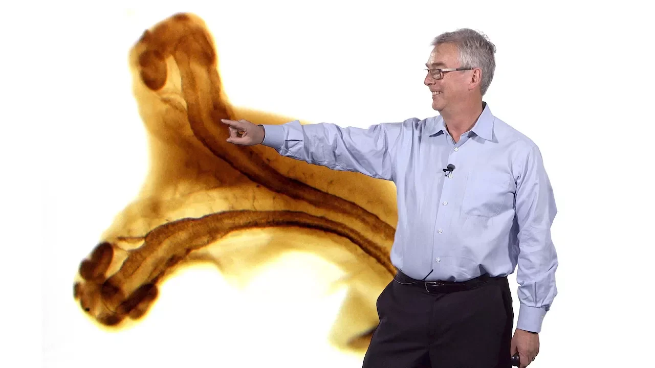
- Why this video: Dr. Richard Harland walks through the precise molecular mechanisms behind embryonic induction. This lecture is critical for understanding how the Spemann-Mangold organizer coordinates development by producing key BMP antagonists like Noggin, Chordin, and Follistatin. These antagonists physically bind to BMP ligands, blocking their interaction with ectodermal receptors and allowing the default neural fate to proceed.
- Why this video: This lecture traces the historical and experimental discoveries of embryology from the legendary Spemann-Mangold organizer tissue grafts to the modern molecular cloning of Noggin. It details the molecular-level interactions of neural induction and validates the "default model" of neural ectoderm differentiation.
Knowledge Checkpoint
- Detail the physical migration of epiblast cells through the primitive streak that establishes the ectoderm, mesoderm, and endoderm.
- Explain the "default model" of neural ectoderm differentiation.
- Describe how BMP signaling promotes an epidermal fate, and how Noggin, Chordin, and Follistatin act as antagonists to induce a neural fate.
- Relate the historical tissue graft experiments of Spemann and Mangold to modern molecular signal identification.
Module 2: Neural Tube Formation (Neurulation)
Here we trace the physical and structural folding of the flat neural plate into the neural tube (primary and secondary neurulation). You will analyze how morphogen gradients—specifically the ventral-to-dorsal gradient of Sonic Hedgehog (Shh) secreted by the notochord and floor plate, in opposition to the dorsal-to-ventral gradient of BMPs secreted by the ectoderm and roof plate—pattern the neural tube. This module also examines the origin of the neural crest, focusing on the cellular mechanics of the Epithelial-Mesenchymal Transition (EMT) that allows these highly multipotent cells to migrate and form the peripheral nervous system.
Recommended Videos
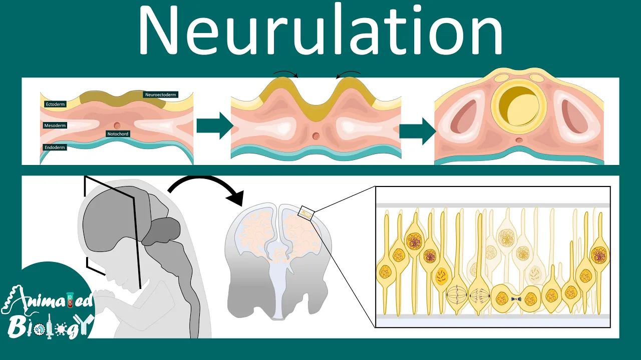
- Why this video: Utilizing high-quality 3D animations and clinical correlations, this video details how the neural ectoderm thickens, folds, and fuses into the neural tube. It serves as a strong foundation for structural neurulation while addressing key clinical pathologies such as spina bifida and anencephaly resulting from neuropore closure failures.

- Why this video: This video breaks down the patterning of the neural tube along the dorsal-ventral axis. It details how the roof plate (secreting BMPs) and the floor plate (secreting Sonic Hedgehog/Shh) generate overlapping concentration gradients that assign unique cellular identities to spinal cord interneurons and motor neurons.
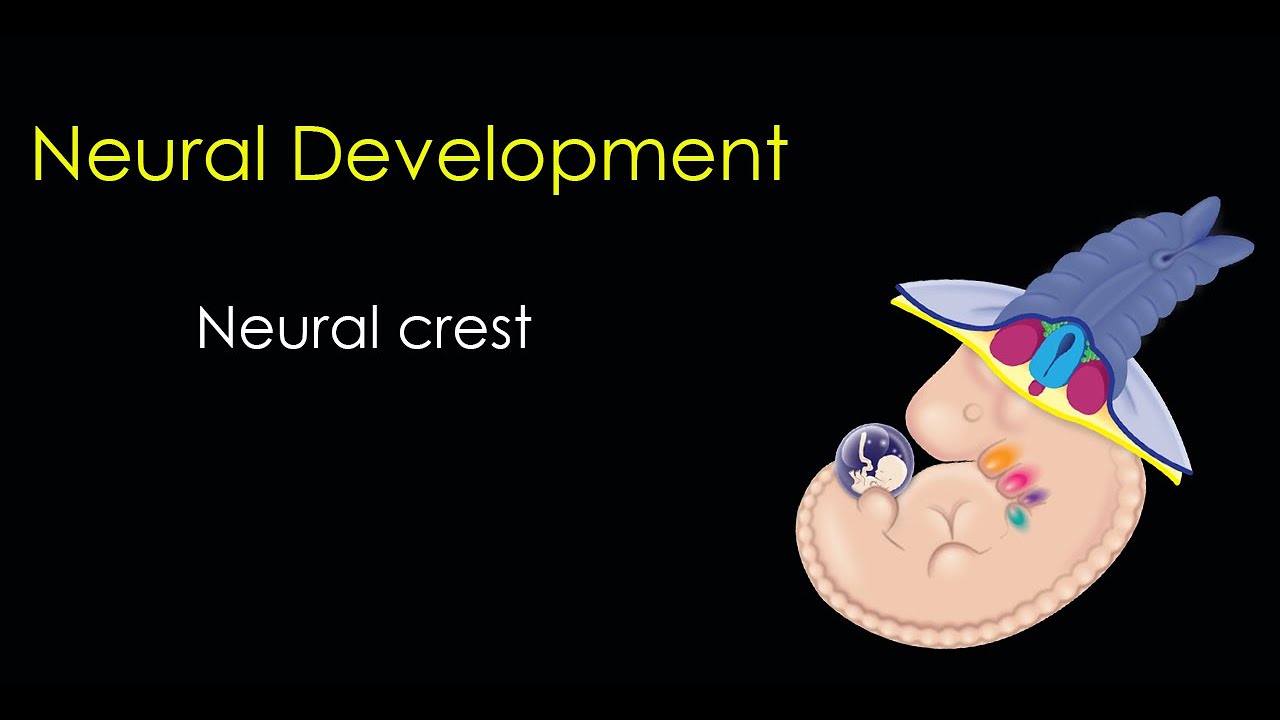
- Why this video: Focusing on neural crest cells, this video explores their birth at the border of neural and non-neural ectoderm. Crucially, it highlights the transition of these cells from stationary epithelial sheets to mobile, migratory mesenchymal cells (EMT) that disperse throughout the body to form Schwann cells, melanocytes, and the peripheral sensory/autonomic ganglia.
Knowledge Checkpoint
- Differentiate between primary and secondary neurulation mechanics and embryonic locations.
- Explain how Sonic Hedgehog (Shh) and BMP gradients establish concentration thresholds that specify motor neuron vs. sensory interneuron progenitor domains.
- Detail the cellular hallmarks of Epithelial-Mesenchymal Transition (EMT), including changes in cadherin expression and cell polarity.
- Identify key derivatives of neural crest cells in the peripheral nervous system and peripheral tissues.
Module 3: Neurogenesis and Neuronal Migration
This module details how neural stem and progenitor cells divide in the ventricular zone and migrate outward to assemble the nervous system. We will study the asymmetric and symmetric divisions of radial glial cells regulated by Notch signaling pathways. You will trace the "inside-out" sequence of cortical development, where early-born neurons form the deepest layers of the cortex (Layers V and VI) and later-born waves of migrating neurons climb past them to form superficial layers (Layers II-IV). We also emphasize the mechanistic differences between radial migration (used by glutamatergic excitatory neurons) and tangential migration (used by GABAergic inhibitory interneurons migrating from the ganglionic eminences).
Recommended Videos

- Why this video: Dr. Pasko Rakic, the pioneer of the radial unit hypothesis, describes how radial glial fibers function as critical structural "highways" for migrating neurons. This historical talk outlines the scaffolding mechanisms that allow cells born in the ventricular zone to reach their precise terminal locations in the cortex.
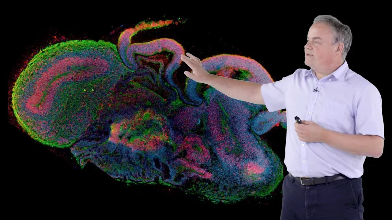
- Why this video: Dr. Knoblich provides a highly concise overview of human cortical layering. He outlines the sequence in which deep cortical layers are established first, followed by superficial layers, verifying the classic "inside-out" model of cerebral cortex development.
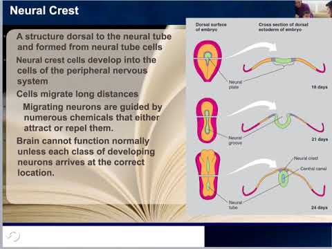
- Why this video: This video introduces the core differences between radial migration (moving perpendicularly from the ventricular zone toward the outer pial surface) and tangential migration (moving parallel to the ventricular walls, typical of inhibitory interneurons originating in the subpallium/ganglionic eminences).
- Why this video: This detailed lecture bridges embryology and pathology. It offers an in-depth analysis of cellular migration mechanics and explores the severe developmental consequences of migration disruption, such as lissencephaly ("smooth brain"), heterotopia, and polymicrogyria.
Knowledge Checkpoint
- Explain how asymmetric vs. symmetric division of radial glia is determined, and describe the role of Notch/PAR3 signaling in maintaining progenitor pools.
- Map out the temporal "inside-out" pattern of cortical layering, indicating which layers are populated first.
- Contrast radial migration and tangential migration regarding their trajectory, structural support, and the cell types that utilize them (e.g., pyramidal cells vs. interneurons).
- Identify the anatomical structures (e.g., ganglionic eminences) where cortical inhibitory interneurons originate.
Module 4: Growth Cones and Axon Guidance
This module examines how newly formed neurons project axons over long distances with millimeter precision. We dissect the structural components of the axonal growth cone—the central core, lamellipodia, and highly dynamic, actin-rich filopodia. We will analyze the molecular interactions of the four major guidance cue families: Netrins (acting through DCC and Unc5 receptors), Slits (interacting with Robo receptors), Semaphorins (signaling via Plexins and Neuropilins), and Ephrins (interacting with Eph receptors). Finally, you will study how these receptor-ligand interactions alter intracellular signaling cascades to direct cytoskeletal remodeling of actin filaments and microtubules, driving growth cone steering.
Recommended Videos

- Why this video: Derived from MIT's introductory biology course, this segment details the role of growth cone cytoskeletal dynamics. It highlights how actin polymerization drives the leading edge of filopodia and lamellipodia, while microtubule stabilization solidifies the progressive axonal shaft behind the steering tip.

- Why this video: Dr. Letourneur delivers an excellent narrated walkthrough of live growth cones in vitro. The video highlights how the growth cone samples the environment through filopodia, and how localized contact with extracellular matrix substrates triggers rapid actin and microtubule assembly to alter growth direction.

- Why this video: This video focuses on the molecular pathways operating during axon guidance. It provides a detailed, step-by-step molecular explanation of midline axon crossing, demonstrating how Netrin-DCC interactions attract the axon toward the midline, and how a subsequent upregulation of Robo receptors makes the growth cone sensitive to Slit repulsion, driving the axon away from the midline.

- Why this video: Produced by Stanford University, this video highlights visual systems to explain how axonal projections navigate toward target tissues. It explores how retinal ganglion cells deploy surface receptor profiles to sense molecular gradients (such as semaphorins, netrins, and ephrins) for accurate topographic mapping in the brain.
Knowledge Checkpoint
- Sketch an axonal growth cone and label the central domain, lamellipodia, and filopodia, detailing their primary cytoskeletal components (F-actin vs. microtubules).
- Explain how a single ligand, like Netrin, can trigger attraction via DCC homodimers but repulsion when DCC heterodimerizes with Unc5.
- Detail the molecular "switch" that allows commissural axons to cross the midline only once, highlighting the roles of Netrin, Slit, Robo, and DCC.
- Describe the differences between contact-dependent guidance (e.g., Ephrins/Eph receptors) and long-range diffusible guidance cues (e.g., Netrins).
Module 5: Synaptogenesis and Activity-Dependent Pruning
Once axons reach their appropriate targets, they must build and refine their synaptic connections. This final module details the molecular cross-talk that occurs during synapse formation, examining cell-adhesion molecules—specifically the trans-synaptic pairing of presynaptic Neurexins and postsynaptic Neuroligins. We will study the structural organization of the Neuromuscular Junction (NMJ) as a classic model of synaptogenesis. Finally, you will explore how excess neural connections are trimmed away postnatally through activity-dependent pruning, detailing how microglia selectively engulf weaker synapses based on patterns of neuronal activity and immune-tagging pathways (such as classical complement proteins).
Recommended Videos

- Why this video: This video abstract breaks down the structural molecular logic of synapse stabilization. It explains how Neurexin-Neuroligin binding complexes initiate dendritic filopodia stabilization, cluster postsynaptic receptors, and recruit presynaptic vesicle docking machinery.
- Why this video: Dr. Beth Stevens presents her lab’s research showing that microglia act as major agents of synaptic pruning. She explains how the brain uses classical immune system complement cascade molecules (like C1q and C3) to "tag" weaker, less active synapses for engulfment and elimination by microglial cells.
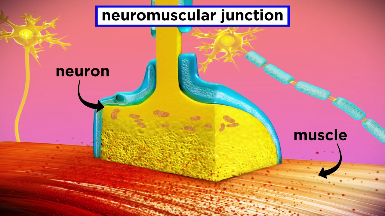
- Why this video: This video focuses on the structure of the Neuromuscular Junction (NMJ). It illustrates how the presynaptic motor neuron terminal align with postsynaptic acetylcholine receptor hot-spots in junctional folds to form a highly efficient synapse.

- Why this video: Dr. Jeff Lichtman uses connectomic imaging data to show how synaptic connections change over development. He presents evidence that axonal branches actively compete at the NMJ, demonstrating how stronger active branches win out while weaker, inactive inputs undergo elimination.
Knowledge Checkpoint
- Detail how trans-synaptic adhesion complexes involving Neurexins (presynaptic) and Neuroligins (postsynaptic) organize intracellular scaffolding proteins.
- Describe the primary structural features of a mature Neuromuscular Junction (NMJ), including junctional folds and postsynaptic acetylcholine receptor clustering.
- Outline the "use-it-or-lose-it" competition hypothesis: how firing patterns determine whether a synapse is potentiated or eliminated.
- Explain how the classical complement proteins C1q and C3 tag inactive synapses for elimination, and how microglial complement receptors recognize these tags.
Course Map
This map outlines the flow of developmental events covered in the modules, emphasizing how molecular induction patterns early structures to guide later cellular migration, projection, and pruning.
Key People Index
The following researchers contributed significantly to our understanding of the pathways covered in this curriculum:
- Hans Spemann & Hilde Mangold: Discovered the embryonic organizer (Spemann-Mangold Organizer), demonstrating that dorsal mesoderm tissue can induce host ectoderm to form a secondary neural axis.
- Dr. Richard Harland (UC Berkeley): Cloned the Noggin gene, providing critical molecular evidence that default neural fate is induced through active BMP inhibition.
- Dr. Pasko Rakic (Yale University): Formulated the Radial Unit Hypothesis, identifying radial glial cells as the primary migratory tracks for newly generated cortical neurons.
- Dr. Jurgen Knoblich (IMBA): Contributed to the understanding of asymmetric cell division of neural progenitors, polarity protein complexes, and the development of 3D brain organoids.
- Dr. Thomas Jessell (Columbia University): Identified the crucial role of Sonic Hedgehog (Shh) concentration gradients in ventral patterning of the spinal cord and progenitor cell specification.
- Dr. Jeff Lichtman (Harvard University): Championed Connectomics, detailing how activity-dependent synaptic competition shapes and prunes axonal inputs during postnatal development.
- Dr. Beth Stevens (Harvard/Broad Institute): Discovered that microglia prune synapses in an activity-dependent manner by utilizing classical complement cascade proteins (C1q, C3).
Final Self-Assessment
Test your mastery of the complete embryonic neurodevelopmental pathway by answering or completing the following prompts:
- Draw a timeline of early neural development, placing the following events in order: Gastrulation, Neural Induction, Neural Tube Closure, Progenitor Division, Cortical Migration, Axon Pathfinding, and Synaptic Pruning.
- Write a short paragraph detailing how the mesoderm-derived notochord acts as a molecular "instructor" for both the initial ectodermal default neural commitment and subsequent dorsal-ventral spinal cord patterning.
- Explain why a complete knockout of the Noggin, Chordin, and Follistatin genes in a model organism would prevent neural plate formation.
- Contrast Epithelial-Mesenchymal Transition (EMT) in migrating neural crest cells with the radial glial climbing mechanism used by migrating cerebral cortical neurons.
- Outline the structural defects you would expect in a cortex that has lost the ability to execute "inside-out" migration (e.g., due to a deficiency in the signaling protein Reelin).
- Distinguish between the origins and ultimate destinations of excitatory glutamatergic neurons and inhibitory GABAergic neurons during cortical assembly.
- Explain how a growing axon can navigate past a repulsive midline barrier using a dynamic receptor switch involving Netrin-DCC attraction and Slit-Robo repulsion.
- Detail how cytoskeletal elements (actin and microtubules) are systematically assembled or disassembled on opposite sides of a growth cone to initiate a physical turn toward an attractive gradient.
- Explain how the presynaptic Neurexin-postsynaptic Neuroligin interaction coordinates the physical localization of synaptic vesicles on the presynaptic side and neurotransmitter receptors on the postsynaptic side.
- Describe the process of synaptic pruning, from the initial patterns of activity-dependent synaptic competition to the final phagocytosis of complement-tagged synapses by microglial cells.