Neurodegeneration: Proteins & Cell Death
Learning Goal: Deconstruct the cellular and molecular pathology of neurodegenerative diseases, focusing on how protein misfolding, mitochondrial dysfunction, and neuroinflammation drive cell death in Alzheimer's and Parkinson's.
- Prerequisites: Basic high school level biology (understanding of cell structure, DNA-to-protein transcription/translation, and general chemistry).
- Estimated Total Study Time: 20 hours
Module 1: Foundations of Neuronal Cell Biology
This module establishes the necessary baseline for studying neurodegenerative pathology. You will explore the specialized structures of neurons, how they generate electrical signals to communicate, the fundamentals of three-dimensional protein structures, and the critical functions of intracellular organelles (such as the endoplasmic reticulum, mitochondria, and lysosomes) that maintain neuronal homeostasis.
Recommended Videos

- Why this video: This video uses a highly intuitive "city" analogy to break down the complex organization of eukaryotic animal cells. It provides an excellent foundational review of organelles that are highly relevant to neurodegenerative diseases—specifically the nucleus (genomic maintenance), endoplasmic reticulum (protein synthesis), and lysosomes (waste management).
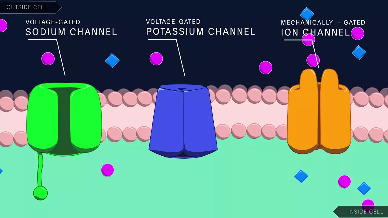
- Why this video: Neurons are electrically excitable cells. This video provides a precise, biophysical breakdown of how voltage-gated sodium and potassium channels coordinate to generate action potentials. Understanding the resting membrane potential and chemical synaptic transmission is vital for appreciating why metabolic or structural stress damages neuronal connectivity.
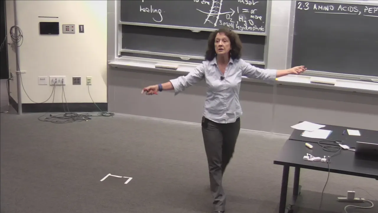
- Why this video: This rigorous MIT lecture explains the hierarchical organization of proteins (primary, secondary, tertiary, and quaternary structures). It covers the thermodynamic forces driving folding, such as hydrogen bonding and hydrophobic interactions, which is crucial background knowledge before examining how proteins misfold and aggregate.
Knowledge Checkpoint
- Diagram a neuron, labeling the dendrites, soma, axon hillock, axon, and synaptic terminal.
- Explain how the opening of voltage-gated and channels governs the depolarization and repolarization phases of an action potential.
- Describe the difference between a protein's primary amino acid sequence and its folded secondary structures (-helices and -sheets).
- Identify the main functions of the rough endoplasmic reticulum, Golgi apparatus, and lysosomes in the lifecycle of a membrane-bound protein.
Module 2: Protein Misfolding and Aggregation Mechanics
In this module, you will transition from normal protein biology to pathophysiology. You will examine how genetic mutations or environmental stressors cause proteins to lose their thermodynamic, native 3D shapes. This triggers hydrophobic sections to face outwards, leading to the formation of toxic oligomers, protofibrils, and insoluble amyloid aggregates. You will also look at how cellular waste management systems fail under this structural burden.
⚠️ Curriculum Gap Alert: While the video pool covers autophagy thoroughly, there is a lack of high-fidelity video content detailing the precise biochemical steps of chaperone-mediated protein folding and the specific E1-E2-E3 ubiquitin ligase enzymatic cascade of the Ubiquitin-Proteasome System (UPS). For deep mastery of these topics, please consult a cellular biology textbook (e.g., Lehninger Principles of Biochemistry).
Recommended Videos

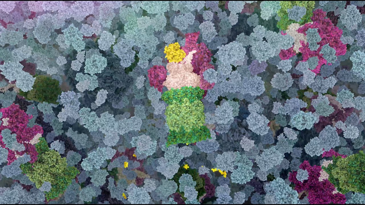
- Why this video: This video offers an excellent molecular description of how normal, soluble cellular proteins undergo conformational changes to become highly stable, insoluble aggregates rich in -sheet structures. Although centered on prions, the thermodynamic principles of self-propagating misfolding apply directly to amyloid-beta, tau, and -synuclein.
- Why this video: This high-quality animation visualizes the physical mechanisms of the proteasome. You will watch how ubiquitin tags are recognized, how target proteins are unfolded and threaded into the catalytic core, and how they are broken down into short peptides.

- Why this video: Direct from the pioneer who discovered the molecular machinery of autophagy, this Nobel Lecture outlines how cells segregate large protein aggregates and damaged organelles into double-membrane autophagosomes for lysosomal degradation. It is an invaluable deep dive into the master cellular clearance pathway.
Knowledge Checkpoint
- Explain how a protein mutation or environmental stressor can thermodynamically favor a misfolded state, exposing its internal hydrophobic residues.
- Describe how soluble monomeric proteins assemble into transient oligomers, and how these transition into insoluble fibrillar aggregates.
- Explain how the ubiquitin-proteasome system (UPS) selectively targets single proteins for degradation, and what prevents it from clearing large, cross--sheet fibrillar aggregates.
- Describe the structural steps of macroautophagy: from the formation of the phagophore to autophagosome-lysosome fusion.
Module 3: Mitochondrial Dysfunction and Oxidative Stress
Mitochondria are the primary energy source of neurons, but they are also a major source of cellular stress. This module details how the Electron Transport Chain (ETC) produces ATP, how electrons leak to generate Reactive Oxygen Species (ROS), and how high levels of ROS damage cellular lipids, proteins, and DNA. You will also learn about mitophagy, the quality control pathway that clears damaged mitochondria before they can trigger apoptosis.
⚠️ Curriculum Gap Alert: The video pool does not contain a step-by-step visual walkthrough of how electron transport chain complex failures directly initiate Bax/Bak-mediated outer mitochondrial membrane permeabilization (MOMP). To supplement this, independently search for the "Intrinsic pathway of apoptosis" and "Mitochondrial membrane potential depolarization."
Recommended Videos

- Why this video: This short video provides a clear, high-level summary of physiological electron leakage in the ETC. It explains that about 0.2% to 2% of electrons escape normal transport and react with molecular oxygen to produce superoxide radicals (), laying the groundwork for understanding oxidative stress.
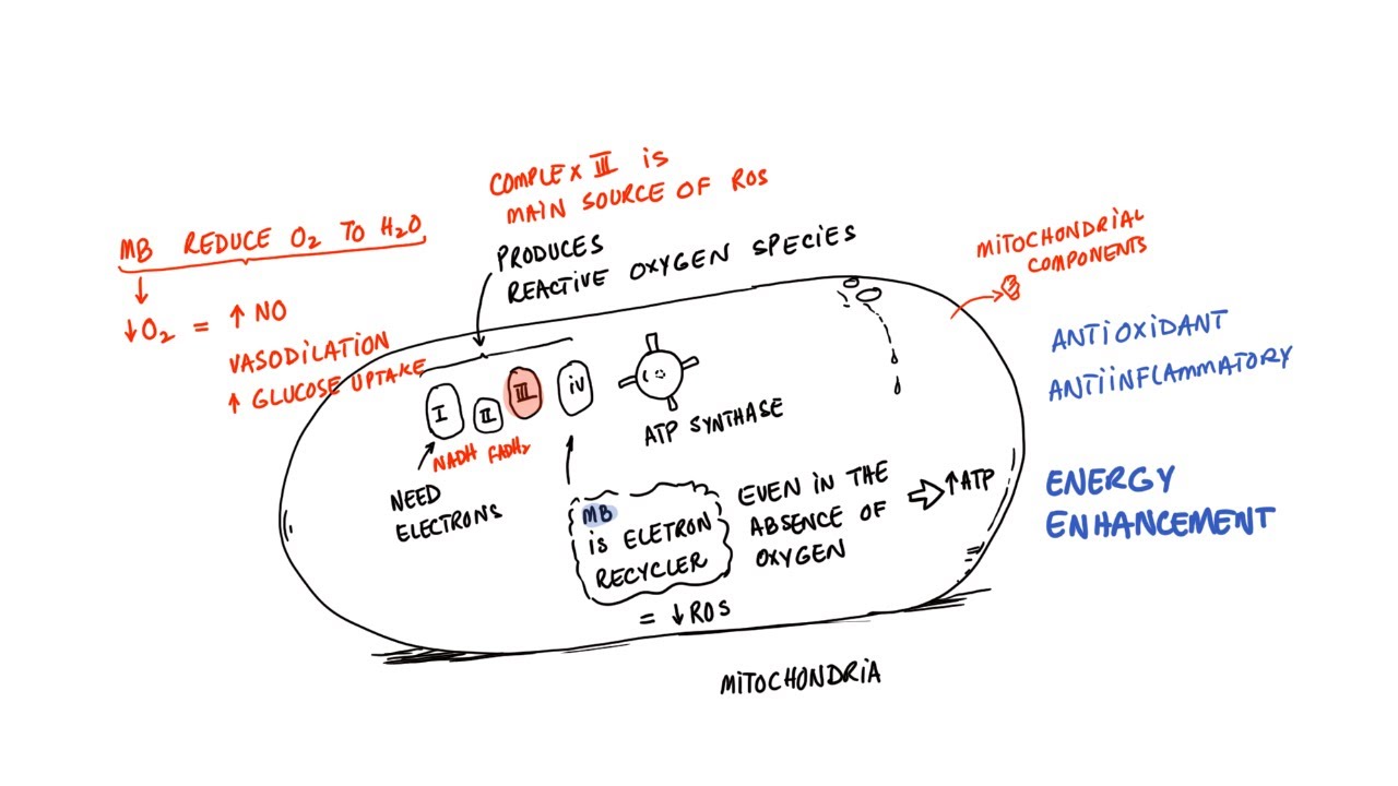
- Why this video: This lecture explains that ETC Complex III is a major site of physiological and pathological ROS production. It helps you understand what happens when electron flow is blocked, leading to a cascade of oxidative damage.
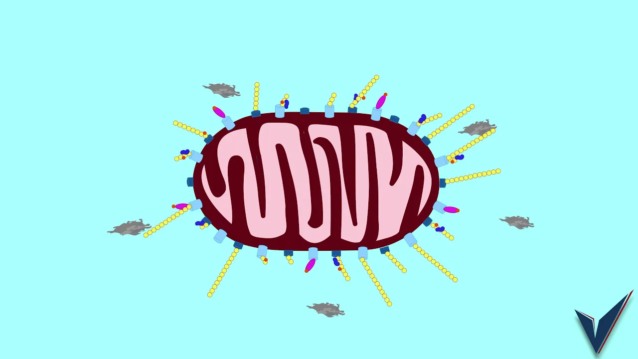
- Why this video: This animation clearly illustrates the molecular details of mitophagy. It shows how healthy mitochondria maintain a polarized membrane, and how a loss of membrane potential prevents the degradation of PINK1, triggering a Parkin-mediated ubiquitin cascade that flags the organelle for autophagic clearance.
Knowledge Checkpoint
- Explain how electrons leak from ETC Complexes I and III to react with molecular oxygen, forming the superoxide radical ().
- Define "mitochondrial membrane potential" () and describe how it is generated by the proton gradient across the inner mitochondrial membrane.
- Outline the step-by-step molecular mechanism of PINK1 and Parkin activation when a mitochondrion depolarizes.
- List three ways in which unchecked Reactive Oxygen Species (ROS) damage a post-mitotic neuron.
Module 4: Neuroinflammation and Glial Activation
The brain's immune system is tightly regulated by non-neuronal cells. This module focuses on the transition of microglia (the resident macrophages of the central nervous system) and astrocytes from protective, homeostatic states to chronic, pro-inflammatory phenotypes. You will learn how these activated glial cells release inflammatory cytokines, creating a self-sustaining cycle of neuroinflammation that actively drives neuronal death.
⚠️ Curriculum Gap Alert: The video pool lacks specific molecular details on microglial polarization states (M1 vs. M2) and the intracellular signaling pathways (such as NF-κB and NLRP3 inflammasome activation) that drive reactive astrogliosis. We recommend researching "Microglial M1/M2 polarization pathways" and "Reactive astrogliosis signaling cascades" on Google Scholar to supplement this module.
Recommended Videos
- Why this video: This video introduces the dynamic, memory-like properties of microglia. It explains "microglial priming," where a primary inflammatory trigger primes these cells to respond with a hyper-exaggerated, chronic neuroinflammatory response to subsequent minor stimuli.

- Why this video: This clip discusses how astrocytes help maintain the Blood-Brain Barrier (BBB). It helps you understand how the loss of astrocyte homeostatic support impairs the brain's microenvironment, allowing peripheral inflammatory elements to influence the central nervous system.

- Why this video: This video highlights the connection between systemic inflammation and glial activation. It explains how peripheral inflammatory cytokines can cross or affect the BBB, triggering a neuroinflammatory cascade in microglia and astrocytes.
Knowledge Checkpoint
- Define the physiological roles of microglia (synaptic pruning, debris clearance) versus their pathologically activated states.
- Explain "microglial priming" and describe how primed microglia behave differently than resting microglia.
- Describe how astrocytes support the Blood-Brain Barrier (BBB) and explain the consequences of reactive astrogliosis on neuronal health.
- Identify how pro-inflammatory cytokines (like TNF- or IL-) released by glia can directly damage neurons.
Module 5: Molecular Pathology of Alzheimer's Disease
This module integrates your knowledge of misfolding, mitochondrial dysfunction, and neuroinflammation to deconstruct Alzheimer's Disease (AD). You will look at the amyloid cascade hypothesis, tracking how Amyloid Precursor Protein (APP) is cleaved by enzymes to form extracellular amyloid-beta plaques. You will also examine how this extracellular pathology triggers intracellular tau hyperphosphorylation, leading to microtubule collapse, neurofibrillary tangles (NFTs), synaptic loss, and cell death.
Recommended Videos
- Why this video: This animation provides a clear molecular walkthrough of Alzheimer's pathology. It visualizes the enzymatic cleavage of APP by -secretase and -secretase to release Amyloid-Beta () peptides, and shows how these self-assemble into extracellular plaques. It also links this process to tau hyperphosphorylation and neurofibrillary tangles.
- Why this video: This video explains how cellular damage scales up to gross anatomical changes. It visualizes how widespread neuronal death leads to cerebral atrophy, narrowing of the gyri, and widening of the sulci.

- Why this video: This video focuses on tau protein. It explains tau's physiological role in stabilizing microtubules (the primary transport tracks of axons) and how hyperphosphorylation causes tau to detach, form intracellular tangles, and disrupt axonal transport.
Knowledge Checkpoint
- Contrast the non-amyloidogenic pathway (cleavage of APP by -secretase) with the amyloidogenic pathway (cleavage of APP by - and -secretases).
- Describe the chemical structure of and explain why it is more prone to aggregation than .
- Explain how hyperphosphorylation of tau protein causes it to detach from microtubules, and describe the impact of this detachment on axonal transport.
- Outline how extracellular oligomers are thought to trigger intracellular kinase pathways that hyperphosphorylate tau.
Module 6: Molecular Pathology of Parkinson's Disease
This final module focuses on Parkinson's Disease (PD). You will investigate the selective vulnerability of dopaminergic neurons in the substantia nigra pars compacta. You will also look at the aggregation of -synuclein into Lewy Bodies, how lysosomal and proteasomal clearance pathways fail, and how genetic mutations in the PINK1/Parkin genes directly link mitochondrial dysfunction to familial forms of PD.
Recommended Videos
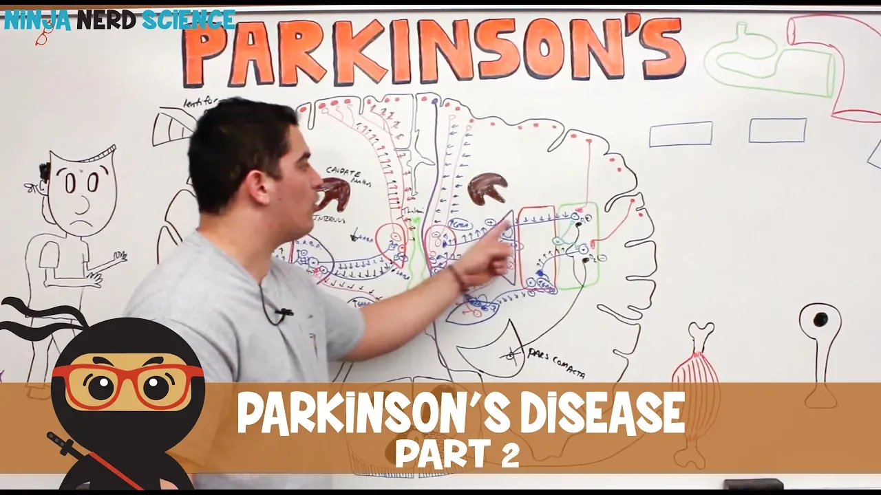
- Why this video: This video explains how lysosomal degradation pathways break down -synuclein, and how mutations in genes like LRRK2 or parkin disrupt this clearance. This failure leads to the accumulation of -synuclein in the cytoplasm.

- Why this video: This video links cellular pathology to clinical motor symptoms. It explains how losing dopaminergic projections from the substantia nigra to the striatum disrupts the basal ganglia circuits, leading to movement deficits like bradykinesia and rigidity.
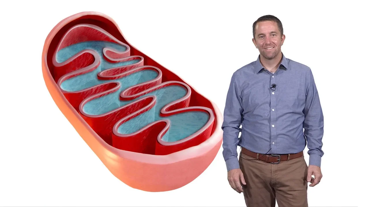
- Why this video: This clip highlights how mitochondrial quality control pathways are linked to familial Parkinson's disease. It explains that mutations in PINK1 or Parkin disrupt mitophagy, causing damaged mitochondria to accumulate, release ROS, and trigger apoptosis in dopaminergic neurons.
Knowledge Checkpoint
- Describe the physiological role of -synuclein at the presynaptic terminal, and how it misfolds from soluble monomers into oligomers and Lewy Bodies.
- Explain why dopaminergic neurons in the substantia nigra pars compacta are uniquely vulnerable to oxidative and metabolic stress.
- Detail how mutations in the PINK1 (kinase) or Parkin (ubiquitin ligase) genes lead to hereditary, early-onset Parkinson's Disease.
- Trace the path of motor control disruption starting from dopaminergic cell loss in the substantia nigra to downstream signaling in the striatum.
Course Map
This flowchart shows the recommended learning path and the dependencies between modules:
Key People Index
- Dr. Yoshinori Ohsumi (Tokyo Institute of Technology): Awarded the 2016 Nobel Prize in Physiology or Medicine for his discoveries of mechanisms for autophagy. His work elucidated the precise gene cascade (ATG genes) regulating autophagosome formation.
- Dr. Randy Schekman (UC Berkeley): Nobel Laureate in Physiology or Medicine (2013) for discoveries of machinery regulating vesicle traffic. He has contributed significantly to identifying cell-to-cell spread of toxic misfolded proteins in neurodegeneration.
- Dr. Stanley Appel (Houston Methodist): A leading neurologist and pioneer in neuroimmunology. His clinical and basic research has helped demonstrate that neuroinflammation is a primary driver of disease progression in ALS, AD, and PD.
- Dr. Friederich Lewy (1885–1950): The German-American neurologist who, in 1912, first described the abnormal intracellular protein clumps in Parkinson's disease that we now call "Lewy Bodies."
Final Self-Assessment
Use this final checklist to assess your understanding of the entire curriculum:
- Protein Structure: I can explain the thermodynamic forces (hydrophobic effect, hydrogen bonding, electrostatic interactions) that stabilize a protein’s tertiary structure.
- Aggregate Progression: I can explain the structural differences between soluble monomers, oligomers, protofibrils, and amyloid fibrils.
- Ubiquitin Cascade: I can describe the step-by-step enzymatic reaction (E1 activation, E2 conjugation, E3 ligation) that tags a protein with ubiquitin for degradation.
- Autophagy Pathways: I can distinguish between macroautophagy, microautophagy, and chaperone-mediated autophagy.
- ETC & ROS: I can trace how electrons leak from the respiratory chain to form superoxides, and outline how the cell neutralizes them using superoxide dismutase (SOD) and catalase.
- Mitophagy Cycle: I can explain how PINK1 accumulates on the outer mitochondrial membrane of depolarized mitochondria and recruits Parkin to initiate mitophagy.
- Glial Signaling: I can describe how chronic exposure to extracellular aggregates (like or -synuclein) activates microglia through Pattern Recognition Receptors (PRRs).
- Amyloid Cleavage: I can diagram the competitive cleavage pathways of APP by -, -, and -secretases.
- Tau Pathology: I can outline the molecular mechanism linking hyperphosphorylation to microtubule disassembly and the formation of paired helical filaments of tau.
- PD Selective Vulnerability: I can explain how high cytosolic dopamine levels, combined with oxidative stress and pacemaker activity, make dopaminergic neurons highly susceptible to cell death.