Synaptic Transmission: Action Potentials & LTP
Learning Goal: Map the molecular cascade of synaptic transmission, focusing on action potential propagation, neurotransmitter release, receptor activation, and the mechanisms of long-term potentiation (LTP).
- Prerequisites: Basic cellular biology (understanding of the lipid bilayer, protein structure, and ATP hydrolysis).
- Estimated Total Study Time: 12 hours
Module 1: Foundations of Neuronal Membrane Potential
This module establishes how resting neurons construct and maintain their electrical properties. You will master the physics of electrochemical gradients, the mathematical principles behind equilibrium potential, and the dynamic biochemical engine driving it all: the -ATPase pump.
Recommended Videos

Why this video: Dr. Najeeb delivers an incredibly rigorous, deeply conceptual lecture detailing the formation of the diffusion potential. He walks you through how selectively permeable membrane pathways—specifically potassium leak channels—allow ions to diffuse down their chemical concentration gradients, creating an electrical charge separation. This is the definitive academic starting point for understanding how the resting potential settles between and .
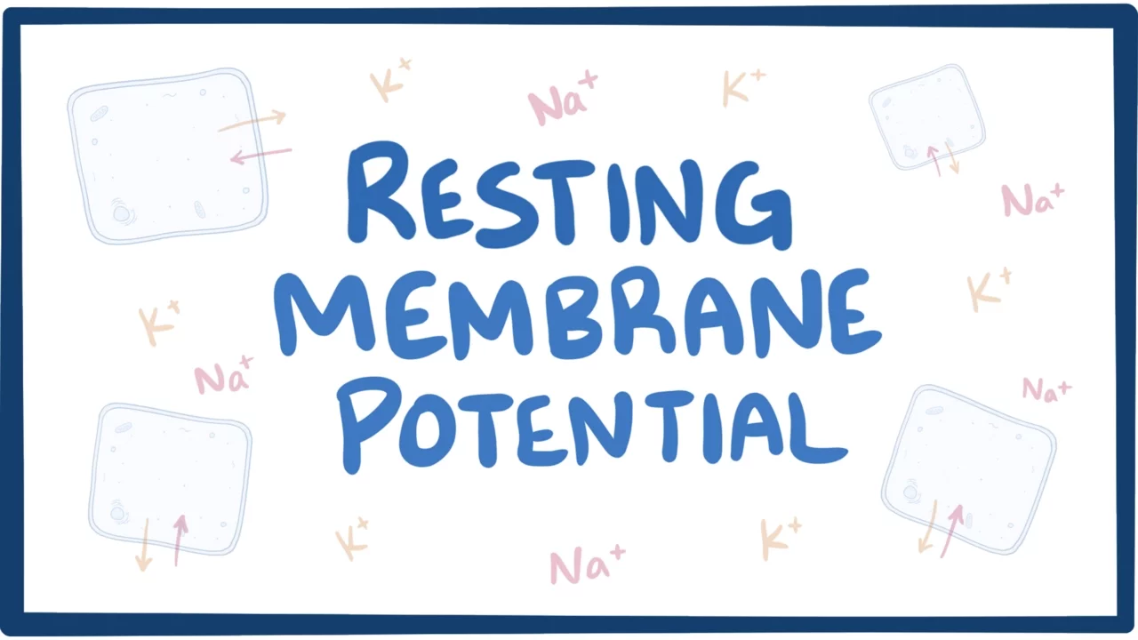
Why this video: This video offers a clear visual breakdown of the balance between chemical concentration gradients and electrostatic forces. It explains how to calculate membrane potential using the Nernst and Goldman-Hodgkin-Katz equations, factoring in both relative ion concentrations and membrane permeability.
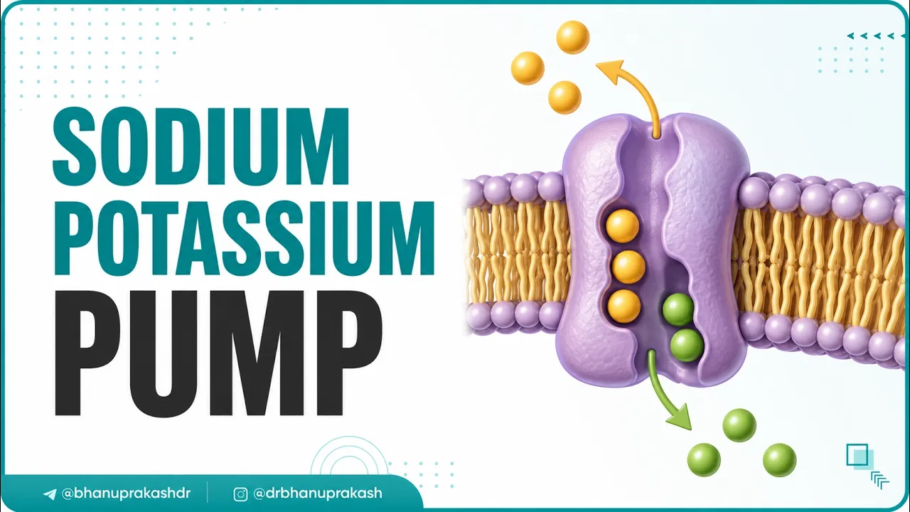
Why this video: This brief, high-yield animation shows the conformational states of the -ATPase pump. You will see how ATP hydrolysis drives the transport of three ions out of the cell and two ions into the cell, maintaining the electrochemical gradient against their respective concentration slopes.

Why this video: This lecture clarifies the thermodynamics behind ion movement. It explains how chemical gradients (entropy-driven diffusion) and electrical gradients (electrostatic interaction) combine to form a single electrochemical potential gradient. This gradient serves as the potential energy source for neuronal firing.
Knowledge Checkpoint
- Explain how the unequal distribution of and across the plasma membrane is established.
- Describe why the resting membrane potential is highly sensitive to changes in extracellular concentration but less sensitive to .
- Calculate how the net charge inside the cell changes with each cycle of the active -ATPase pump.
- Define "equilibrium potential" and describe the physical state of an ion when the membrane reaches this potential.
Module 2: The Action Potential and Propagation
Once the resting membrane potential is established, the neuron can use it to transmit rapid electrical signals. This module covers the rapid voltage changes that occur during an action potential, the transitions of voltage-gated ion channels, and how myelin sheaths increase signal velocity.
Recommended Videos
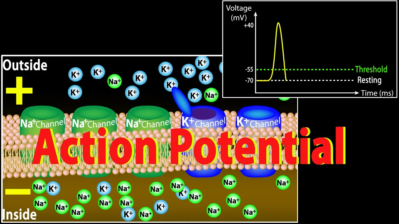
Why this video: This animation breaks down the action potential curve into distinct phases: depolarization, repolarization, and hyperpolarization. It aligns each phase with the opening and closing of voltage-gated and channels, making it easy to visualize the underlying ion currents.
Why this video: This video focuses on the structural mechanics of voltage-gated channels. It explains the dual-gate mechanism of the voltage-gated sodium channel—featuring both an activation gate and an inactivation gate—and details how the channel cycles through closed, open, and inactivated states.
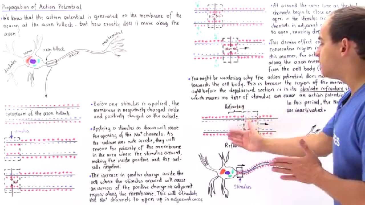
Why this video: This lecture explains how an action potential travels down an unmyelinated axon. It details how local positive current spreads to adjacent membrane segments, depolarizing them to threshold and triggering a cascade of channel openings that keeps the signal moving forward.

Why this video: This video explores saltatory conduction in myelinated axons. It explains how myelin sheaths act as electrical insulators, allowing the charge to jump between the Nodes of Ranvier and significantly increasing signal propagation speed.
Knowledge Checkpoint
- Detail the conformational states of the voltage-gated sodium channel during depolarization, peak, repolarization, and resting states.
- Explain why an action potential travels in only one direction down an axon, highlighting the role of the absolute refractory period.
- Compare the metabolic energy required to propagate an action potential down an unmyelinated axon versus a myelinated axon of the same length.
- Explain how myelin prevents charge leakage and why voltage-gated channels are clustered primarily at the Nodes of Ranvier.
Module 3: The Presynaptic Terminal and Neurotransmitter Release
This module details how an electrical signal is converted into a chemical message. When an action potential reaches the presynaptic terminal, it triggers calcium influx, vesicle docking, and membrane fusion, releasing neurotransmitters into the synaptic cleft.
Curriculum Gap Alert: High-quality videos showing the step-by-step structural biochemistry of the SNARE complex (such as the coiled-coil interactions of syntaxin, SNAP-25, and synaptobrevin) and the exact calcium-binding mechanism of synaptotagmin-1 are limited. To master these details, pay close attention to the structural biology lectures below.
Recommended Videos

Why this video: This advanced lecture addresses key concepts around SNARE proteins. It explains how v-SNAREs (synaptobrevin) on vesicle membranes interact with t-SNAREs (syntaxin and SNAP-25) on the target membrane. It shows how these proteins form a four-helix bundle that pulls the vesicle and cell membranes together for fusion.

Why this video: This specialized neurobiology video highlights the role of calcium ions in neurotransmitter release. It explains how calcium entering through voltage-gated calcium channels binds to synaptotagmin, which acts as a sensor to trigger exocytosis.
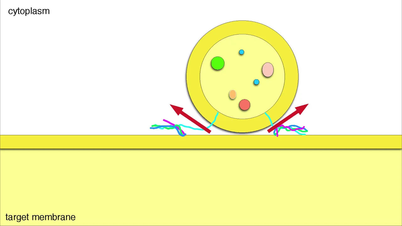
Why this video: This detailed video shows the cellular process of vesicle transport and docking. It highlights how Rab GTPases coordinate with tethering proteins to guide vesicles to their target sites before the SNARE complex assembles.

Why this video: This tutorial integrates these molecular steps into a clear, chronological sequence: action potential arrival, voltage-gated channel opening, localized calcium influx, vesicle fusion, and transmitter release.
Knowledge Checkpoint
- List the three synaptic proteins that form the core SNARE complex and identify which are located on the vesicle versus the target membrane.
- Describe how synaptotagmin acts as a calcium sensor and how it triggers membrane fusion.
- Explain why localized calcium domains are critical for fast, synchronous neurotransmitter release.
- Describe what happens to synaptic transmission if SNARE proteins are cleaved by bacterial toxins, such as botulinum or tetanus toxin.
Module 4: Postsynaptic Receptor Activation & Signal Integration
Once neurotransmitters cross the synaptic cleft, they bind to postsynaptic receptors. This module covers the difference between fast-acting ionotropic receptors and slow-acting metabotropic (GPCR) receptors. It also explains how postsynaptic potentials are integrated through spatial and temporal summation.
Recommended Videos
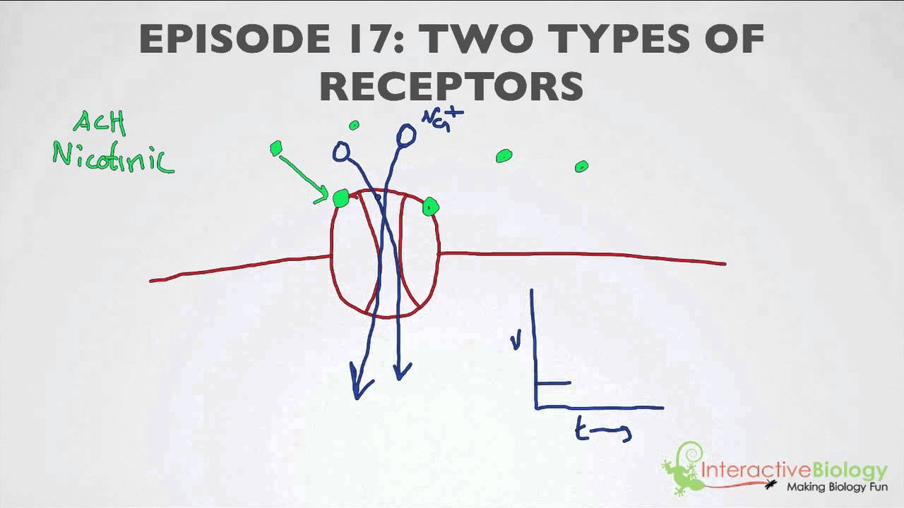
Why this video: This video contrasts ionotropic and metabotropic receptors. It explains how ionotropic receptors function as ligand-gated ion channels for fast synaptic transmission, while metabotropic receptors initiate slower, G-protein-coupled intracellular signaling cascades.

Why this video: This animation details the G-protein activation cycle. It shows how ligand binding triggers GDP-to-GTP exchange on the G-alpha subunit, causing it to dissociate from the beta-gamma complex and modulate effector enzymes or ion channels.

Why this video: This lecture explains how neurons process incoming inputs. It details how ligand-gated channels produce local graded responses (EPSPs and IPSPs) and how these signals are integrated at the postsynaptic membrane.

Why this video: This video focuses on summation. It uses clear diagrams to differentiate between spatial summation (inputs from multiple physical synapses firing at the same time) and temporal summation (high-frequency inputs from a single synapse firing in rapid succession).
Knowledge Checkpoint
- Compare the activation times and signaling mechanisms of ionotropic and metabotropic receptors.
- Describe the activation cycle of a heterotrimeric G-protein, including the roles of GDP, GTP, and the dissociation of G-alpha and G-beta-gamma subunits.
- Explain how a ligand-gated channel generates an Excitatory Postsynaptic Potential (EPSP) versus an Inhibitory Postsynaptic Potential (IPSP).
- Explain why a single EPSP is usually not enough to fire an action potential, and how spatial and temporal summation help reach threshold at the axon hillock.
Module 5: Molecular Mechanisms of Long-Term Potentiation (LTP)
This module explores the molecular basis of synaptic plasticity, learning, and memory. You will study how high-frequency stimulation triggers calcium entry through NMDA receptors, activates downstream kinases, recruits AMPA receptors, and drives retrograde signaling to strengthen synaptic connections.
Recommended Videos
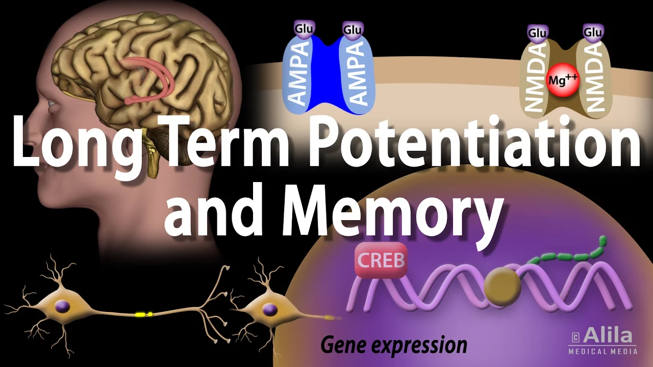
Why this video: This animation provides a clear overview of early-phase and late-phase LTP. It details how strong depolarization removes the block from NMDA receptors, allowing calcium to enter and trigger the insertion of additional AMPA receptors into the postsynaptic membrane.
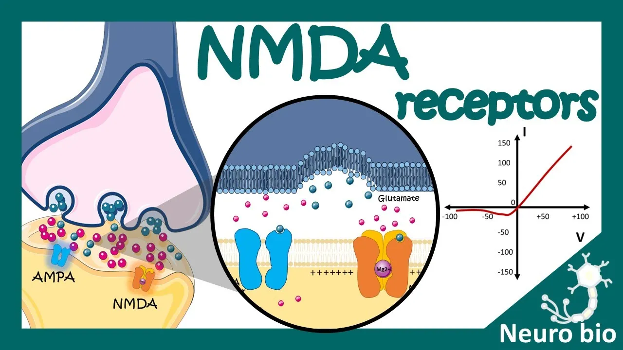
Why this video: This video focuses on the NMDA receptor's role as a molecular coincidence detector. It explains why the receptor requires both glutamate binding and postsynaptic depolarization to conduct ions, making it key to detecting paired activity.

Why this video: This video details the intracellular signaling cascade initiated by calcium entry. It covers the activation of Calmodulin, CaMKII autophosphorylation, and the cellular pathways that transport new AMPA receptors to the postsynaptic density.

Why this video: This animation covers retrograde signaling. It shows how postsynaptic activity triggers the synthesis of endocannabinoids, which travel backward across the synapse to bind presynaptic CB1 receptors and modulate neurotransmitter release.
Knowledge Checkpoint
- Explain why the NMDA receptor functions as a "coincidence detector," referencing its voltage-dependent block.
- Describe the molecular steps that connect calcium influx through NMDA receptors to the insertion of new AMPA receptors.
- Explain the role of autophosphorylated CaMKII in maintaining synaptic strength during early-phase LTP.
- Define "retrograde messenger" and describe how molecules like endocannabinoids or nitric oxide act backward to influence presynaptic release probability.
Course Map
This flowchart shows the recommended learning order and the conceptual dependencies between modules.
Key People Index
- Timothy Bliss & Terje Lømo (1973): Discovered Long-Term Potentiation (LTP) in the mammalian hippocampus. They demonstrated that high-frequency stimulation of the perforant path produced a long-lasting increase in synaptic efficacy, establishing the experimental model for how synapses store information.
- Dr. Najeeb: A renowned medical educator whose lectures provide deep, foundational breakdowns of cellular electrophysiology, particularly resting membrane potential and neuromuscular transmission mechanics.
Final Self-Assessment
This comprehensive self-assessment covers the molecular and physiological concepts across all five modules.
- Define the ionic concentrations that establish the resting membrane potential and explain how the -ATPase pump maintains them.
- Explain how an action potential threshold is reached and describe the feedback loop that drives rapid depolarization.
- Trace the structural changes in voltage-gated sodium channels that prevent backward signal propagation during an action potential.
- Explain how myelination and saltatory conduction increase signal speed down an axon.
- Describe the assembly of the SNARE complex and identify the roles of synaptobrevin, syntaxin, SNAP-25, and synaptotagmin-1.
- Compare the activation timecourses, structures, and downstream targets of ionotropic versus metabotropic receptors.
- Differentiate between spatial and temporal summation and describe how they influence firing decisions at the axon hillock.
- Explain the physical mechanism of the voltage-dependent magnesium block in NMDA receptors and how it is cleared.
- Detail the signaling pathway from postsynaptic calcium entry to CaMKII activation and subsequent AMPA receptor insertion.
- Explain how retrograde messengers are synthesized and how they modulate presynaptic neurotransmitter release.