Bioarchaeology: Bones, Diet & Ancient Health
Learning Goal: Apply bioarchaeological methods to reconstruct the health, diet, and social structures of ancient populations through the analysis of skeletal remains and paleopathology.
- Prerequisites: None (an interest in biological anthropology, history, and basic human anatomy is recommended).
- Estimated Total Study Time: 20 Hours
Module 1: Introduction to Bioarchaeology & Skeletal Anatomy
This module establishes the core definition, historical evolution, and theoretical frameworks of bioarchaeology. You will master the foundational elements of osteology, learning to identify the differences between the axial and appendicular skeletons, and discover how bones act as dynamic, biological archives of past life histories.
Recommended Videos

Why this video: This video introduces bioarchaeology as the contextual, scientific study of human skeletal remains from archaeological sites. It explains how skeletons act as an interactive record of an individual's life experience, health, and adaptations, bridging the gap between history and biology.
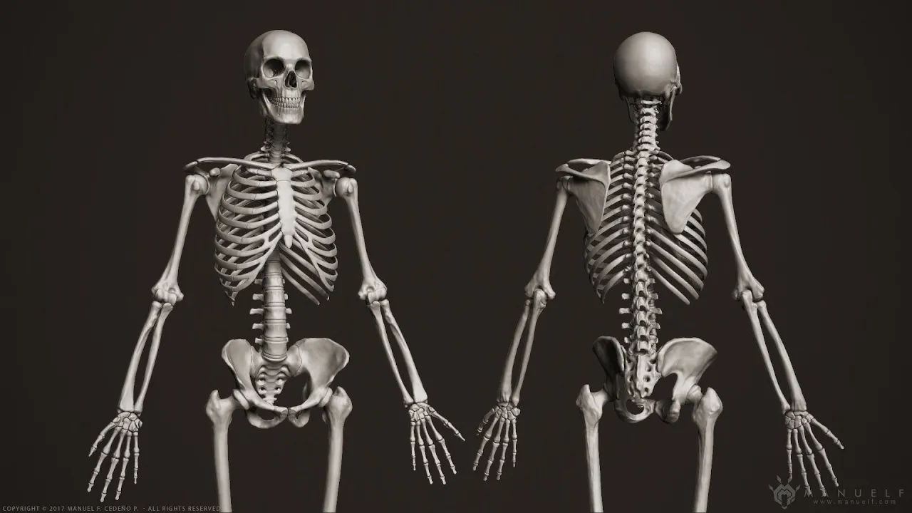
Why this video: Professor Dave delivers a breakdown of human skeletal anatomy (osteology), separating the skeletal system into the axial and appendicular skeletons. This physical anatomical knowledge is vital before you can begin interpreting modifications, traumas, or pathologies in the skeletal record.
Why this video: This video offers an academic lecture overviewing bioarchaeology's problem-oriented, interdisciplinary nature. It highlights how practitioners utilize multiple lines of evidence to reconstruct demographic profiles and investigate systemic social behaviors of ancient societies.
Knowledge Checkpoint
- Define "bioarchaeology" and trace its evolution from 19th-century descriptive anatomy to modern biocultural frameworks.
- List the primary anatomical elements of the human axial skeleton versus the appendicular skeleton.
- Explain how bone functions as dynamic, living tissue that remodels in response to environmental and physical stressors.
Module 2: Reconstructing Profile: Age, Sex, and Stature
Determining the biological profile of an individual is a fundamental step in bioarchaeological analysis. In this module, you will learn the primary morphological features used to estimate biological sex from pelvic and cranial structures, methods for tracking aging through tooth eruption and epiphyseal fusion, and the basic methodology for stature estimation.
Recommended Videos
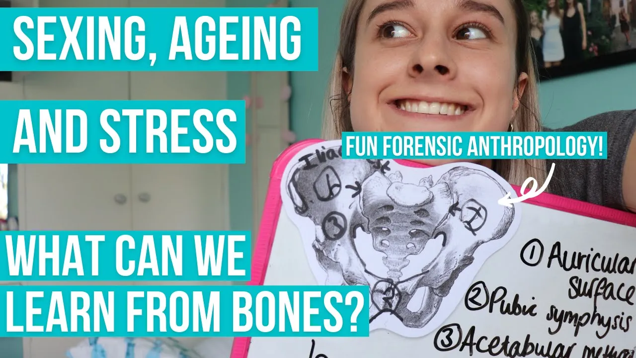
Why this video: This video focuses on the pelvis and the skull as the primary skeletal indicators of age and sex. You will learn why these particular regions show marked sexual dimorphism and how they evolve across different developmental milestones of a human lifespan.

Why this video: This guide outlines the six key skeletal indicators used by anthropologists to estimate age-at-death. It details infant/subadult age estimation methods, such as dental development and suture fusion patterns, alongside adult degeneration metrics.

Why this video: This practical laboratory tutorial demonstrates how to determine biological sex using real human pelvic bones. It isolates three diagnostic elements: the sub-pubic angle, the greater sciatic notch, and the pelvic inlet.
Why this video: Using a real-world case, this excerpt illustrates how stature can be calculated by measuring long bones (e.g., measuring the femur in millimeters) and plugging these lengths into mathematical regression formulas to estimate physical height in life.
Pedagogical Note on Stature Estimation
Coverage Gap Identified: While the forensic excerpt above demonstrates the practical application of calculating stature from a single long bone, our core video pool lacks a detailed mathematical tutorial breaking down the precise regression equations (e.g., the Trotter-Gleser formulas) across different populations.
To master this area independently, we recommend searching for: "Trotter-Gleser stature estimation regression formulas biological anthropology".
Knowledge Checkpoint
- Identify three pelvic features used to distinguish female remains from male remains.
- Explain the difference in reliability between estimating age-at-death for a subadult (using dental eruption and epiphyseal fusion) versus an adult (using joint degeneration).
- Detail how long bone lengths are used in regression equations to reconstruct the stature of an individual.
Module 3: Paleopathology: Reading Disease and Trauma in Bone
This module explores how paleopathologists identify ancient diseases and trauma patterns. You will examine metabolic bone disorders, infectious diseases, and trauma indicators that leave permanent markings on human bone, while learning to critically differentiate between chronic conditions and acute causes of death.
Recommended Videos

Why this video: This medical lecture covers metabolic bone disorders from a clinical perspective. It explains the biological impact of mineral deficiencies (resulting in rickets and osteomalacia) and organic matrix issues (such as scurvy), laying the physiological groundwork for understanding how these diseases alter bone structure.
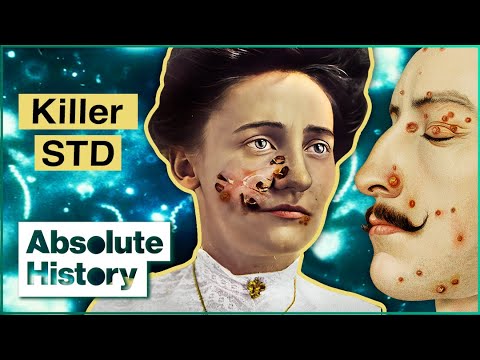
Why this video: This segment demonstrates how paleopathologists identify chronic infectious diseases like syphilis by examining diagnostic markers, including pitted cranial lesions (caries sicca) and "saber shin" deformities of the tibiae.
Why this video: This practical archaeological investigation covers abnormal bone pathologies, demonstrating how chronic conditions like Paget's disease alter bone density, causing distinct thickening and weakening patterns that are clearly visible during analysis.

Why this video: This seminar addresses a vital limitation in paleopathology: only chronic infections that a host survives long enough to provoke an immune response will manifest on the bone. Acute, rapid-killing infections leave no trace on the skeleton.
Pedagogical Note on Metabolic Disease Markers
Coverage Gap Identified: While clinical overviews of rickets and scurvy are provided above, specialized videos demonstrating dry bone indicators of childhood iron-deficiency anemia and metabolic stress—specifically cribra orbitalia (porous lesions in the eye orbits) and porotic hyperostosis (cranial bone porosity)—are limited in our core video pool.
To study these skeletal paleopathologies, search independently for: "Cribra orbitalia and porotic hyperostosis skeletal paleopathology tutorial".
Knowledge Checkpoint
- Differentiate between the skeletal presentation of rickets (mineralization failure) and scurvy (collagen matrix synthesis failure).
- Explain why chronic illnesses are easier to diagnose on ancient human skeletons than rapid, acute infections.
- Describe the classic diagnostic bone markers associated with venereal or congenital syphilis (e.g., caries sicca, saber shin).
Module 4: Ancient Diets: Isotopes and Dental Pathology
This module explores the dietary reconstructions of ancient populations. You will investigate how food leaves clear chemical signatures in bone collagen and tooth enamel, and discover how to read direct indicators of physiological stress and dental disease on ancient teeth.
Recommended Videos
Why this video: This video introduces stable isotope analysis of carbon and nitrogen in bone and teeth. You will learn how the ratio of Carbon-13 to Carbon-12 distinguishes between different plant groups ( versus plants), while Nitrogen-15 ratios reconstruct trophic levels and marine food sources.

Why this video: Dr. Eades explains the chemical foundations of mass spectrometry as used in stable isotope analysis. He clarifies how stable isotopes do not decay over archaeological timescales, remaining locked in biological tissues for thousands of years.
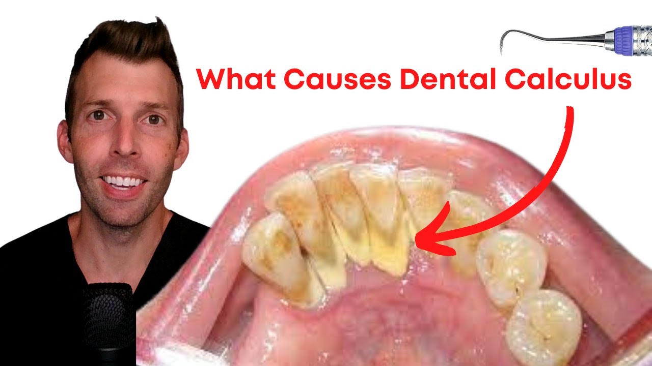
Why this video: This video describes how dental calculus (calcified tartar) mineralizes from bacterial biofilm in saliva. In bioarchaeology, mineralized dental calculus serves as a micro-vault, preserving ancient food starches, phytoliths, and oral microbiomes.
Why this video: This segment details how physiological stress can disrupt childhood dental development. You will learn to recognize linear enamel hypoplasia (LEH)—horizontal lines running across teeth—which mark periods of systemic illness or malnutrition during childhood.
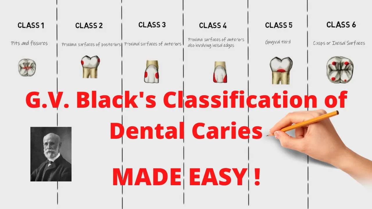
Why this video: A comprehensive review of G.V. Black's classification of dental caries. Although clinical, this structural breakdown of how and where carious lesions develop helps bioarchaeologists categorize caries found in ancestral remains.

Why this video: This video shows what dental abscesses look like on dry bone. It describes how untreated caries penetrate deep into tooth sockets, forming distinct osteolytic lesions in the mandible or maxilla that caused significant systemic pain during life.
Knowledge Checkpoint
- Explain how carbon isotope ratios () differentiate diets rich in wheat and barley from those based on maize or millet.
- Describe the process of dental calculus formation and list three types of micro-particles it can trap in the archaeological record.
- Define "linear enamel hypoplasia" (LEH) and describe how it acts as an archaeological indicator of childhood stress.
- Sketch or describe how a dental abscess manifests on the alveolar bone of an ancient skull.
Module 5: Social Bioarchaeology: Status, Identity, and Culture
Bioarchaeology reaches its peak when biological metrics are integrated to answer questions about culture, status, identity, and gender. This final module synthesizes osteology, paleopathology, and isotopic science to reconstruct ancient labor systems, social stratification, and body-modification practices.
Recommended Videos
Why this video: This segment details how daily mechanical activities alter skeletal remains. You will learn about anatomical markers of physical labor, such as compressed lumbar spine joints from repetitive heavy lifting and squatting facets on ancient ankle bones.

Why this video: This video explores artificial cranial deformation (head binding), a practice of reshaping infant skulls using tightly bound boards or bands. It explains how these permanent alterations acted as visual symbols of royalty, identity, and group status in ancient societies.

Why this video: This case study of the Tiwanaku civilization demonstrates how cranial deformation patterns served as clear physical indicators of social affiliation, signaling ethnicity and elite lineage in South American antiquity.
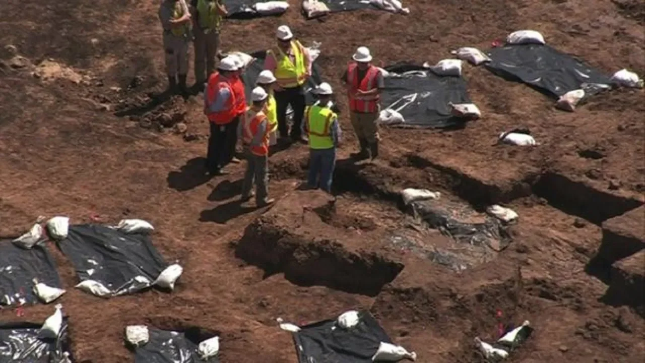
Why this video: This news piece highlights musculoskeletal stress markers (MSMs) on human remains from a historic convict lease mass grave. It shows how systemic social inequality, exploitation, and forced labor translate directly into structural changes at bone insertion points.
Pedagogical Note on Muscle Insertion Changes
Coverage Gap Identified: While the videos above provide clear evidence of occupational and structural stress, our video pool lacks a detailed anatomical close-up showing entheseal changes (alterations at the site of tendon and ligament attachments) or osteoarthritic joint wear on dry bone.
To explore entheseal changes further, search independently for: "Entheseal changes muscle insertion markers skeletal anthropology".
Knowledge Checkpoint
- Define "musculoskeletal stress markers" (MSMs) and explain how bone remodeling at tendon attachment sites documents physical activity.
- Describe how and when artificial cranial deformation is performed on a child, and discuss its primary social functions in historical civilizations.
- Connect osteological evidence of labor (such as ankle squatting facets or compressed vertebrae) to broader socio-economic structures in an ancient population.
Course Map
This dependency diagram shows the recommended order of study. Ensure you master the underlying osteology and biological profiling methods before analyzing disease, diet, and social status.
Key People Index
These researchers, pioneers, and historical classification authorities are central to the methods taught in this curriculum:
- Jane Buikstra (PhD): Highlighted as a founding figure in Module 1. She redefined "bioarchaeology" in 1977 as the study of human remains from archaeological contexts within an anthropological, population-centered framework.
- Grahame Clark: Coined the term "bio-archaeology" in 1972 (originally referencing animal remains) before it was adapted to human osteology.
- G.V. Black: The "Father of Operative Dentistry" (Module 4), who designed the structural classification of dental caries used today to assess tooth decay.
- Paul Budd (PhD): A researcher at Durham University featured in Module 4 for pioneering stable isotope analysis of tooth enamel to map geographical origins and ancient migration patterns.
Final Self-Assessment
Complete this comprehensive self-assessment to verify your mastery of the curriculum's learning goals:
- Explain how bioarchaeology differs from traditional forensic anthropology in its research questions, context, and population scope.
- Correctly assign a bone fragment to either the axial or appendicular skeleton.
- Describe the morphological differences in the greater sciatic notch and sub-pubic angle between biological males and females.
- Outline how teeth can estimate age-at-death in children (eruption stages) versus adults (wear patterns).
- Contrast the physical appearance and biological causes of rickets versus scurvy on skeletal remains.
- Detail why chronic systemic infections like syphilis leave distinct skeletal lesions, whereas acute infections like cholera leave none.
- Reconstruct an ancient individual's trophic level using nitrogen isotope ratios ().
- Explain how dental calculus can be processed in a laboratory to extract ancient dietary starch granules and micro-pathogens.
- Differentiate between a developmental dental pathology (linear enamel hypoplasia) and an acquired dental pathology (caries).
- Describe two osteological markers that indicate intense manual labor (e.g., squatting facets, spinal compression).
- Analyze how artificial cranial deformation represents identity, class, and social status in ancient human societies.