PETase Evolution: Library Design & Kinetics
Learning Goal: This curriculum provides a comprehensive guide to designing and executing a directed evolution campaign to improve the thermal stability and catalytic efficiency of a PET-degrading enzyme (PETase). It covers wild-type enzyme structures and plastic degradation biology, mutant library generation via error-prone PCR (epPCR), the setup and execution of high-throughput colorimetric microplate screening assays, recombinant protein expression and purification in E. coli, and rigorous kinetic ( and ) and thermal shift (DSF) characterizations.
- Prerequisites: Basic biochemistry, introductory molecular biology (DNA replication, transcription, translation), and basic laboratory algebra (concentration and dilution calculations).
- Estimated Study Time: 36 hours (including reading, video-guided study, conceptual exercises, and mock experimental planning).
Module 1: Foundations of Enzymes and Plastic Biodegradation
This module establishes the core biochemistry of enzymes and examines the ecological and industrial significance of PET-degrading enzymes (PETase). You will learn how enzymes dramatically lower activation energy to accelerate chemical reactions and study the specific discovery and mechanism of PETase from Ideonella sakaiensis in breaking down polyethylene terephthalate (PET) plastic.
Recommended Videos

- Why this video is valuable: It delivers a clear, foundational review of enzyme catalysis, explaining how biological catalysts lower activation energy without shifting reaction equilibria. This is essential for understanding how directed evolution modifies the activation energy barrier for PET hydrolysis.

- Why this video is valuable: This high-quality animation visualizes the physical mechanics of enzyme active sites and the classic induced-fit model of substrate binding, helping you picture how plastic polymer chains interact with the PETase active site.

- Why this video is valuable: Provides deep real-world context on the global plastic waste crisis and explores how biotechnology can utilize microbial enzymes to break down highly stable crystalline plastics. It highlights why engineering PETase is critical for industrial viability.

- Why this video is valuable: Focuses specifically on Ideonella sakaiensis PETase, detailing how it cleaves the complex ester links of PET into soluble oligomers (MHET, BHET) and monomer units (terephthalic acid and ethylene glycol).
Knowledge Checkpoint
- Draw a reaction coordinate diagram illustrating how an enzyme lowers activation energy () while leaving overall free energy change () unaltered.
- Describe the chemical structure of Polyethylene Terephthalate (PET) and identify the specific ester bonds targeted by PETase.
- Explain why native PETase from Ideonella sakaiensis requires engineering before it can be effectively used in high-temperature, industrial-scale bioreactors.
Module 2: Recombinant Protein Expression in E. coli
To evolve an enzyme, you must express it in a laboratory host. This module covers plasmid vector selection (focusing on the widely used pET system), modern cloning workflows, bacterial transformation protocols, and the molecular mechanism of IPTG-induced recombinant protein expression within E. coli BL21 host strains.
Recommended Videos

- Why this video is valuable: Explains how recombinant plasmids are constructed and delivered into host bacteria via heat shock, laying down the molecular biology fundamentals needed to handle wild-type and mutated PETase genes.

- Why this video is valuable: High-yield animation explaining why IPTG (a non-hydrolyzable lactose mimic) is used to trigger high-level transcription of target genes placed downstream of the T7/lac promoter.

- Why this video is valuable: Details the lac promoter repressor mechanism and illustrates the exact molecular dance that occurs when IPTG is introduced, preventing repressor binding and unlocking transcription.

- Why this video is valuable: Breaks down how expression host strains (like E. coli BL21) are specialized for protein production. It discusses codon optimization and rare tRNA supply, which is critical when expressing eukaryotic or non-native bacterial genes like PETase in E. coli.
Knowledge Checkpoint
- List the three essential components of a standard cloning plasmid vector (origin of replication, selectable marker, and multiple cloning site).
- Explain why IPTG is used for induction instead of natural lactose, focusing on metabolic stability.
- Differentiate between a cloning strain (e.g., DH5) and an expression strain (e.g., BL21(DE3)) of E. coli, explaining why we separate plasmid propagation from protein expression.
Module 3: Directed Evolution and Mutant Library Generation
This module introduces the Nobel-prize-winning concepts of directed evolution. You will learn how to mimic natural selection in the laboratory by generating vast genetic diversity using error-prone PCR (epPCR)—intentionally introducing random mutations into the PETase gene to create a "mutant library".
Recommended Videos

- Why this video is valuable: A primary-source lecture by Professor Frances Arnold outlining the birth, logic, and massive impact of directed evolution. It provides inspiration and high-level conceptual context directly from the field's pioneer.

- Why this video is valuable: Directly explains the molecular mechanism of error-prone PCR (epPCR). It highlights how altering reaction conditions—such as adjusting magnesium concentration, introducing manganese ions, or using biased dNTP ratios—forces non-proofreading DNA polymerases to make errors.
- Why this video is valuable: Walkthrough of how directed evolution connects random mutagenesis to microtiter plate screening, acting as an exceptional framework for building your experimental campaign.
Knowledge Checkpoint
- State the three steps of the directed evolution cycle and explain why it is an iterative process.
- Detail how error-prone PCR alters standard PCR parameters to decrease replication fidelity (e.g., addition, unbalanced dNTP pools, Taq polymerase lack of proofreading).
- Calculate the target mutation rate (e.g., 1-2 amino acid substitutions per enzyme molecule) and explain why too many mutations per gene typically lead to inactive protein variants.
Module 4: High-Throughput Screening (HTS) Assays
Once genetic diversity is generated, you must screen the library. Because solid PET films degrade too slowly for rapid screening, directed evolution campaigns use soluble ester proxy substrates. This module details how to design and execute a high-throughput 96-well microplate colorimetric assay using p-nitrophenyl esters (such as pNPA or pNPL) as proxies for esterase activity, and how to configure plate-based thermal assays to isolate heat-tolerant mutants.
Curriculum Gap Alert: While our video pool contains high-quality technical demonstrations of high-throughput structures, systematic multi-well pipetting, and proxy colorimetric reactions, there is no single "all-in-one" video showing a PETase-specific pNPL screening protocol. We have carefully bridged this gap below by synthesizing these concepts into a complete, actionable experimental protocol.
Recommended Videos
- Why this video is valuable: Directly references the implementation of 96-well microplate activity screens for PETase-like enzymes, proving that using proxy ester substrates at variable temperatures is the gold standard for high-throughput engineering.

- Why this video is valuable: Breaks down high-throughput screening (HTS) infrastructure, showing how liquid handling, automated pipetting, 96/384-well plates, and microplate readers allow you to query thousands of library variants quickly.
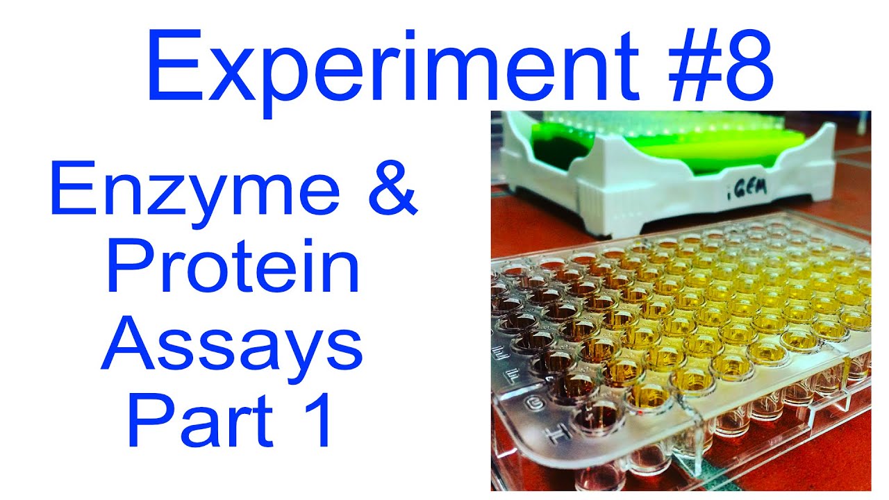
- Why this video is valuable: Provides a clear lab-bench demonstration of how to map out and systematically load enzyme lysates and controls across a 96-well microplate using multichannel pipettes.

- Why this video is valuable: Explores the underlying biochemistry of colorimetric p-nitrophenyl assays. It details how the enzyme hydrolyzes a colorless ester substrate to release p-nitrophenol (pNP), which deprotonates in alkaline buffers to form a yellow anion measurable at 405 nm.
Step-by-Step Colorimetric PETase Screening Protocol
Because PETase is structurally a cutinase/esterase, it readily cleaves soluble p-nitrophenyl esters. Below is the protocol for utilizing p-nitrophenyl butyrate (pNPB) or p-nitrophenyl laurate (pNPL) to screen your epPCR mutant library in a high-throughput fashion:
              epPCR PETase Library in E. coli BL21
                            │
           ┌────────────────┴────────────────┐
   [Active Screening]               [Thermal Screening]
           │                                 │
 Direct 96-Well Assay              Pre-Heat Plate (e.g. 50°C)
 with pNPL Substrate                      for 30 Mins
           │                                 │
 Measure Absorbance (405nm)         Cool, then add pNPL Substrate
           │                                 │
           └────────────────┬────────────────┘
                            │
                  Identify Top Hits!
Step 1: Culturing and Lysis in 96-Well Deep-Well Plates
- Inoculate individual colonies of transformed E. coli BL21 (each harboring a distinct PETase epPCR variant) into a 96-deep-well plate containing of LB medium + antibiotic per well. Reserve Well A1 for WT-PETase (positive control) and Well H12 for empty vector (negative control).
- Incubate overnight at with vigorous shaking.
- Dilute cultures 1:10 into a fresh deep-well plate, grow to , and induce with IPTG at overnight.
- Harvest cells by centrifugation of the plate ( for 15 mins). Decant supernatant.
- Lyse pellets by adding of lysis buffer ( Tris-HCl, , NaCl, lysozyme, and Triton X-100) per well. Shake at for 30 minutes, freeze at , and thaw to complete physical lysis. Centrifuge to pellet cell debris, leaving clear lysate containing the soluble PETase variants.
Step 2: Running the Activity Assay
- Transfer of clear lysate from each well into a new flat-bottom, transparent 96-well microplate.
- Prepare the substrate master mix: Tris-HCl buffer () supplemented with p-nitrophenyl laurate (pNPL) (dissolved first in acetonitrile or DMSO to a final solvent concentration ).
- Dispense of substrate master mix into each well using a multichannel pipette.
- Place the microplate immediately into a plate reader and measure absorbance at in kinetic mode at for 10 minutes.
- Calculate the slope () for each well. A higher slope indicates a mutant with improved catalytic activity.
Step 3: Running the Dual-Plate Thermal Stability Challenge
To specifically find variants with improved thermal stability, run a parallel plate assay:
- Prepare a duplicate 96-well plate containing of the same lysates.
- Seal the plate and incubate it in a thermal cycler or dry bath at an elevated "challenge" temperature (e.g., , at which WT-PETase rapidly inactivates) for 30 minutes.
- Cool the plate back down to room temperature.
- Add of the pNPL substrate master mix.
- Measure residual activity () at in the plate reader.
- Compare activity of the challenged plate vs. the unchallenged plate. Variants that retain high levels of activity after the heat challenge are selected as hits with improved thermal stability!
Knowledge Checkpoint
- Explain why solid-state PET cannot be used directly inside a microplate reader for high-throughput enzyme screening.
- Write out the chemical reaction that occurs when a PETase mutant processes p-nitrophenyl laurate (pNPL) and state what wavelength is used to measure the product.
- In a thermal stability screening campaign, why must the cell lysates be returned to room temperature before adding the proxy substrate?
Module 5: Recombinant Protein Purification
After identifying your top mutant hits via screening, you must isolate and purify them to characterize their improved properties accurately. This module teaches cell lysis methodologies and the step-by-step procedure of Immobilized Metal Affinity Chromatography (IMAC) using polyhistidine (His-tag) separation, followed by purity validation using SDS-PAGE.
Recommended Videos

- Why this video is valuable: Reviews physical and chemical cell lysis methodologies (grinding, high pressure, lysozyme treatment, and sonication), helping you choose the best extraction method to harvest stable target proteins.

- Why this video is valuable: Clear, animated guide to His-tagged affinity chromatography. It explains the molecular mechanics of histidine coordinate bonds with matrix-bound Nickel ions (-NTA) and how imidazole is used to elute the purified target protein.

- Why this video is valuable: Provides a detailed, bench-level tutorial of the real-world setup, equilibration, sample loading, washing, and elution phases of gravity-flow chromatography.

- Why this video is valuable: A thorough, step-by-step visual demonstration of gel preparation, sample loading, running, and staining of SDS-PAGE to evaluate protein purity.
Knowledge Checkpoint
- Explain how a sequence of 6 to 8 consecutive histidines (His-tag) fused to the N- or C-terminus of your PETase enzyme allows it to bind specifically to a -charged IMAC resin.
- Why is a low concentration of imidazole (e.g., ) typically included in the binding/wash buffer, and why is a high concentration (e.g., ) used for elution?
- Draw the layout of an SDS-PAGE gel verifying your purification process, indicating where you expect to find the molecular weight marker, lysate, flow-through, wash, and eluted target protein.
Module 6: Enzyme Kinetics and Thermal Stability Characterization
In this final module, you will analyze your purified PETase mutants quantitatively. You will perform Michaelis-Menten kinetic assays to calculate key catalytic parameters ( and ) and measure physical thermal denaturation () using Differential Scanning Fluorimetry (DSF) to confirm stability improvements.
Recommended Videos

- Why this video is valuable: Establishes the relationship between substrate concentration and enzymatic reaction velocity, defining critical parameters like and and laying the math foundation for your kinetic assays.

- Why this video is valuable: Delivers an advanced explanation of the turn-over number () and the specificity constant (), showing you how to prove whether your engineered PETase is actually more efficient.
- Why this video is valuable: Walks step-by-step through calculating and values using the double-reciprocal Lineweaver-Burk plot transformation ( vs. ).

- Why this video is valuable: Explains the molecular mechanism of Differential Scanning Fluorimetry (DSF, also known as thermal shift assay). It details how hydrophobic dyes like Sypro Orange bind to hydrophobic regions exposed during denaturation, allowing you to measure the melting temperature () of your PETase variants.
Knowledge Checkpoint
- Define and in terms of enzyme-substrate interaction and reaction rate.
- Describe the linear relationship of a Lineweaver-Burk plot, identifying where and are represented on the x and y axes.
- Explain how a higher melting temperature () measured via DSF directly relates to improved structural stability in your evolved PETase variants.
Course Map
This flowchart shows the critical path and prerequisites required to navigate your PETase directed evolution campaign successfully.
Key People Index
- Dr. Frances Arnold (Caltech): Recipient of the 2018 Nobel Prize in Chemistry for her pioneering work in directed evolution. She demonstrated that biological processes can be harnessed in laboratory environments to generate enzymes with customized, non-natural, or highly optimized activities.
- Kary Mullis: Inventor of the Polymerase Chain Reaction (PCR) technique (Nobel Prize in Chemistry 1993), which forms the bedrock of mutant library generation through error-prone PCR modifications.
Final Self-Assessment
Test your comprehension across the entire curriculum by verifying your ability to explain or execute each item in this self-assessment:
- Explain the active site differences between wild-type I. sakaiensis PETase and engineered thermotolerant variants.
- Diagram the molecular components of a pET-derived expression vector (T7 promoter, lac operator, ribosome binding site, multiple cloning site).
- Define the specific reaction conditions of epPCR that decrease DNA polymerase proofreading.
- Design a 96-well plate layout including proper positive (WT-PETase), negative (empty vector), and background controls.
- Explain why p-nitrophenyl esters function as successful proxies for PET cleavage kinetics.
- Describe the physical steps of purifying a His-tagged enzyme via gravity-flow chromatography.
- Calculate the specific activity of a purified enzyme sample given absorbance readings, extinction coefficient, pathlength, and protein concentration.
- Sketch a Michaelis-Menten plot and demonstrate how to derive its Lineweaver-Burk reciprocal transformation.
- Interpret a Differential Scanning Fluorimetry (DSF) melt curve to identify the inflection point representing the enzyme's melting temperature ().
- Formulate a complete directed evolution loop to explain how you would iterate on a first-generation "hit" to achieve even higher thermal stability.