Building mRNA-LNPs: From DNA to Expression
Course Overview & Learning Objectives
Learning Goal: Design, prepare, and verify a functional mRNA-lipid nanoparticle (LNP) delivery system. This curriculum covers the complete pipeline: in silico sequence design, wet-lab plasmid preparation and linearization, in vitro transcription (IVT) with co-transcriptional capping, microfluidic encapsulation using controlled fluid dynamics, physical characterization of the resulting nanoparticles, and in vitro expression verification in mammalian cells.
- Prerequisites: Basic college-level molecular biology and general chemistry.
- Estimated Study Time: 32 Hours (including practical exercises, computational design sessions, and recommended reading).
Module 1: Foundations of Molecular Biology & mRNA Therapeutics
This module establishes the core biological framework of mRNA therapeutics. You will review the flow of genetic information (the Central Dogma), analyze how synthetic mRNA functions as a transient therapeutic blueprint inside eukaryotic cells, and study the critical physiological barriers that require lipid nanoparticle packaging.
Recommended Videos
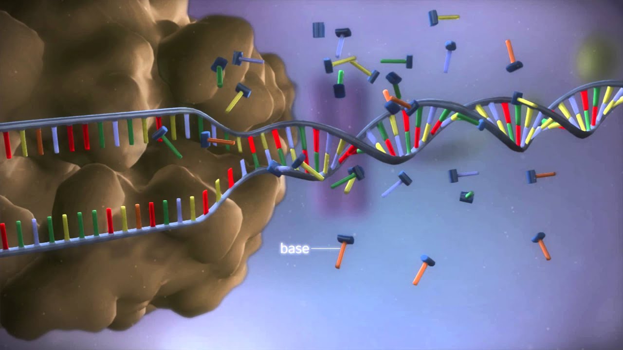
Why this video
This 3D molecular animation provides a spatial, molecular-scale visualization of transcription and translation. Observing RNA polymerase build an RNA strand and watching ribosomal subunits decode mRNA codons in real-time establishes a concrete mental model of cellular protein expression machinery.
Knowledge Checkpoint
- Understand the spatial orientation and movement of RNA polymerase along the DNA template strand.
- Identify the structural transition of mRNA as it leaves the nucleus and enters the ribosomal complex.
- Explain how tRNA molecules align amino acids based on mRNA codon sequences during translation elongation.

Why this video
This video transitions from basic biology to the medical application of synthetic transcripts. It details how exogenously delivered mRNA bypasses genomic integration, utilizing host ribosomes to produce target therapeutic proteins while addressing the inherent instability and transient nature of RNA molecules.
Knowledge Checkpoint
- Differentiate between traditional protein replacement therapies and mRNA-based intracellular translation.
- Explain the clinical safety advantages of mRNA's temporary expression profile and its inability to integrate into host genomic DNA.
- List the primary extracellular barriers (e.g., serum nucleases) that degrade unprotected synthetic mRNA.

Why this video
This video explains how host cellular machinery processes synthetic genetic instructions to produce therapeutic proteins or antigens, demonstrating how the immune system subsequently recognizes these self-translated proteins.
Knowledge Checkpoint
- Describe the process of antigen presentation following host cell translation of synthetic mRNA.
- Explain how delivering genetic instructions differs from introducing pre-assembled recombinant proteins.
Module 2: DNA Template Design and Preparation
An mRNA transcript is only as good as its DNA template. In this module, you will learn to perform computational in silico design of plasmid vectors, execute large-scale plasmid purification (Maxiprep), and perform restriction digestions to linearize your circular DNA plasmid. Linearization is critical: it prevents the RNA polymerase from transcribing indefinitely around the plasmid circle, ensuring a clean, uniform length for your mRNA transcripts.
Recommended Videos
![[LIVE] Writing DNA Code! | Learn Real Genetic Engineering - Part 2](https://i.ytimg.com/vi/1ThNnuSuZC0/hqdefault.jpg?v=63dfe3c3)
Why this video
This video provides a practical, screencast-based walk-through of Benchling vector design software. It is highly valuable for mastering sequence assembly, identifying restriction enzyme recognition sites for linearization, performing organism-specific codon optimization, and organizing structural components (such as promoters and UTRs) in a digital environment.
Knowledge Checkpoint
- Import and map a plasmid vector within Benchling or equivalent molecular suite software.
- Locate the exact T7 promoter sequence and annotate the optimal restriction site directly downstream of the poly(A) tail.
- Perform a simulated back-translation of a protein sequence, applying host-specific codon bias optimization.

Why this video
This step-by-step wet-lab guide demonstrates the isolation of transfection-grade, endotoxin-free plasmid DNA from high-volume bacterial cultures. Large-scale mRNA production requires highly pure, high-concentration plasmid DNA templates free of bacterial endotoxins, which can compromise downstream cell viability.
Knowledge Checkpoint
- Explain the biochemical basis of alkaline lysis and how circular plasmid DNA is separated from genomic bacterial DNA.
- Describe the purpose of using anion-exchange chromatography columns in Maxiprep kits.
- State why removing endotoxins during plasmid purification is critical for downstream cell expression assays.

Why this video
This video demonstrates the practical bench-level technique of setting up a restriction enzyme digest. You will learn the exact sequence of adding sterile water, reaction buffers, plasmid DNA, and restriction enzymes to a reaction vessel, which is critical for linearizing the circular plasmid template.
Knowledge Checkpoint
- Calculate the required volume of reaction components (DNA template, enzyme, 10X reaction buffer, and water) for a standard digestion reaction.
- State the correct order of addition for enzyme digestion components to maintain enzyme activity and stability.
- Explain why standard molecular biology protocols recommend incubating digestions at exactly 37°C.
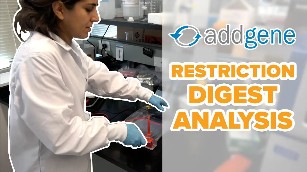
Why this video
After completing a restriction digest, you must verify that the plasmid is completely linearized rather than partially digested or circular. This video demonstrates how to run and analyze an agarose gel, matching expected migration patterns of linearized versus supercoiled plasmid bands.
Knowledge Checkpoint
- Predict the visual migration difference on an agarose gel between a circular supercoiled plasmid, a nicked circular plasmid, and a completely linearized plasmid.
- Diagnose partial digestion issues on an agarose gel by identifying unexpected bands.
- Use a DNA ladder to mathematically estimate the concentration and base-pair size of your linearized DNA template.
Independent Search Suggestion: To complement this module's computational aspect, independently search for
"Benchling plasmid mapping tutorial for mRNA synthesis"to find specific guides on adding synthetic 5' and 3' Untranslated Regions (UTRs) to your templates.
Module 3: In Vitro Transcription (IVT) and mRNA Synthesis
With a pure, linearized DNA template prepared, you are ready to synthesize mRNA. In this module, you will master the biochemical reaction of In Vitro Transcription (IVT) using T7 RNA Polymerase. You will study co-transcriptional capping technologies, learn how to prevent immunogenicity using modified nucleotides, and master column-based purification protocols to clean your synthesized mRNA.
Recommended Videos

Why this video
This video provides a clear, high-level workflow summary of the enzymatic transition from a linearized plasmid template to highly pure, translatable mRNA. It connects the biological principles of T7 RNA polymerase activity with the practical steps of DNA template removal using DNase I.
Knowledge Checkpoint
- Explain why the DNA template must be digested with DNase I post-reaction rather than simply physical filtration.
- Identify the role of NTPs (Nucleoside Triphosphates) as both building blocks and energy sources in the IVT system.
Why this video
This video explains the molecular structure and chemical biology of the 5' Cap. It compares legacy enzymatic capping methods with modern, highly efficient co-transcriptional trinucleotide capping technologies (specifically CleanCap), which yield highly translatable Cap 1 structures directly during the IVT reaction.
Knowledge Checkpoint
- Describe the chemical structural difference between a standard Cap 0 structure and a modern Cap 1 structure.
- Explain how co-transcriptional capping reagents reduce manufacturing steps compared to enzymatic capping workflows.
- State how a 5' Cap structure prevents premature exonuclease degradation inside mammalian target cells.
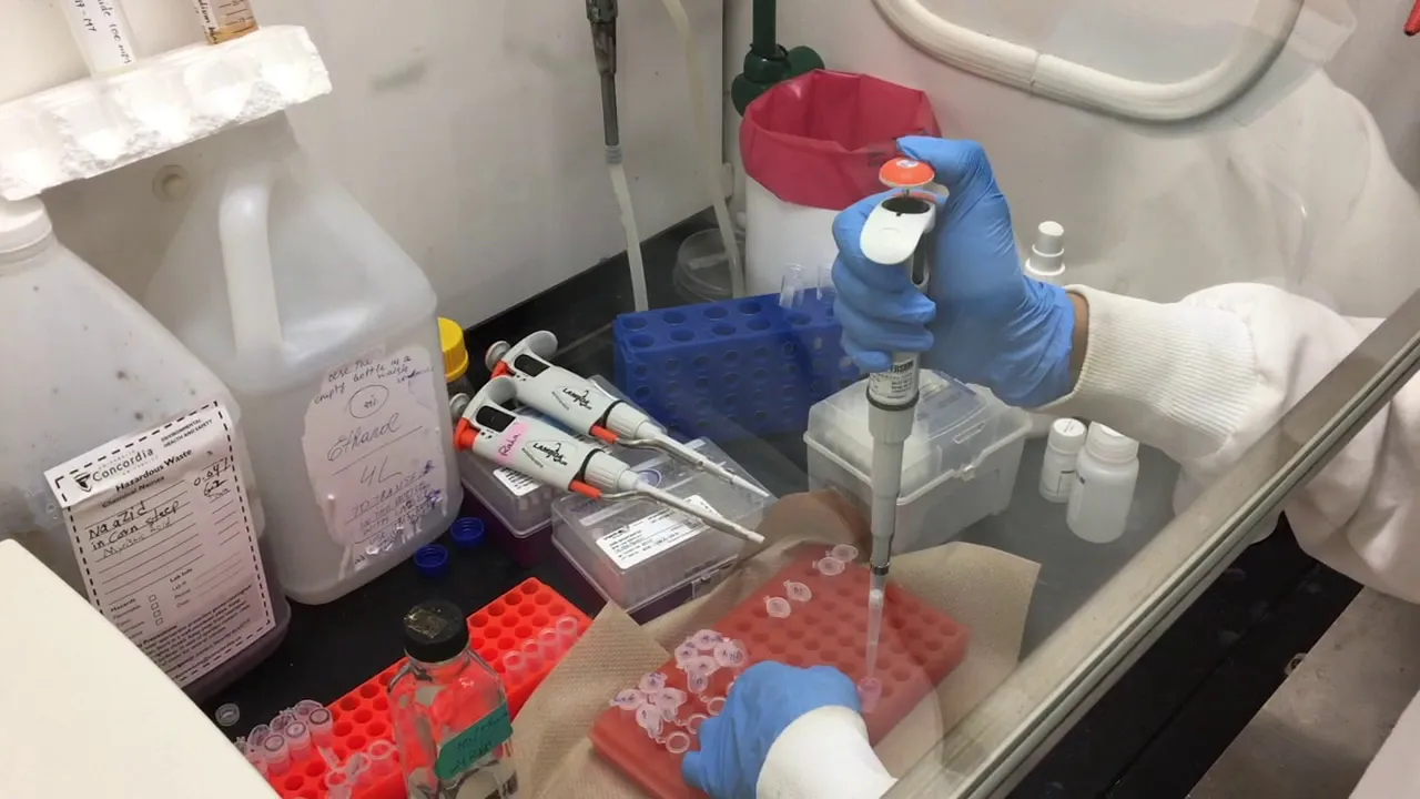
Why this video
Following an IVT reaction, the synthesized mRNA must be isolated from proteins, salts, and unincorporated nucleotides. This video demonstrates the practical steps of column-based RNA purification, showing how ethanol addition modifies binding kinetics to enable selective capture of RNA on a silica matrix.
Knowledge Checkpoint
- State why adding a precise volume of ethanol is required to bind your synthesized RNA to the silica membrane of a spin column.
- Describe the mechanistic purpose of the wash steps and how to ensure complete ethanol removal before final elution.
- Explain why nuclease-free water (DEPC-treated or equivalent) must be used for final elution of the therapeutic mRNA transcript.
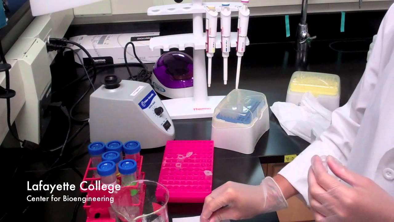
Why this video
This micro-demonstration shows the physical manipulation, correct pipetting, and centrifugation techniques required for silica-column RNA purification kits. It ensures proper bench handling of RNA to prevent common physical contamination or loss.
Knowledge Checkpoint
- Demonstrate correct positioning and handling of spin columns in microcentrifuges to avoid cross-contamination of flow-through.
- Define the centrifugation parameters typically utilized for binding, washing, and eluting RNA from silica membranes.
Practical Gap Alert: Standard IVT reactions must substitute standard UTP with modified nucleotides like N1-methylpseudouridine () to prevent recognition by mammalian toll-like receptors (TLRs). For a complete recipe, search:
"N1-methylpseudouridine IVT reaction setup protocol".
Module 4: Lipid Nanoparticles (LNPs): Components & Chemistry
Therapeutic mRNA is a large, highly anionic (negatively charged) molecule that cannot cross cell membranes on its own. This module introduces the four-component lipid nanoparticle (LNP) delivery system. You will explore the physical chemistry of ionizable lipids, helper lipids, cholesterol, and PEGylated lipids, and learn how their precise stoichiometric ratios dictate physical self-assembly.
                ┌─────────────────────────┐
                │  4-Component LNP Mix    │
                │                         │
                │  • Ionizable Lipid (50%)│
                │  • Helper DSPC (10%)     │
                │  • Cholesterol (38.5%)   │
                │  • PEG-Lipid (1.5%)     │
                └────────────┬────────────┘
                             │
                             ▼
                 pH drops below lipid pKa
            (e.g., pH 4.0 during mixing)
                             │
                             ▼
                ┌─────────────────────────┐
                │ Protonated, Cationic    │
                │ Lipids Electrostatically│
                │ Complex with Anionic    │
                │ Phosphate Backbone of   │
                │ mRNA to form Core       │
                └─────────────────────────┘
Recommended Videos
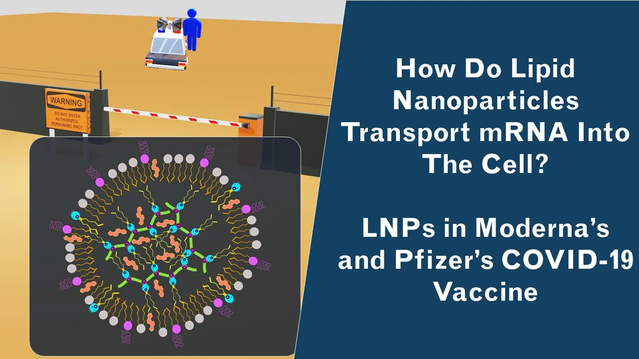
Why this video
This video explains the structural orientation and function of the four key lipid elements. It clearly illustrates how the core of the nanoparticle is packed with ionizable lipids complexed with mRNA, while helper lipids, cholesterol, and PEG-lipids stabilize the outer shell.
Knowledge Checkpoint
- Name the four essential components of a therapeutic lipid nanoparticle formulation.
- Explain how PEGylated lipids control nanoparticle size and prevent agglomeration during storage.
- Describe the mechanical role of cholesterol in stabilizing the hydrophobic domain of the LNP membrane.

Why this video
This short demonstration details the pH-dependent charge transition of ionizable lipids. It illustrates how these molecules transition from a positive charge at acidic pH (to complex with anionic RNA) to a neutral charge at physiological pH (reducing toxicity and enabling intracellular release).
Knowledge Checkpoint
- Define the chemical state of an ionizable lipid at an acidic pH (pH < 4.0) versus a physiological pH (pH 7.4).
- Explain how a neutral surface charge at pH 7.4 minimizes clearance by cellular macrophage systems in the blood.
- Detail how endosomal acidification (pH drop inside the cell) triggers the endosomal escape of the mRNA cargo.

Why this video
This video covers LNP pharmacology and physical properties, demonstrating how modulating physical parameters affects targeted delivery to specific organs and cell types.
Knowledge Checkpoint
- Explain how changing the physical composition of an LNP affects its target tissue specificity.
- Describe the path of an LNP from systemic injection to intracellular uptake.
Stoichiometry Reference Guide: A standard, globally validated molecular formulation is:
- Ionizable Cationic Lipid (e.g., ALC-0315, MC3):
- Helper Phospholipid (e.g., DSPC):
- Cholesterol:
- PEG-Lipid (e.g., ALC-0159):
Always calculate your lipid stock preparation based on these molar ratios relative to the total Nitrogen-to-Phosphate (N/P) ratio of the target mRNA (standard N/P target is typically 6:1).
Module 5: Microfluidic Encapsulation and Formulation
LNP self-assembly is dynamic and depends heavily on fluidic mixing. This module covers the physics of microfluidic mixing. You will learn to optimize fluid parameters like Flow Rate Ratio (FRR) and Total Flow Rate (TFR), study the design of herringbone micromixers, and master Tangential Flow Filtration (TFF) to concentrate and purify your formulated particles.
                ┌──────────────────────────┐
                │      Aqueous Phase       │
                │ (mRNA in Acidic Buffer,  │
                │        pH ~4.0)          │
                └────────────┬─────────────┘
                             │
                             ├──────────► Microfluidic Herringbone Mixer
                             │            (Precise FRR/TFR controls mixing)
                ┌────────────┴─────────────┐
                │      Organic Phase       │
                │ (4-component Lipids in   │
                │        Ethanol)          │
                └──────────────────────────┘
Recommended Videos

Why this video
This webinar is a masterclass in LNP formulation engineering. It demonstrates how to operate microfluidic hardware, control flow rates, and establish correct Flow Rate Ratios (typically 3:1 aqueous to organic) to consistently generate monodisperse nanoparticles.
Knowledge Checkpoint
- Define Flow Rate Ratio (FRR) and state why a 3:1 (Aqueous:Organic) ratio is commonly utilized.
- Explain how increasing the Total Flow Rate (TFR) affects nanoparticle size and polydispersity index (PDI).
- Describe the transition that occurs when lipids dissolved in solvent (ethanol) meet mRNA dissolved in an acidic aqueous buffer.

Why this video
To achieve uniform self-assembly, fluids must mix via chaotic advection rather than slow laminar diffusion. This video demonstrates the design of a staggered herringbone microfluidic mixer, showing how the channel geometry forces fluid streams to fold over themselves to rapidly encapsulate the cargo.
Knowledge Checkpoint
- Explain how staggered herringbone grooves induce chaotic advection in a microfluidic channel.
- State why laminar flow (diffusion-only mixing) results in larger, highly polydisperse, and non-functional nanoparticles.
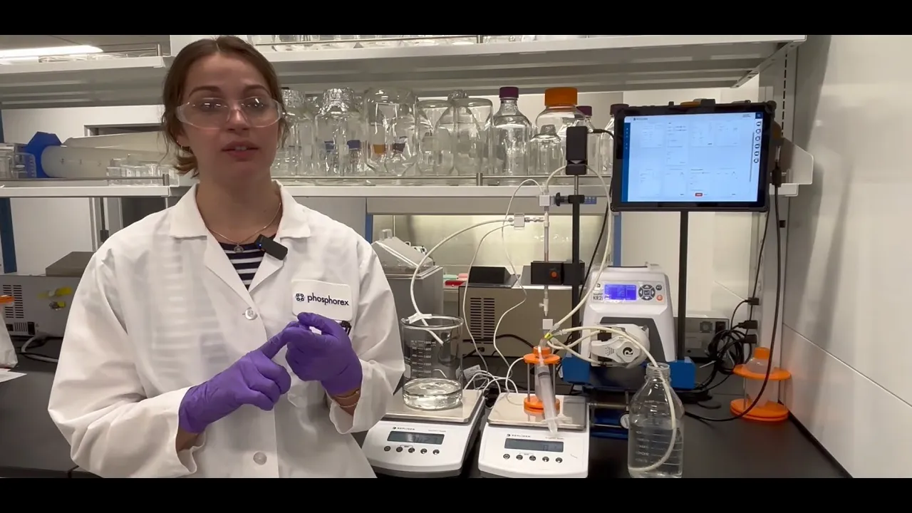
Why this video
After microfluidic mixing, your LNPs reside in a toxic mixture of ethanol and acidic buffer. This practical video demonstrates how Tangential Flow Filtration (TFF) systems use hollow fiber membranes to exchange this solvent for a storage buffer (dialysis) and concentrate the nanoparticles without damaging them.
Knowledge Checkpoint
- Explain why Tangential Flow (cross-flow) filtration prevents membrane clogging (fouling) compared to normal flow (dead-end) filtration.
- Describe the process of diafiltration and how it removes toxic solvents (such as ethanol) from the formulation.
- Monitor inlet, retentate, and permeate pressures to prevent particle shearing and membrane rupture during filtration.
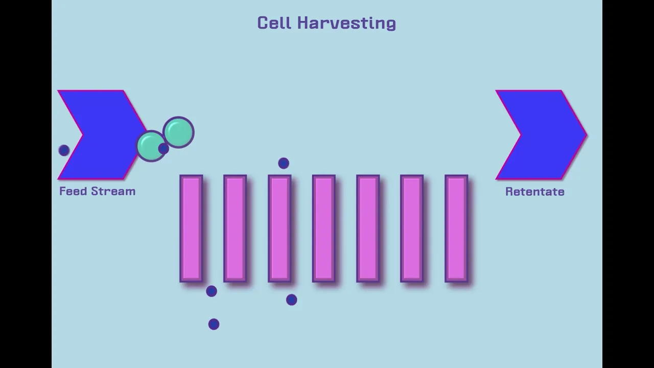
Why this video
This video explains the mechanics of TFF. It clarifies concepts like Molecular Weight Cut-Off (MWCO), shear rates, and the fluid dynamics that occur inside hollow fiber filters during concentration and buffer exchange.
Knowledge Checkpoint
- Select the correct Molecular Weight Cut-Off (MWCO) membrane for a nanoparticle of approximately 80-100 nm.
- Explain how filtrate flux and trans-membrane pressure (TMP) are calculated and maintained.
Module 6: LNP Characterization & Expression Verification
Before testing your LNPs in vitro, you must characterize their physical and biological properties. In this module, you will learn to measure particle size and polydispersity using Dynamic Light Scattering (DLS), calculate encapsulation efficiency using the RiboGreen dye assay, and transfect mammalian cell cultures to verify protein expression via reporter assays.
                ┌──────────────────────────┐
                │    Freshly Purified      │
                │    LNP Formulation       │
                └────────────┬─────────────┘
                             │
     ┌───────────────────────┼───────────────────────┐
     ▼                       ▼                       ▼
┌──────────────┐ ┌──────────────┐ ┌──────────────┐ │ Dynamic Light│ │ RiboGreen │ │ Cell Culture │ │ Scattering │ │ Assay (EE%) │ │ Transfection │ │ (Size & PDI) │ │ │ │ & Reporter │ └──────────────┘ └──────────────┘ └──────────────┘
Recommended Videos
Why this video
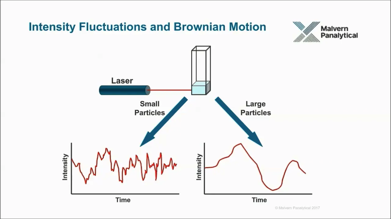
This deep-dive technical video explains the theory and practice of Dynamic Light Scattering (DLS). It describes how DLS measures the Brownian motion of particles to calculate the mean hydrodynamic diameter and Polydispersity Index (PDI), which are critical quality control metrics for your formulations.
Knowledge Checkpoint
- Define the relation between temperature, viscosity, Brownian motion velocity, and the light scattering signal.
- Interpret a DLS size distribution graph, identifying monodisperse versus polydisperse or aggregated populations.
- Define an acceptable Polydispersity Index (PDI) limit for clinical-grade LNP delivery systems (typically < 0.2).
Why this video
This standard protocol webinar explains how to calculate Encapsulation Efficiency (EE%). You will learn how to measure unencapsulated RNA directly, lyse the nanoparticles using Triton X-100 to release and measure the total RNA payload, and calculate the final encapsulation ratio.
Knowledge Checkpoint
- Prepare standard curves of RNA dilutions to enable quantitative concentration calculations.
- Explain why Triton X-100 is used to break down the lipid outer shell of the nanoparticles during the assay.
- Apply the mathematical formula to calculate the final encapsulation efficiency percentage based on raw fluorescence values.
Why this video
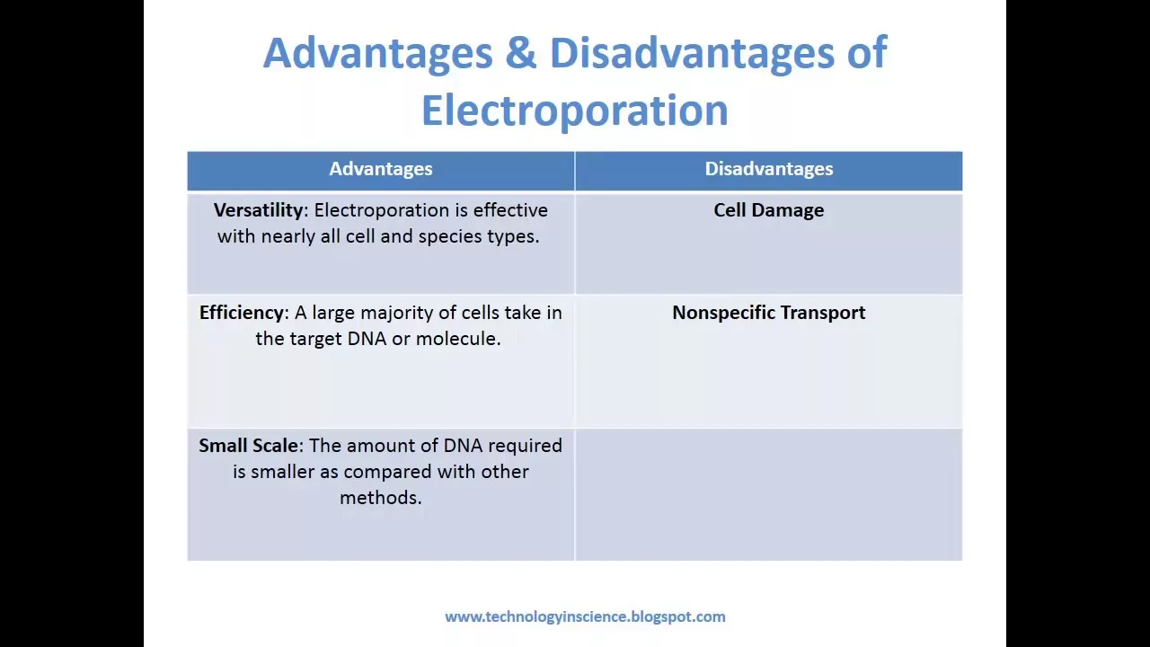
This video explains mammalian cell transfection. It outlines how cellular uptake occurs, details optimization strategies for seeding density and dose, and reviews transfection troubleshooting to ensure cellular survival and high translation rates.
Knowledge Checkpoint
- Explain the cellular pathway of endocytosis through which cells uptake foreign nanoparticles.
- Differentiate between transient transfection and stable genomic transfection.
- List the primary factors (e.g., cell confluence, media composition, cytotoxicity) that must be optimized for robust in vitro expression.
Why this video
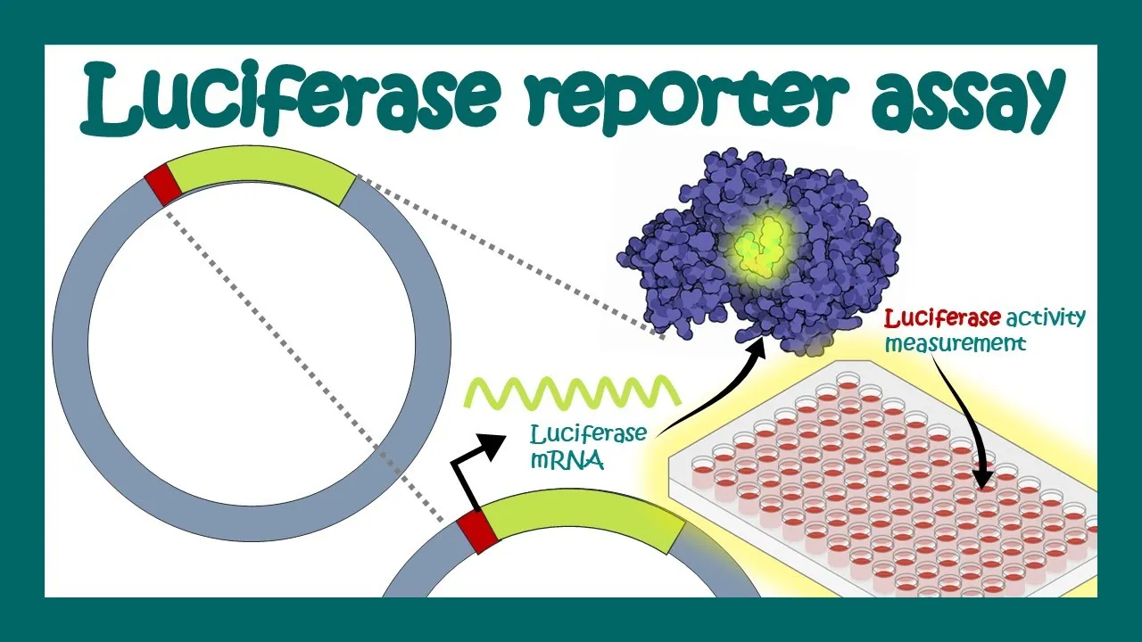
This video explains how to quantify translation. By transcribing mRNA that encodes the Firefly Luciferase reporter enzyme, you can introduce its substrate (luciferin) to transfected cells and measure the resulting light output, which is directly proportional to translation efficiency.
Knowledge Checkpoint
- Describe the enzymatic reaction where luciferase converts substrate luciferin to oxyluciferin, emitting light.
- Explain how relative light units (RLUs) measured on a luminometer correspond to the physical quantity of translated protein.
- List the essential cell lysis steps required prior to running a reporter assay.
Course Map
This map outlines the recommended learning order and dependency structure for the curriculum.
Key People Index
The researchers listed below are key figures in the history of mRNA therapeutics and LNP drug delivery:
- Dr. Katalin Karikó & Dr. Drew Weissman: Pioneered the use of modified nucleosides (such as pseudouridine) to prevent synthetic mRNA from triggering inflammatory toll-like receptors (TLRs). This discovery enabled the development of viable mRNA therapeutics.
- Dr. Pieter Cullis: Led the development of ionizable lipid nanoparticles. His work on pH-dependent self-assembling lipids resolved early cargo delivery and toxicity issues, enabling modern LNP delivery systems.
- Dr. Robert Malone: Published early papers in 1989 demonstrating that mRNA wrapped in cationic liposomes could transfect mammalian cells, establishing the concept of lipid-mediated RNA delivery.
Final Self-Assessment
Complete this comprehensive self-assessment to verify your mastery of the complete DNA-to-LNP expression pipeline.
- Plasmid Verification: I can analyze a restriction digestion gel and confirm that my plasmid template is completely linearized with no supercoiled or circular DNA remaining.
- Sequence Layout: I can design a clean transcription unit in Benchling, positioning the T7 promoter, 5' UTR, open reading frame, 3' UTR, and Poly(A) tail correctly.
- Capping Selection: I can explain the structural difference between Cap 0 and Cap 1 and identify the correct reagents (e.g., CleanCap) to yield Cap 1 co-transcriptionally.
- N1-Methylpseudouridine Chemistry: I can explain how substituting uridine with N1-methylpseudouridine () prevents endosomal TLR receptors from detecting synthetic mRNA as a foreign pathogen.
- Lipid Formulation Ratios: I can calculate the exact mass of each lipid required to yield a standard 50:10:38.5:1.5 molar ratio (Ionizable:DSPC:Cholesterol:PEG) based on a target N/P ratio.
- Fluidic Dynamics: I can operate a microfluidic device, adjusting flow rates to achieve a target Flow Rate Ratio (FRR) and Total Flow Rate (TFR) to control particle size.
- TFF Purification: I can explain the mechanics of Tangential Flow Filtration, monitor trans-membrane pressure (TMP), and complete a multi-volume diafiltration buffer exchange.
- Dynamic Light Scattering: I can run a DLS sizing instrument, interpret the correlation decay curve, and determine if my formulation has a monodisperse size distribution (PDI < 0.2).
- Encapsulation Efficiency: I can perform a dual-well RiboGreen assay, construct an RNA standard calibration curve, and calculate the percentage of encapsulated mRNA.
- In Vitro Validation: I can transfect cell cultures with formulated LNPs, execute a reporter cell assay, and measure expression levels using a luminometer or fluorescence microscope.